1
2
3
4
5

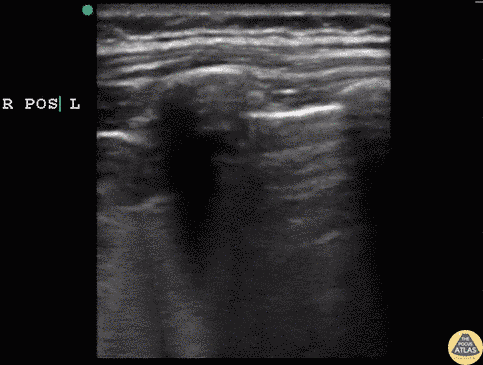
Acute Chest Syndrome
6 y/o sickle cell (HbSS) coughing with left-sided chest pain and 1 day of fever. Lungs without crackles, good air entry bilaterally.
A consolidative process is seen with a hypoechoic region with posterior enhancement greater than 1cm in an area where normal A lines should be present. This is highly suggestive of acute chest syndrome given clinical features.
Dr. Sathya Subramaniam, Pediatric EM Fellow - Kings County/SUNY Downstate
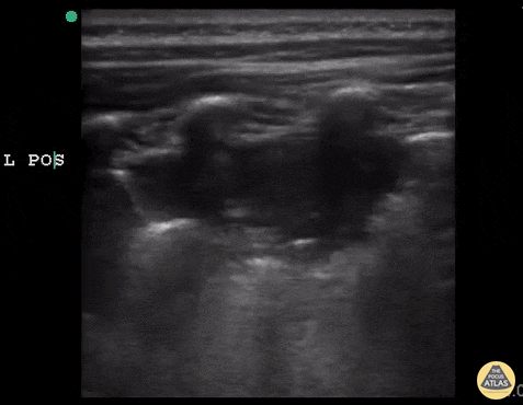
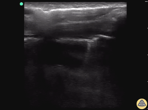
Lung Hepatization (Pneumonia)
5 year old child with sickle cell disease. Coughing and fever for 3 days. On exam not ill appearing but decreased breath sounds over right lung. POCUS completed to evaluate for pneumonia.
Hepatization of the lung clearly demonstrates consolidative process concerning for pneumonia. The beginning of the image demonstrates hepatization in the lung field. The ultrasonographer then slides the probe inferiorly over normal lung past the diaphragm to the liver, demonstrating how similar lung hepatization can appear compared to the actual liver.
Dr. Sathya Subramaniam, Pediatric EM Fellow - Kings County/SUNY Downstate
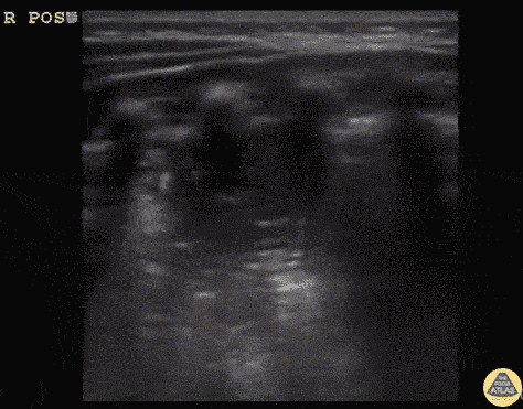
Infant Pneumonia with C-Lines
11 month old unvaccinated infant presenting with cough, fever and tachypnea starting today. Exam with crackles bilaterally in an infant with subcostal retractions and respiratory distress.
Right posterior lung with clear large consolidative process with C lines present.
Dr. Sathya Subramaniam, Pediatric EM Fellow - Kings County/SUNY Downstate
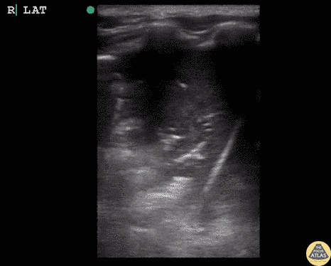
Hydrocarbon Ingestion with C-Lines
5 year old male that drank out of container with gasoline and started coughing and was breathing fast. On exam appeared tachypneic, with air entry bilaterally and subcostal retractions.
POCUS revealed bilateral infiltrates, confirmed with CXR. Infiltrate, similar to C lines seen in other consolidative processes, present in patient post hydrocarbon ingestion. This suggests an aspiration pneumonia.
Dr. Sathya Subramaniam, Pediatric EM Fellow - Kings County/SUNY Downstate
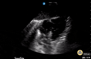
Pneumothorax with Lung Point
18 y/o M stabbed in the back presents to the trauma bay with left-sided chest pain and shortness of breath. E-FAST revealed decrease lung slide and a clear lung point.
While decreased lung slide is highly sensitive, it lacks specificity. Lung point indicates the transition point between normal pleura with normal lung sliding (on the left side of the image) and where there is air disrupting the pleural space with decreased lung sliding (on the right side of the image). Lung point is a highly specific finding indicating a pneumothorax.
Dr. Sathya Subramaniam, Pediatric EM Fellow - Kings County/SUNY Downstate