
Pediatrics
The best PEDIATRIC POCUS clips. This is free and open access medical education content. Right click to download.
Pediatrics
The best Pediatric POCUS clips. This is free and open access medical education content. Right click to download.
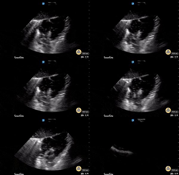
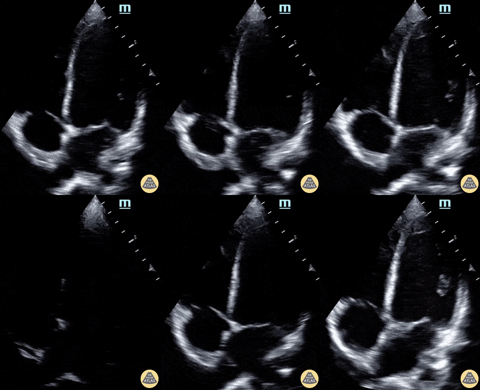
14 year-old male presents to the ED with chest pain two weeks after being having been diagnosed with COVID-19. Labs were notable for elevated CBC, CRP, ESR, and troponin. POCUS revealed moderately decreased function and LV dilation, consistent with the diagnosis of COVID-19 myocarditis.
Paul Khalil, MD and N. Akwesi Poteh, MD at University of Louisville
@khalil3paul
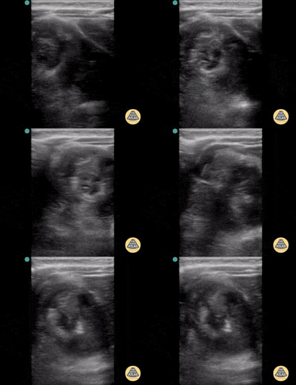
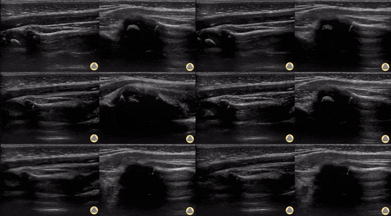
2-year-old female was brought to the ED refusing to use her left arm after her father caught her by the arm to prevent her falling down flight of stairs. POCUS was performed to confirm clinical suspicion of a Nursemaid’s elbow (or radial head subluxation). The left image reveals classic findings of a Nursemaid’s elbow including widening of the synovial fringe and inward extension of the annular ligament. Further, you can appreciate both the supinator muscle, as well as the annular ligament are being pulled into the joint space, a finding known as hook sign. The image on the right is the post-reduction image showing normal alignment and contour of the annular ligament.
Austin Meggitt, MD
Pediatric Emergency Ultrasound Fellow
Denver Health Medical Center @DenverEMed
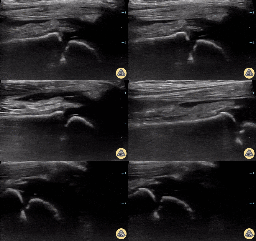
2 year-old male presented with fever, right knee swelling, and elevated CBC, CRP and ESR. POCUS revealed a right knee effusion.
Paul Khalil, MD. Assistant PEM POCUS director at University of Louisville/Norton Children’s
@khalil3paul
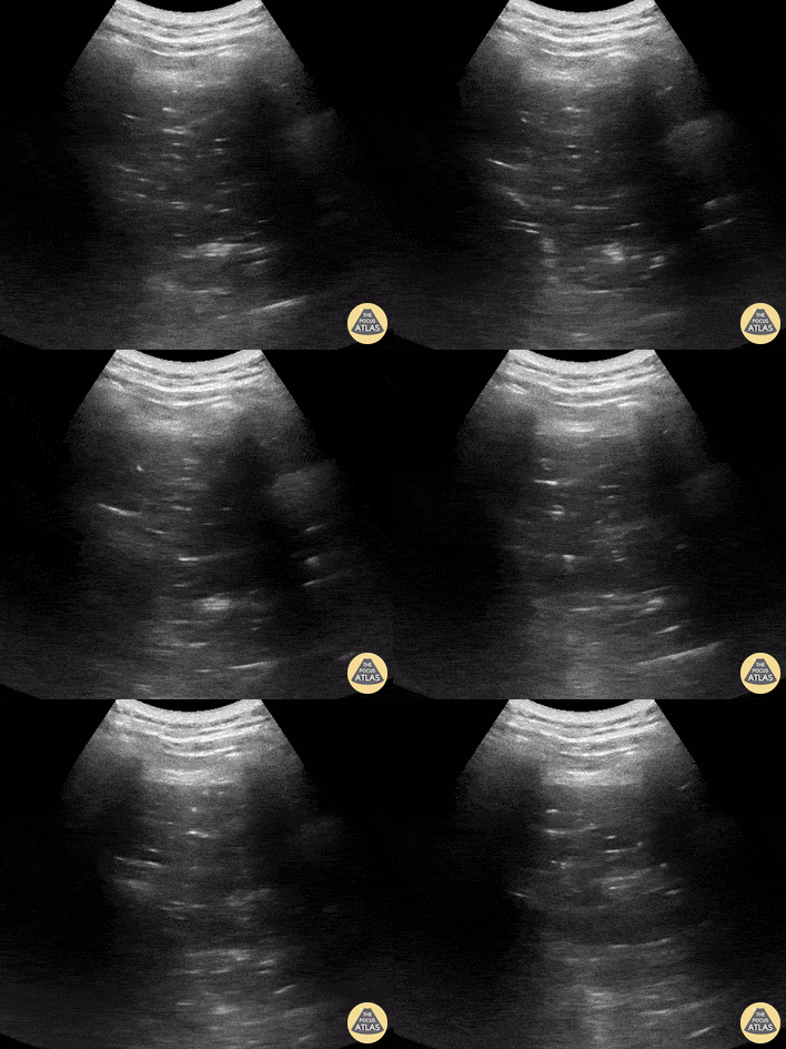
15 year-old male with cerebral palsy presents to the ED with hypoxia. Physical exam notable for left lung with decreased air movement on auscultation. POCUS demonstrates dynamic air bronchograms consistent with suspected diagnosis of pneumonia.
Paul Khalil, MD. Assistant PEM POCUS director at University of Louisville/Norton Children’s
@khalil3paul
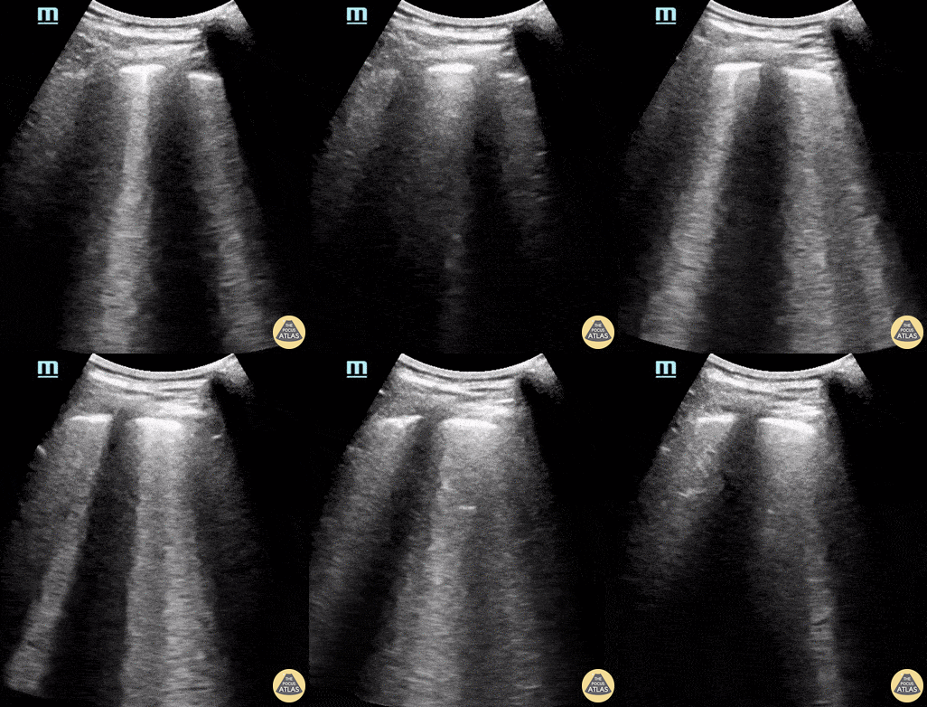
14 year-old female known to be SARS-CoV-2 positive presented with chest pain and shortness of breath. POCUS revealed findings consistent with COVID pneumonia including thickened pleura and presence of multiple B-lines.
Paul Khalil, MD. Assistant PEM POCUS director at University of Louisville/Norton Children’s
@khalil3paul
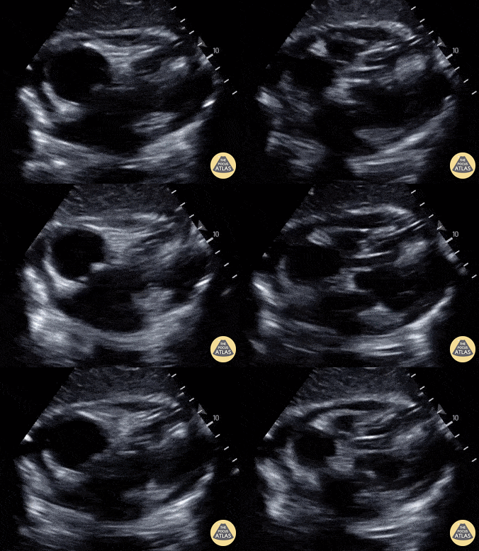
An 8-year-old male presented with a 1 day history of fever and chest pain. Cardiac POCUS shown here revealed a pericardial effusion; he was later started on steroids for the same enabling hospital discharge on hospital day #2.
Amar Singh, MD. University of Louisville
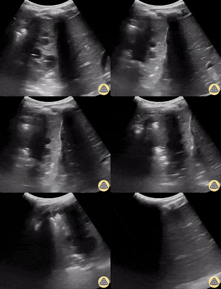
A 15-month-old male presented with cough, fever, and tachypnea of 3-days duration. POCUS revealed findings of right lung consolidation, consistent with pneumonia referred to as hepatization of the lung. Seen here territories above and below the diaphragm show ultrasonographic findings resembling liver parenchyma.
Amar Singh, MD. Pediatrics specialist in Louisville, KY

Decreased color doppler is seen in this pediatric patient with testicular torsion.
Dr. Lianne McLean (@doctorlianne) FRCPC of the PEM POCUS Group: Division of Emergency Medicine in the Hospital for Sick Children (@epocus)
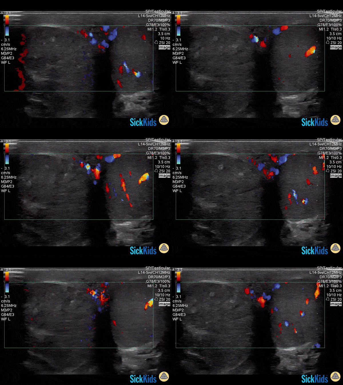
Decreased color doppler is seen in this pediatric patient with testicular torsion.
Dr. Lianne McLean (@doctorlianne) FRCPC of the PEM POCUS Group: Division of Emergency Medicine in the Hospital for Sick Children (@epocus)
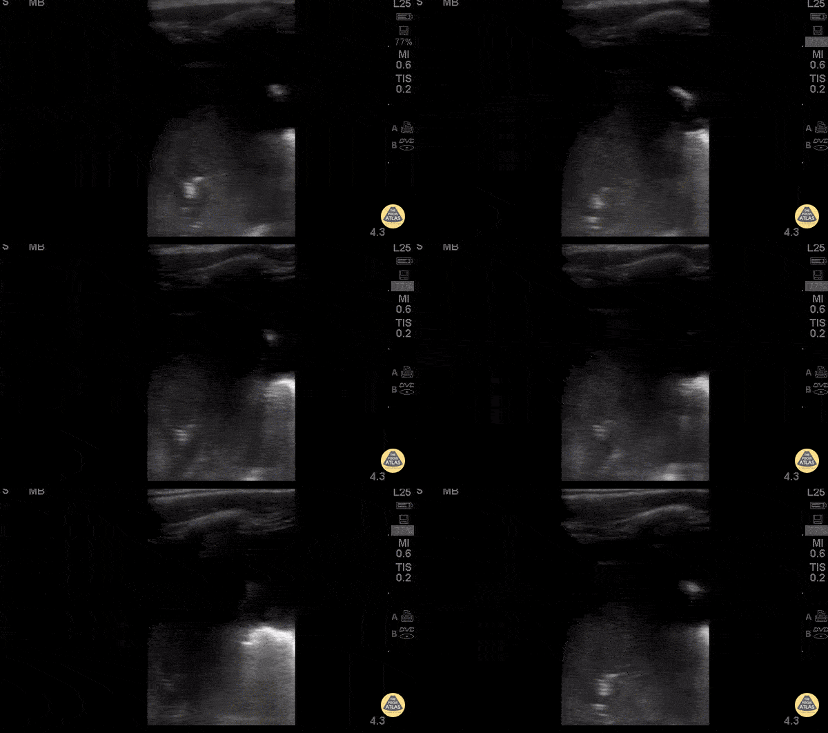
3 yo m previously healthy UTD with vaccines. 5 days of cough and fevers. 3 days of abdominal pain, acutely worsening day of presentation with 1 episode of NBNB emesis.
Febrile tachypneic and hypoxic to 91% on RA.
CXR: white out of right lung.
POCUS: large right sided effusion. Hepatization with air bronchograms on the right lateral view.
Dr. Isaac Gordon - Kings County Pediatric Emergency Medicine
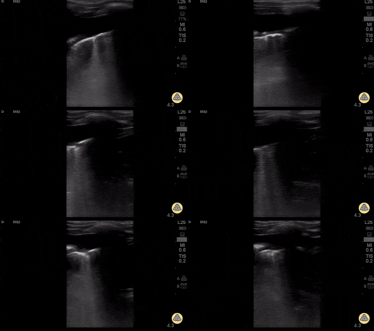
3 yo m previously healthy UTD with vaccines. 5 days of cough and fevers. 3 days of abdominal pain, acutely worsening day of presentation with 1 episode of NBNB emesis.
Febrile tachypneic and hypoxic to 91% on RA.
CXR: white out of right lung.
POCUS: large right sided effusion. Hepatization with air bronchograms on the right lateral view.
Dr. Isaac Gordon - Kings County Pediatric Emergency Medicine
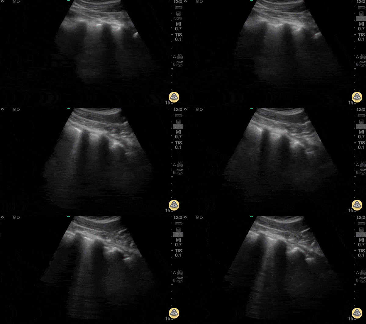
2 years old male with hx of endotracheal intubation secondary to RSV infection presents with 2 days of fever, cough, rhinorrhea and nasal congestion. Denied nausea, emesis, diarrhea, chest pain, syncope, confusion, change in eating patterns and voiding patterns. POCUS demonstrates a focal area of B lines c/w pneumonia (likely viral).
Early PNA B lines: short path reverberation artifacts create by fluid filled alveoli. In the appropriate clinical scenario B lines and pleural consolidation suggest PNA.
Dr. Carolina Camacho Ruiz - Kings County Emergency Medicine
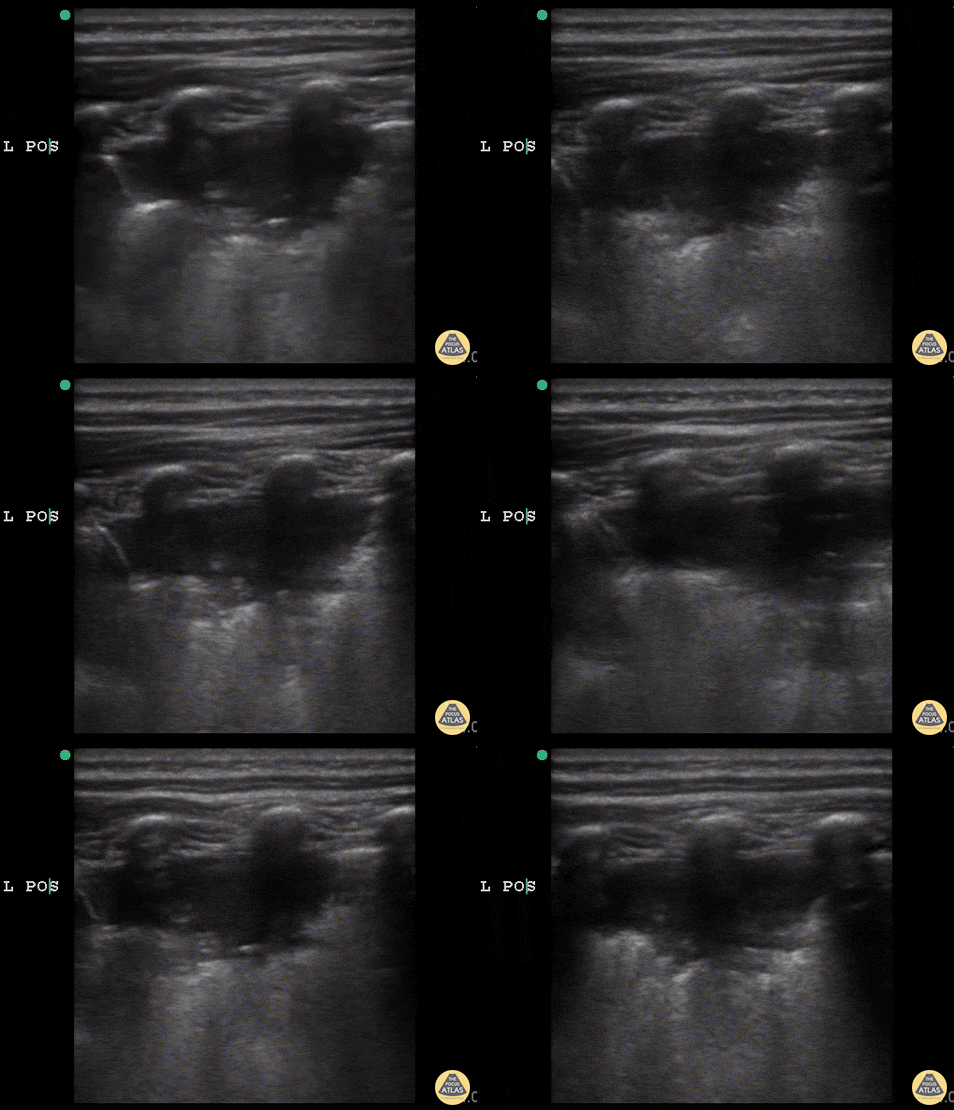
6 y/o sickle cell (HbSS) coughing with left-sided chest pain and 1 day of fever. Lungs without crackles, good air entry bilaterally.
A consolidative process is seen with a hypoechoic region with posterior enhancement greater than 1cm in an area where normal A lines should be present. This is highly suggestive of acute chest syndrome given clinical features.
Dr. Sathya Subramaniam, Pediatric EM Fellow - Kings County/SUNY Downstate
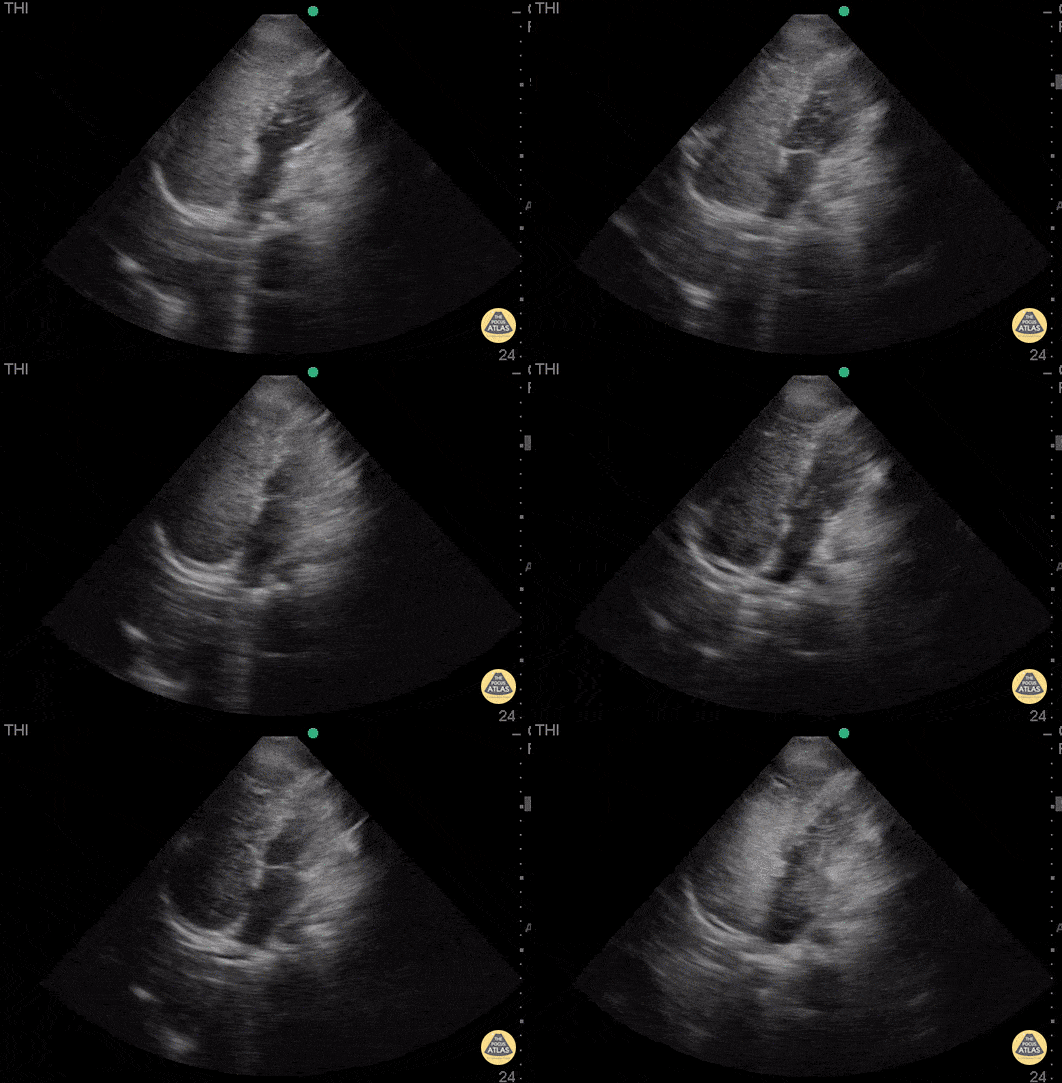
WCUME 2017 submission for "Novel Indication"
Bubble test reveals an atrial septal defect seen as bubbles floating from the right side of the heart to the left side. An obvious defect can be seen.
Dr. Mojtaba Chahardoli - Firoozgar Hospital
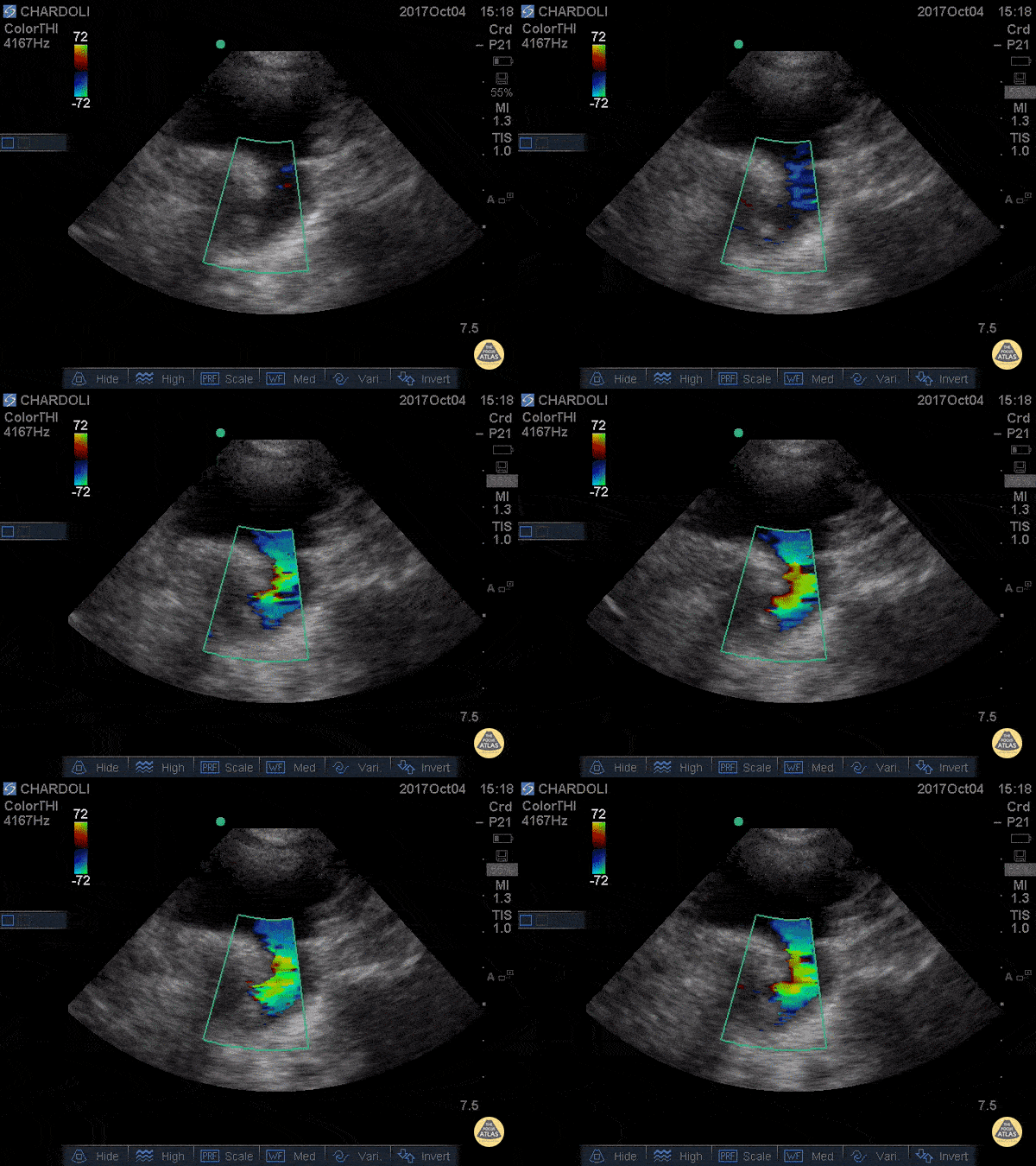
WCUME 2017 submission for "Best POCUS"
8 y/o M with undifferentiated dyspnea. POCUS reveals he has open PDA on aorta from suprasternal view.
Dr. Nima Shekar Riz Fomani - Firoozgar General Hospital
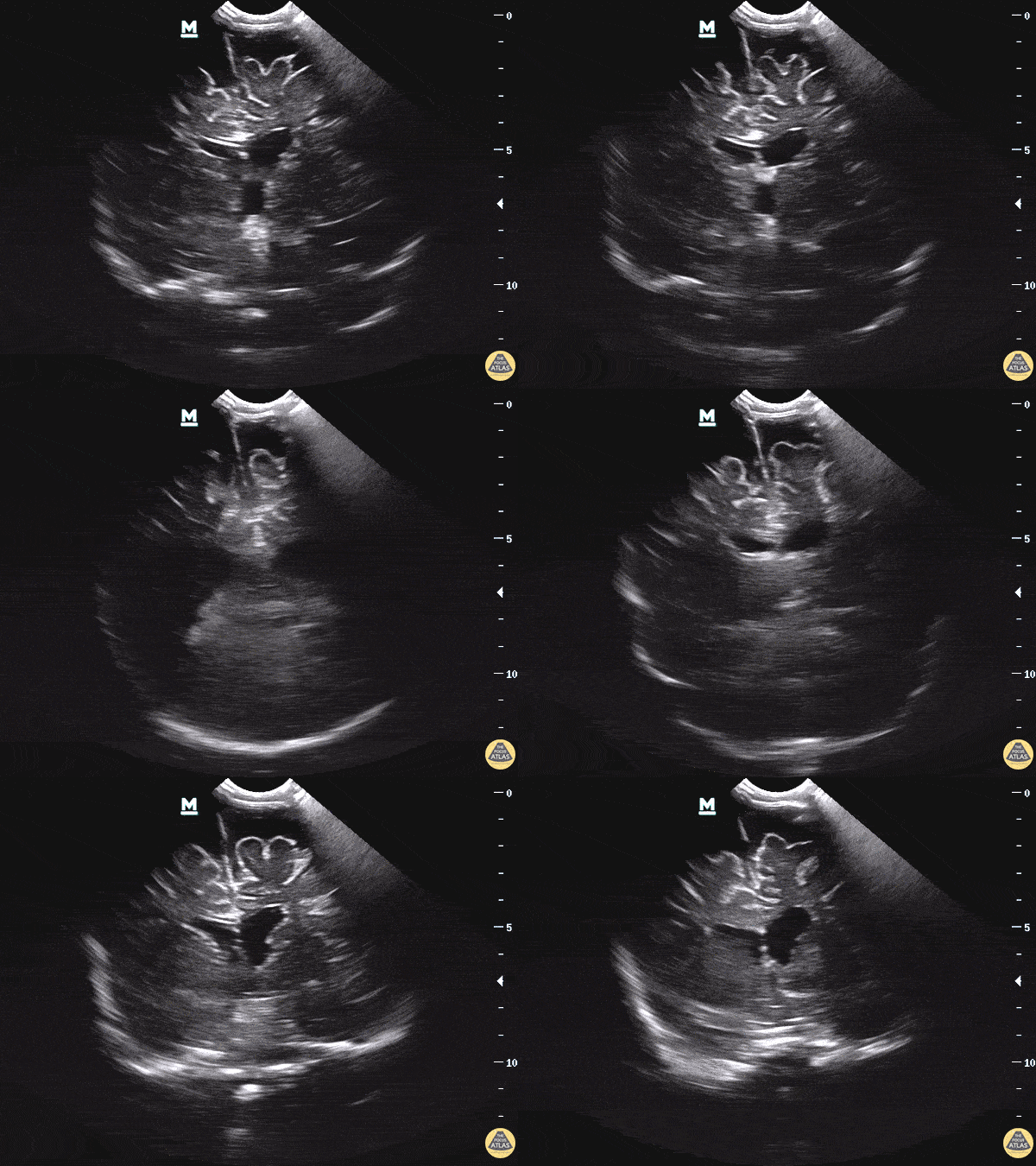
WCUME 2017 winner for "Novel Indication" - See our blog post for a deep dive on this topic!
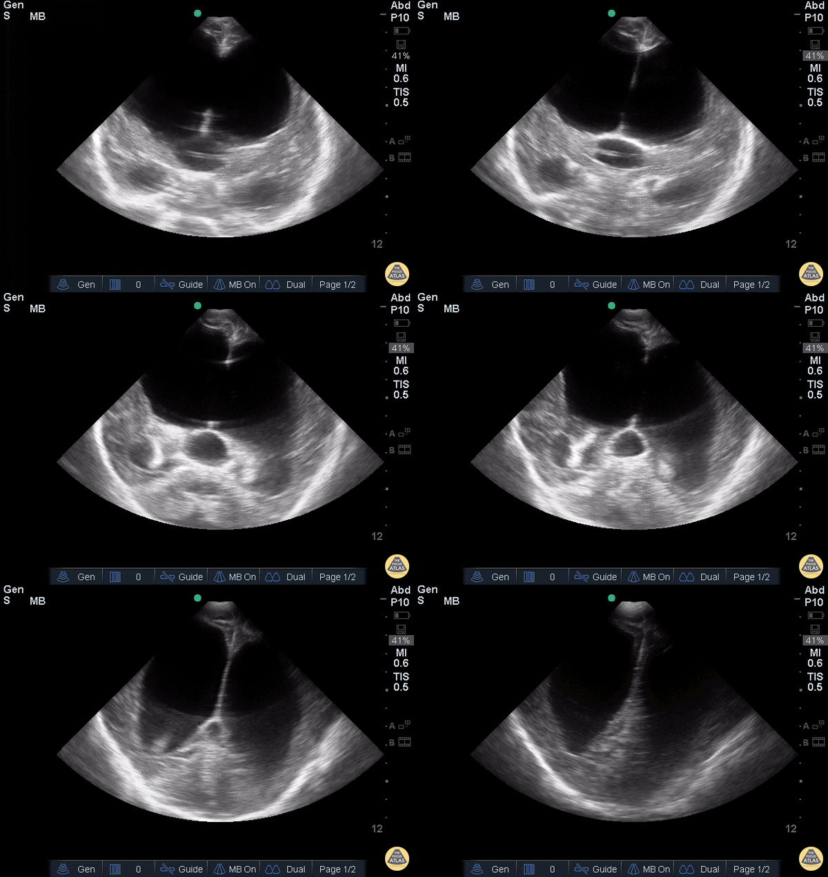
7 month old being followed by PCP for increasing head circumference, scheduled for outpatient MRI next week. Mother found patient was becoming somnolent with a full anterior fontanelle and brought the baby to the ER. POCUS performed immediately revealed unequal ventricle size, L>R, at which time neurosurgery was consulted, later CT, MRI performed as inpatient confirming obstructive hydrocephalus.
If you were in a community ER and CT will take a while, should you just POCUS first? How well can this see blood? Masses?
Dr. Sathya Subramaniam - Childrens Hospital of Philadelphia Pediatric, EM Ultrasound
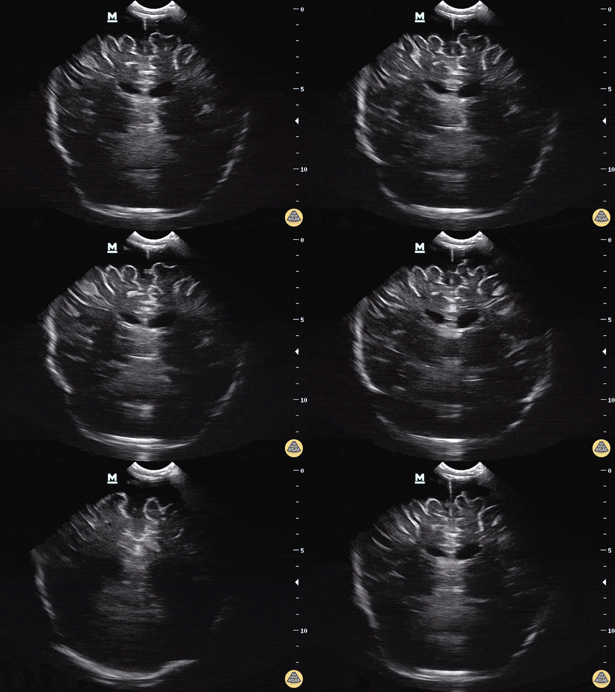
Extra-axial fluid overlying the superior portion of the frontal lobes in the patient from our blog post on the topic.
7 month old being followed by PCP for increasing head circumference, scheduled for outpatient MRI next week. Mother found patient was becoming somnolent with a full anterior fontanelle and brought the baby to the ER. POCUS performed immediately revealed unequal ventricle size, L>R, at which time neurosurgery was consulted, later CT, MRI performed as inpatient confirming obstructive hydrocephalus.
Dr. Sathya Subramaniam - Childrens Hospital of Philadelphia Pediatric, EM Ultrasound
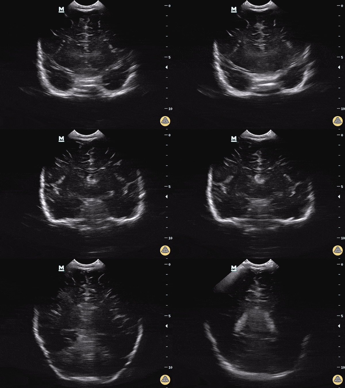
Please see our blog post for further information on this topic.
The coronal plane has the indicator marker to the right and is placed on the anterior fontanelle. The ultrasound beam is swept from the anterior to posterior aspect of the head.
Dr. Sathya Subramaniam - Childrens Hospital of Philadelphia Pediatric, EM Ultrasound
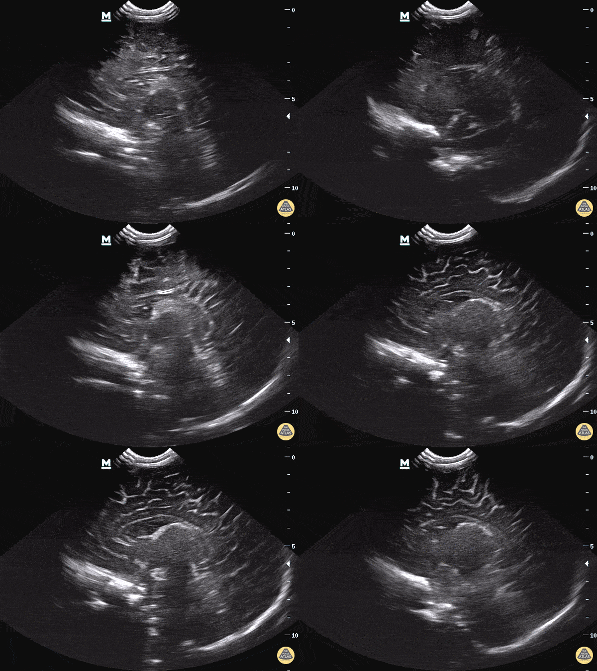
The sagittal plane has the indicator marker facing the anterior aspect of the face and the ultrasound beam is swept in either the left to right or right to left direction of the patient’s shoulders.
Please see our blog post for further information on this topic.
Dr. Sathya Subramaniam - Childrens Hospital of Philadelphia Pediatric, EM Ultrasound
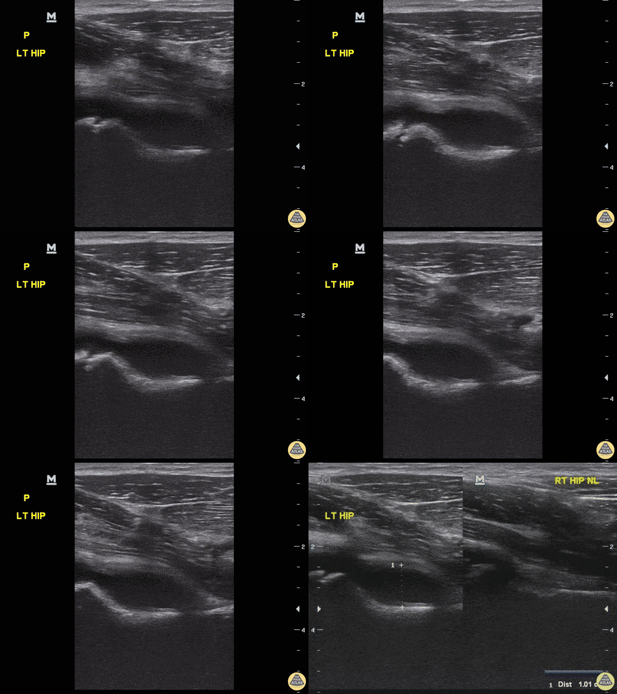
8-year-old female with fever and upper left thigh pain starting last night. Refusing to bear weight and will not flex hip, discomfort with rotating hip.
POCUS performed, revealed effusion. Still image comparing sides confirms effusion. Etiology of effusion remained uncertain.
Dr. Sathya Subramaniam, Pediatric EM Fellow - Kings County/SUNY Downstate
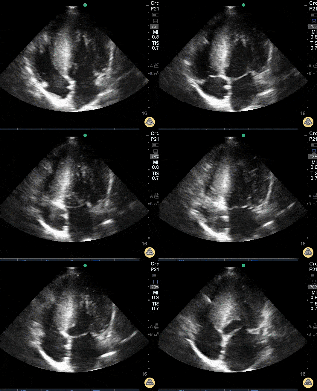
19 y/o came in after a syncopal episode playing video games. Got defibrillated in the field Vfib rate 200s. Bedside echo demonstrated global LVH with septal predominant thickening consistent with HOCM. Father has a history of ICD of “heart condition.”
Dr. Dustin Morrow
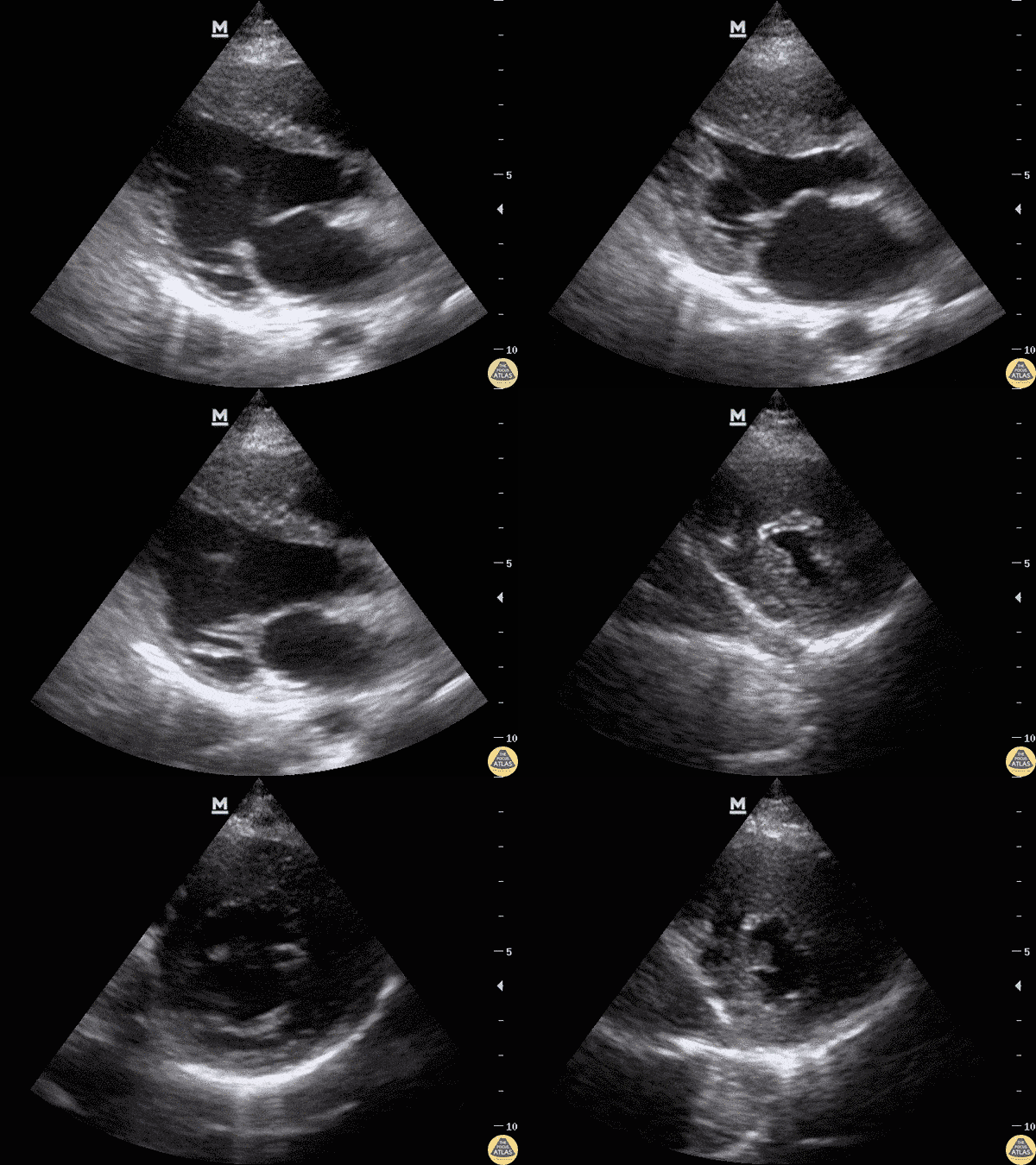
8-year-old with syncope while playing in school. EKG with some non-specific T wave inversions in precodial leads. No murmur, normal heart sounds. POCUS completed for concerning history and EKG changes. Interventricular septal hypertrophy seen on parasternal short and long views (concerning if measurement > 15mm).
Dr. Sathya Subramaniam, Pediatric EM Fellow - Kings County/SUNY Downstate
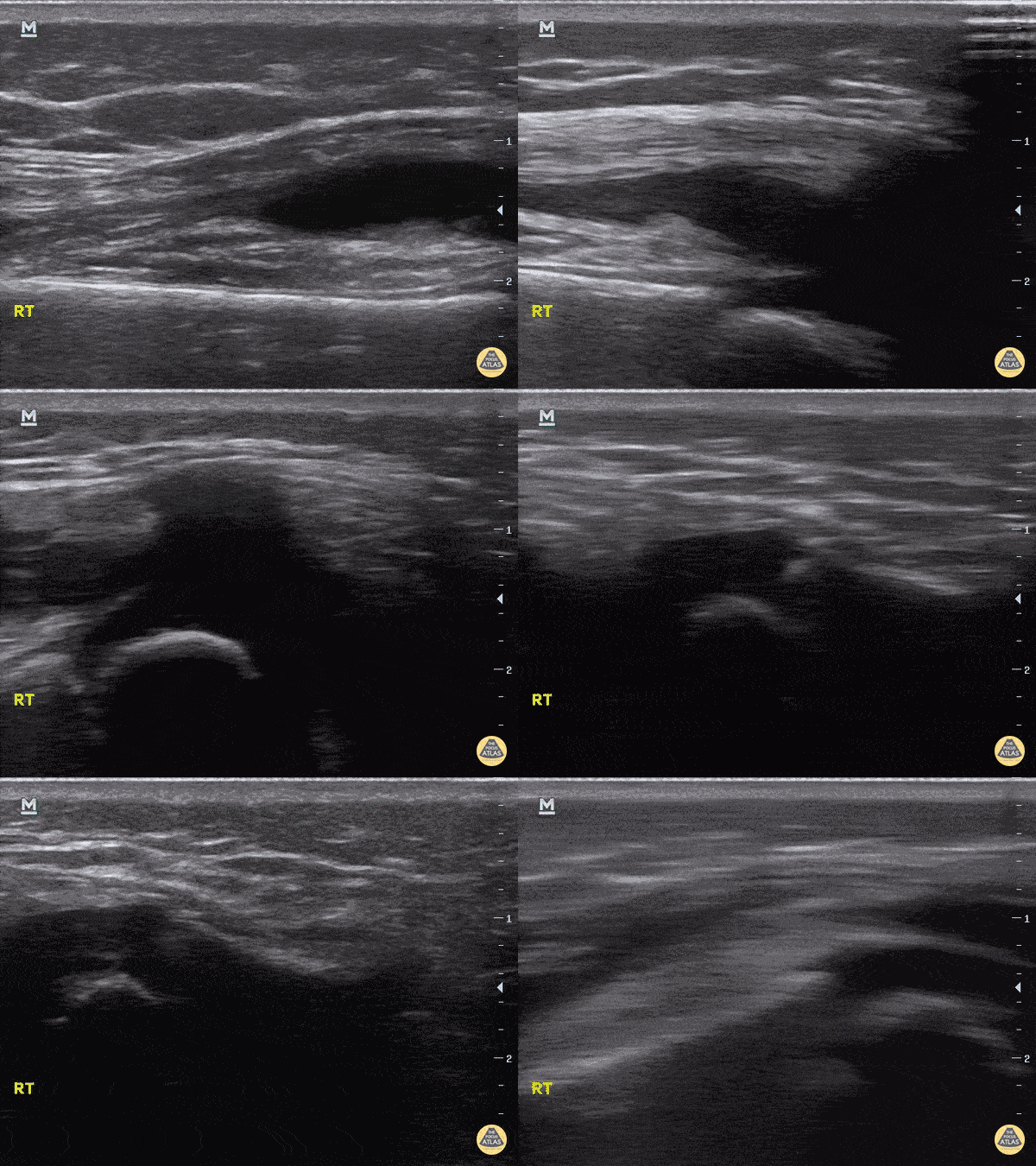
20 month old with right sided limp for 2 days, no fevers, swelling of knee, able to partially range without discomfort, knee aspirate after POCUS purulent, admitted for wash out. Septic vs Lyme's differential.
Aspirate yielded 923000 WBC from the cell count, but nothing grew from the cultures. Discharged home on 4 weeks of abx for Lyme, as his titers were positive on Elisa and Western blot.
Dr. Sathya Subramaniam - Children's Hospital of Philadelphia
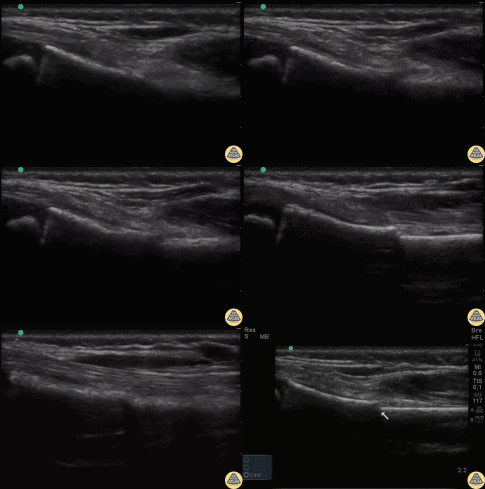
7 year old fallen off monkey bars. Tender over right distal radius with mild swelling.
POCUS reveals a discontinuity in the hyperechoic cortex of the child's distal radius with minimal displacement. This is suggestive of a buckle fracture or minimally displaced distal radius fracture.
Dr. Sathya Subramaniam, Pediatric EM Fellow - Kings County/SUNY Downstate
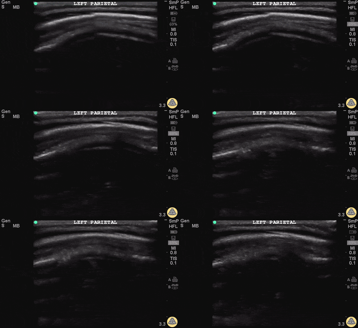
10 month old F presents one day after unwitnessed height level fall. Left parietal hematoma without step-off found on physical exam. Otherwise well appearing with normal vital signs. POCUS found a defect in cortex in area of the hematoma. CT head confirmed non-displaced skull fracture of the left parietal bone.
Patient was observed and did not require neurosurgical intervention. More research is needed into the value of POCUS for pediatrics skull fractures and how it can fit into our PECARN decision rules.
Dr Iain Jeffery - Brooklyn Hospital Emergency Medicine
Dr. Tian Liang and Dr Jeffery Rallo - Kings County Department of Pediatrics Emergency Medicine
Tags
- air bronchograms
- Airway
- b-lines
- bladder
- c lines
- Congenital Defects
- cranial
- fracture
- hepatization
- hydrocephalus
- hydronephrosis
- hypertrophic cardiomyopathy
- musculoskeletal
- myocarditis
- pleural effusion
- pneumonia
- pneumothorax
- prune belly syndrome
- pulmonary
- renal
- shoulder
- skull fracture
- supracondylar
- Testicular Torsion
- trauma