
Aorta
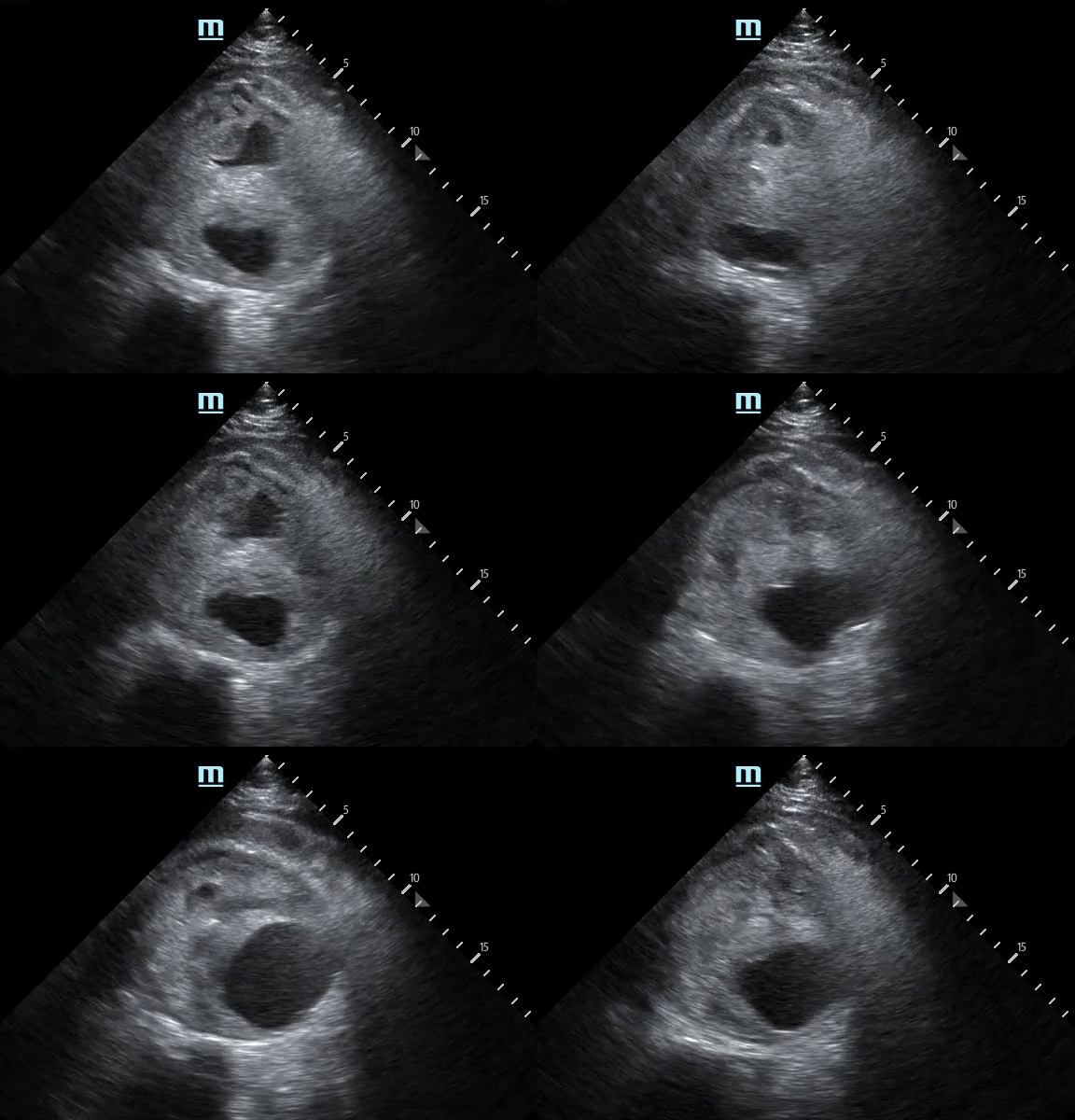
AAA Rupture: Not Your Typical Flank Pain
A 61-year-old male was brought in via EMS after a syncopal episode. He was diaphoretic and hypotensive, complaining of severe right flank pain.
While IV access was being obtained a bedside ultrasound was performed demonstrating a large abdominal aortic aneurysm with significant heterogeneous intraluminal clot. There is also appreciable focal hypoechoic disruption of the wall of the aneurysm consistent with rupture.
The patient was resuscitated in the ED and taken emergently to the OR to surgical repair.
Michael Macias, MD
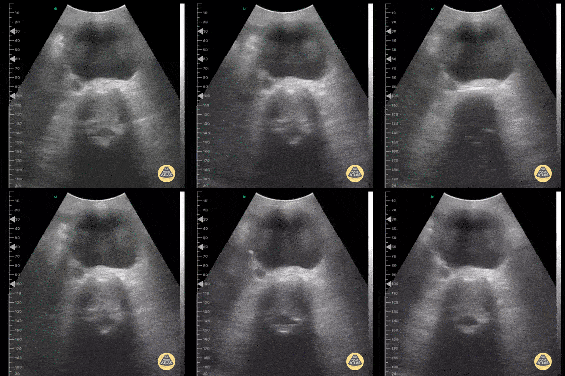
Abdominal Aortic Aneurysm
This image was taken from a pocket wireless device in the mesogastric region. We can see the short-axis aorta with its increased diameter and neighboring anatomical references such as the dorsal spine and inferior vena cava laterally.
Image courtesy of Dr. Renato Tambelli

Abdominal Aortic Dissection Flap
Patient with abrupt onset chest pain radiating to back. Normal ECG and Troponin. POCUS revealed dissection flap within abdominal aorta.
Nishant Cherian
Emergency Medicine Registrar
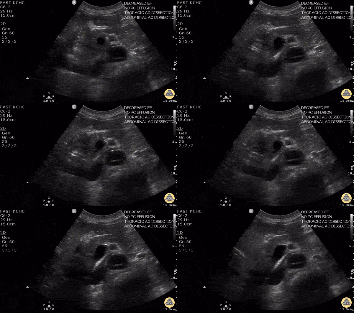
Aortic Dissection
50 y/o M w/ hx of HTN p/w sudden onset upper back pain. POCUS found dissection flap in the descending aorta in both parasternal long view and abdominal aorta. The diagnosis of aortic dissection was quickly confirmed by CT. Given the importance of timely diagnosis with aortic dissection, POCUS allowed rapid and non-invasive diagnosis of a potentially tricky diagnosis, and facilitated expedited treatment and transfer to a cardiothoracic surgery center.
Dr. Robert Allen - Kings County Emergency Medicine
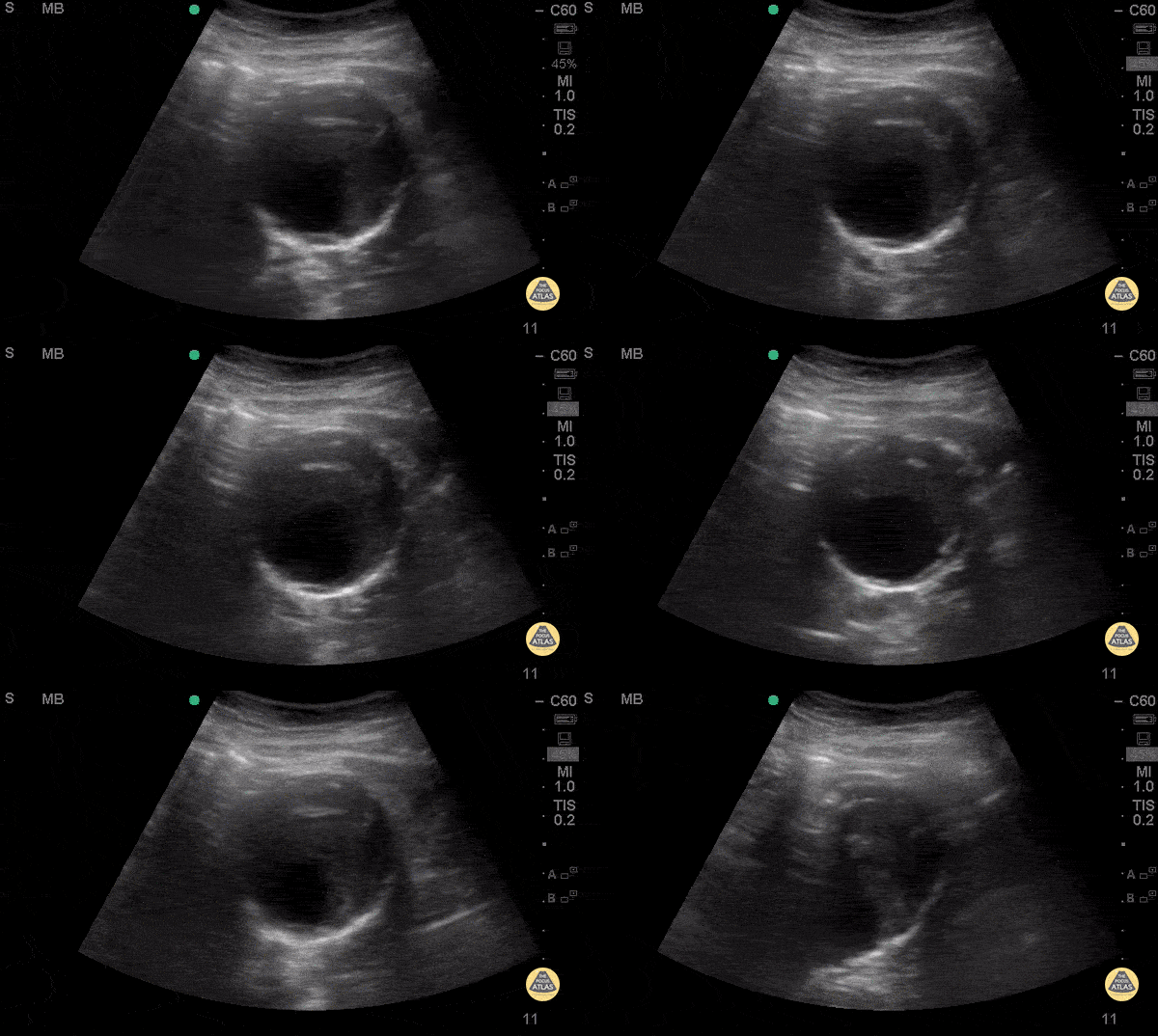
Mural Thrombus in AAA
74 y/o F hx stage 4 lung cancer, AAA, presented with chest pain and SOB x 1 day. POCUS shows the aorta is larger than the normal diameter of 3 cm, representing an abdominal aortic aneurysm, and measuring approximately 5.6 cm at its largest point. On cross section you are able to see a large mural thrombus with decreased diameter of blood flow through the center.
Juliana Jaramillo MD - Kings County Emergency Medicine
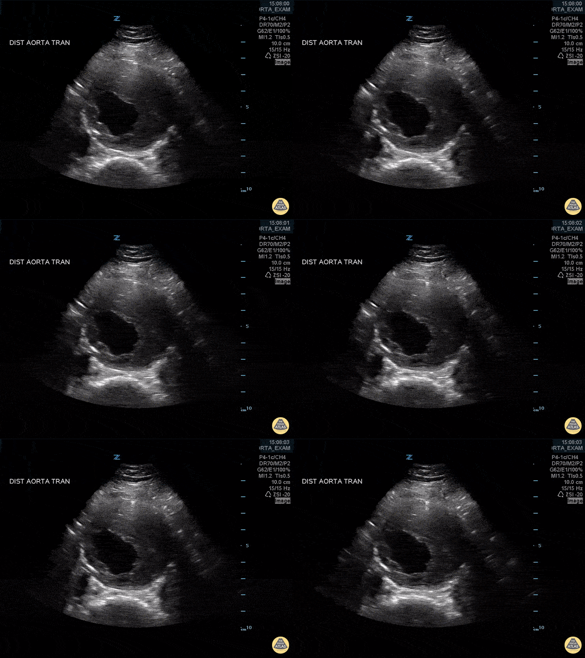
Abdominal Aortic Aneurysm with Thrombus
Approximately 6 cm abdominal aortic aneurysm with intramural thrombus.
Frances Russell, MD, RDMS
Assistant Professor of Emergency Medicine Division Chief, Ultrasound Fellowship Director, Ultrasound

Mural Thrombus in AAA - Doppler
74 y/o F hx stage 4 lung cancer, AAA, presented with chest pain and SOB x 1 day. POCUS shows the aorta is larger than the normal diameter of 3 cm, representing an abdominal aortic aneurysm, and measuring approximately 5.6 cm at its largest point. On cross section you are able to see a large mural thrombus with decreased diameter of blood flow through the center.
Juliana Jaramillo MD - Kings County Emergency Medicine
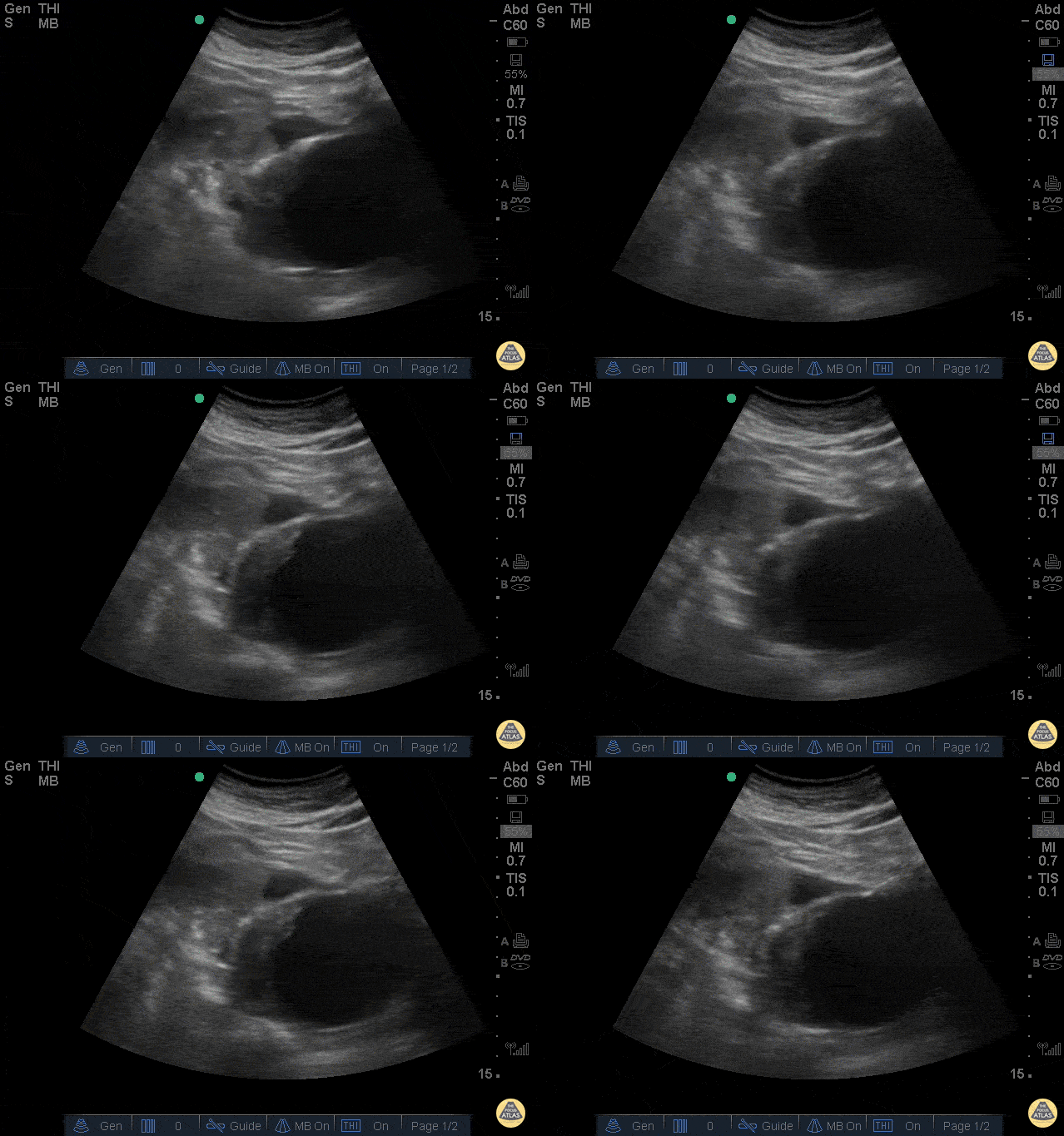
Ruptured AAA
The clip above is from a patient who presented with abdominal pain and syncope. His vitals were notable for tachycardia and hypotension. The image demonstrates a very large abdominal aortic aneurysm with anterior free fluid suggestive of rupture. The patient was taken emergently to the operating room for endograft repair and did well.
Jason Tanguay, DO
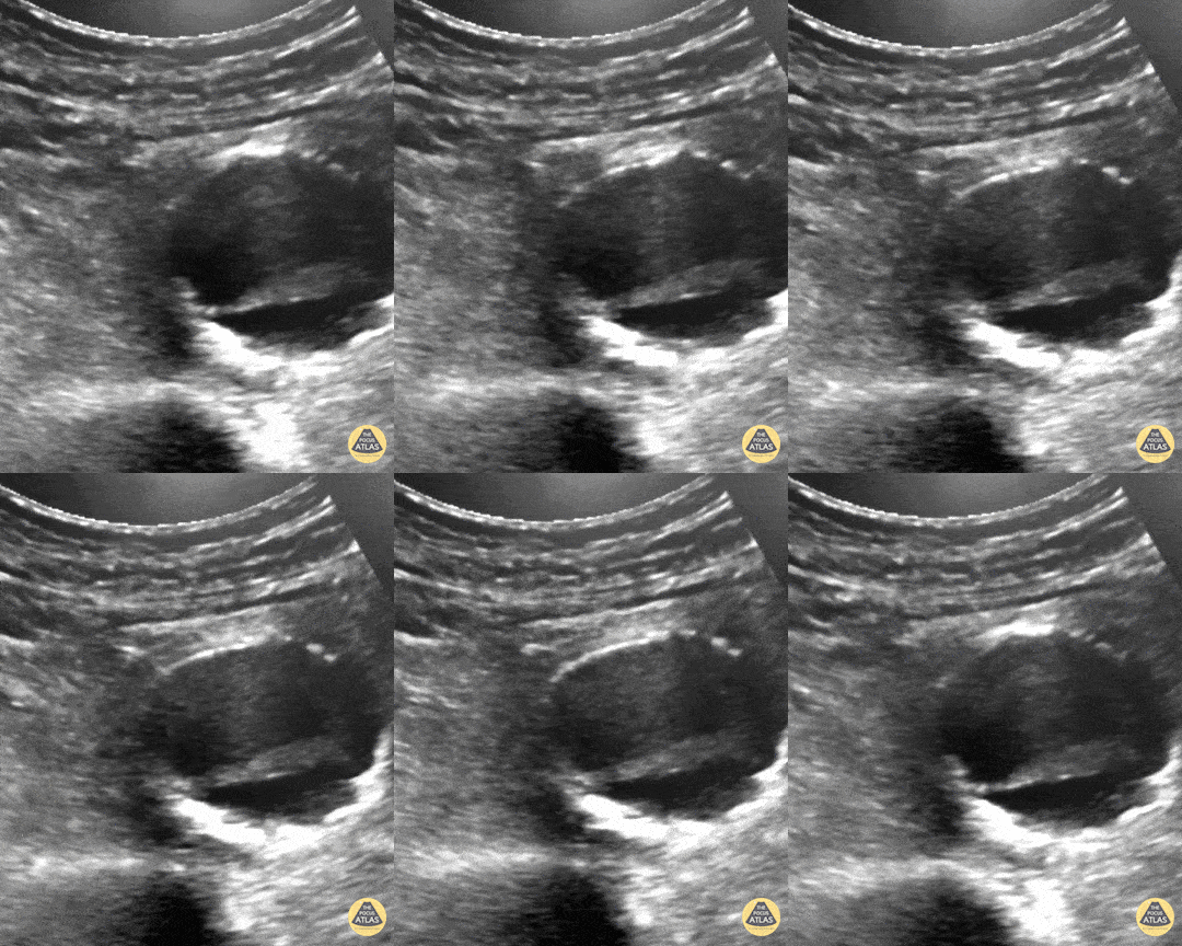
Pulsating Aortic Aneurysm
You may think this a dissection but it's actually an aortic aneurysm filled with thrombus, "with holes" making it a very "happy flappy." CT Confirmed.
Dr. Vincent Rietveld - Amsterdam, The Netherlands

Abdominal Aortic Aneurysm
AAA is defined as a localized balloon-like dilatation of the abdominal aorta greater than 3cm. Risk factors include male sex, increased age, and tobacco use. AAAs should be closely monitored for changes in size. Due to the risk of rupture, elective surgery is recommended when the dilatation is greater than 5-5.5cm, or it is growing in size by greater than 1cm/year. The classic triad of a ruptured AAA include pulsatile abdominal mass, hypotension and pain.
This AAA has an intramural thrombus. Some studies have claimed that POCUS has a ~100% sensitive for increased diameter. 3cm from outer wall to outer wall defines an aneurysm. Slow, graded compression is key to move the bowel out of the way in any abdominal study.
Sukh Singh, MD
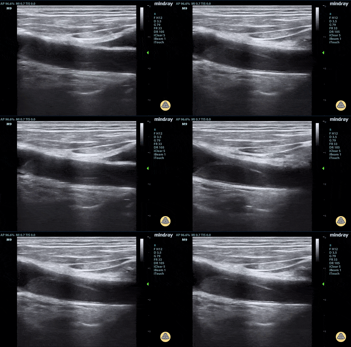
Aortic dissection flap tamponade
Elderly fellow who had a headache while bike riding, with some leg weakness. No chest or back pain. Stable for hours then came to hospital, suddenly hypotension and drowsy in ER
POCUS RUSH Exam performed lead to rapid diagnosis of Aortic Dissection with tamponade.
A dissection flap can clearly be visualized.
Claire Heslop - Pediatric Emergency Medicine - University of Toronto Hospital for Sick Children


