
Biliary
Biliary
Note: These search bar results are only for the Biliary image archive. To search the entire site, click here.
Recently Added
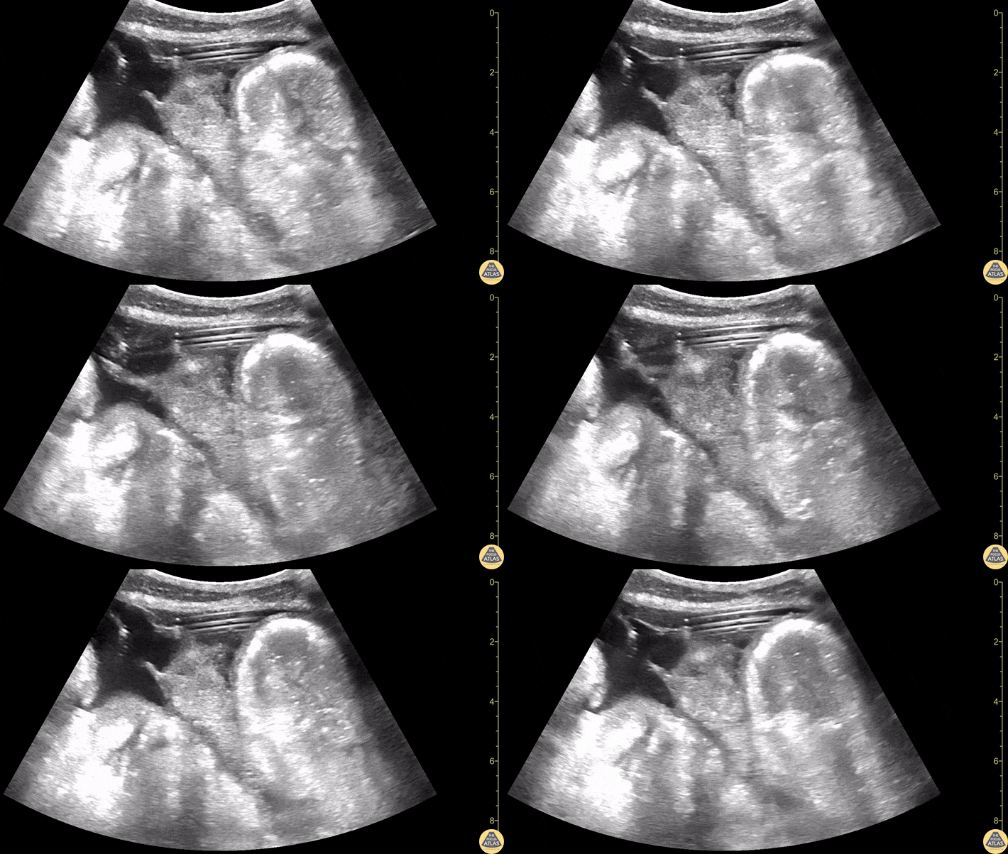
Drainage tube within a located liver fluid collection.
Contributors: Dimitri Livshits, DO; Jane Belyavskaya, MD; Chris Hanuscin, MD
Kings County/SUNY Downstate
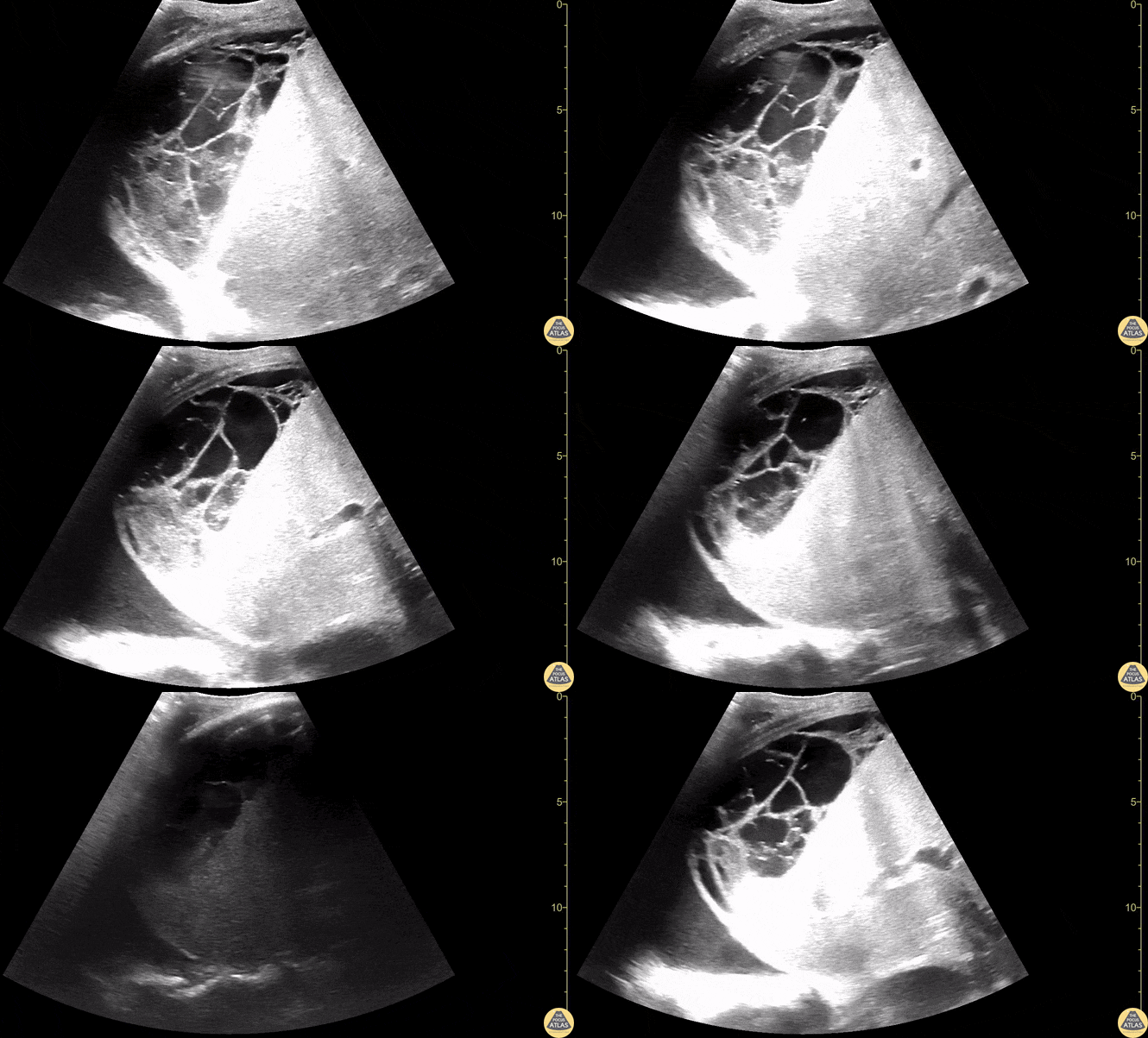
Located collection within the liver consistent with likely liver abscess.
Contributors: Dimitri Livshits, DO; Jane Belyavskaya, MD; Chris Hanuscin, MD
Kings County/SUNY Downstate
![Thick Walled Gallbladder in Dengue Fever (Short Axis) [2/2]](https://images.squarespace-cdn.com/content/v1/58118909e3df282037abfad7/1728883282342-87F9ZQFLQ314C6WAFAAC/image-asset.gif)
A 28 year old female comes to the ER with fever, myalgia, hypotension, and abdominal pain.
This case illustrates one of the ultrasound findings in Dengue Fever in the Leakage Syndrome Stage. We can see a large, thick-walled gallbladder and the presence of pericholecystic fluid. This clip demonstrates the gallbladder in short-axis view.
Contributor: Renato Tambelli (@R_Tambelli @JediPocus)
![Thick Walled Gallbladder in Dengue Fever (Long Axis) [1/2]](https://images.squarespace-cdn.com/content/v1/58118909e3df282037abfad7/1728882762911-WS6YZE9IZ5WVP4XD8BX1/image-asset.gif)
A 28 year old female comes to the ER with fever, myalgia, hypotension, and abdominal pain.
This case illustrates one of the ultrasound findings in Dengue Fever in the Leakage Syndrome Stage. We can see a large, thick-walled gallbladder and the presence of pericholecystic fluid. This clip demonstrates the gallbladder in long-axis view.
Contributor: Renato Tambelli (@R_Tambelli @JediPocus)
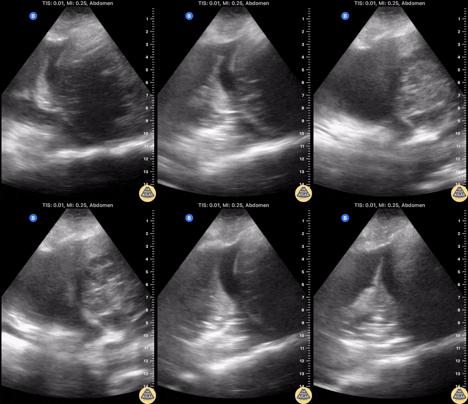
A elderly female with severe sepsis with E. coli bacteremia initially thought to be from urinary source developed worsening hypoxia overnight.
Using a Butterfly US, a moderate-sized right pleural effusion with associated ~6cm liver abscess is seen. These images helped mobilize pulmonology and IR to place a chest tube and hepatic drain, which led to resolution of hypoxemia and sepsis. Hepatic drain cultures grew E coli. Although one may appreciate a diaphragmatic defect on the image, the pleural fluid remained sterile.
POCUS in this case led to successful identification of the true sepsis source and associated pleural effusion as a complication of pyogenic liver abscess.
Contributors: Eric Reid, MD
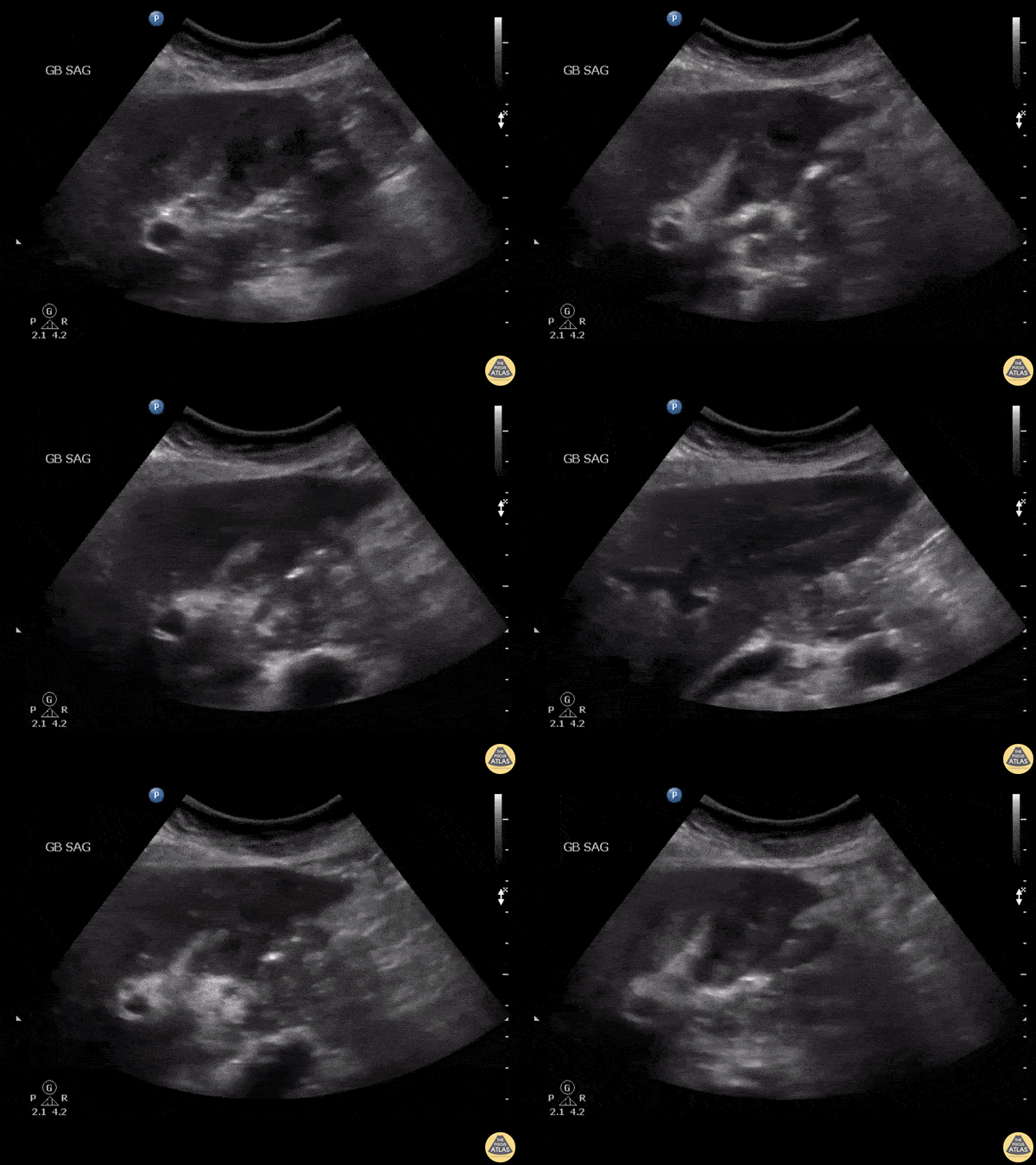
Patient presented with right upper quadrant pain. Clip shown demonstrated what appeared to be sludge however it was non-mobile and color doppler demonstrated internal flow. Further work up revealed gallbladder malignancy.
Image courtesy of Robert Jones DO, FACEP @RJonesSonoEM
Director, Emergency Ultrasound; MetroHealth Medical Center; Professor, Case Western Reserve Medical School, Cleveland, OH
View his original post here
Normal Anatomy
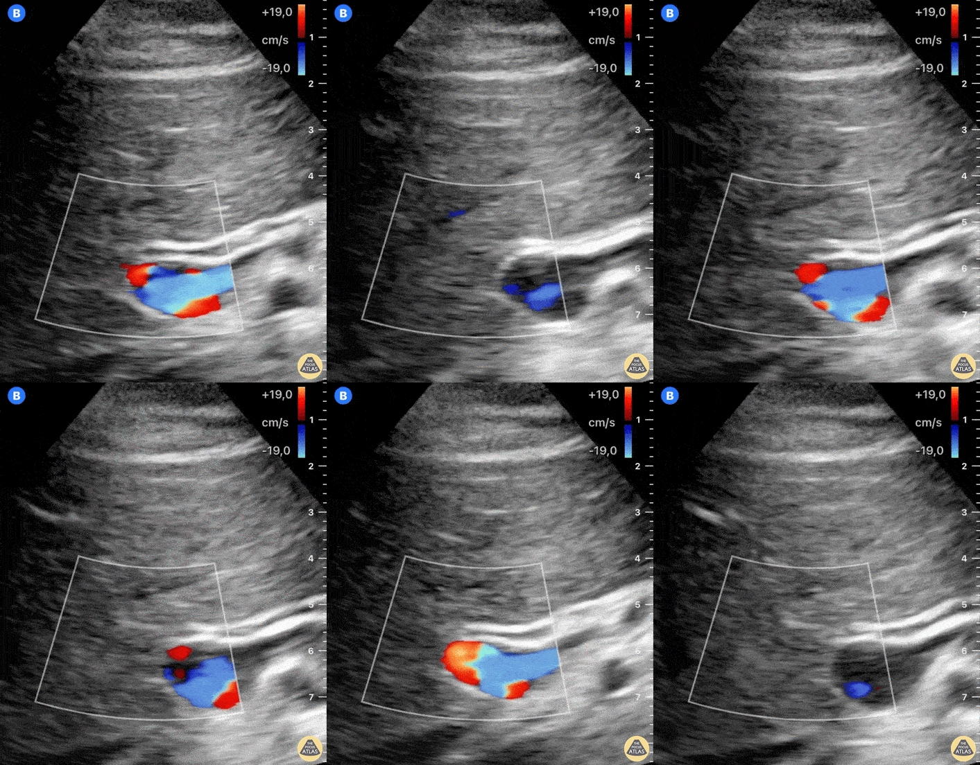
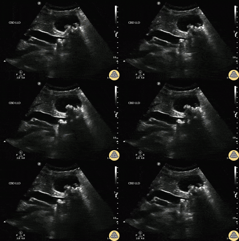
Often, a left lateral position delivers a clear image of the common bile duct (CBD) on its entire length, as is the case in this clip. Notice the normal hyperechoic walls and its small diameter. The CBD inner wall diameter should be < 6 mm in healthy adults, although it can be enlarged in post-cholecystectomy patients.
Dr. Felipe Urriola P. Emergency Unit, Puerto Aysen Hospital. Chilean Patagonia.
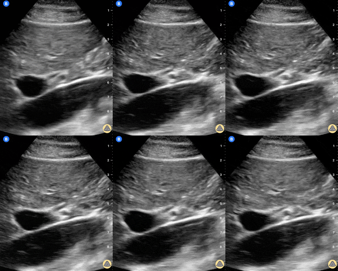
The large, pulsatile inferior vena cava lies at the bottom of the screen. Anterior to it and from left to right we see the large portal vein and small, round hepatic artery. Just above the hepatic artery lies another tubular structure which presents hyperechoic walls and an anechoic lumen; this is the common bile duct.
Dr. Felipe Urriola P. Emergency Unit, Puerto Aysen Hospital. Chilean Patagonia.
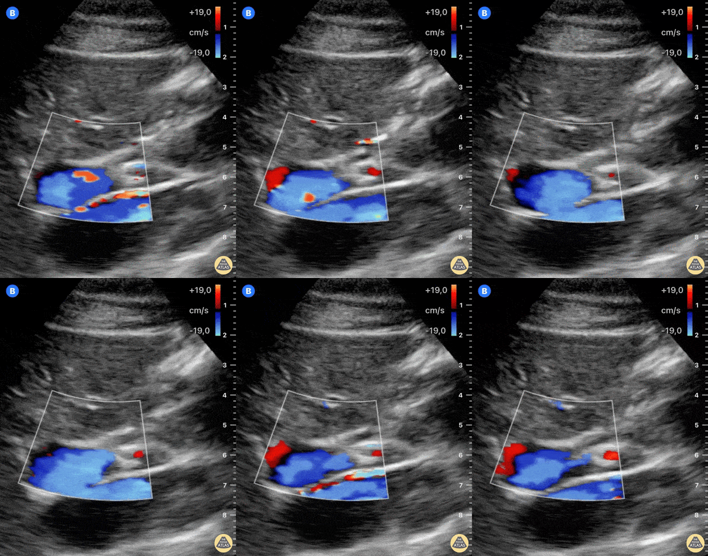
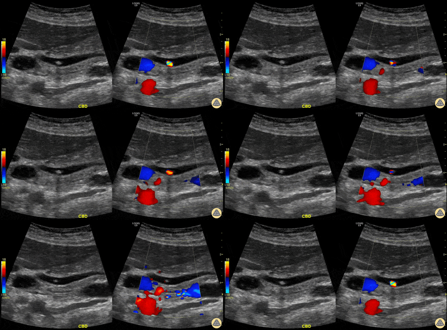
Both the adjustment of depth and color-doppler allow for better identification of structures. The large, pulsatile inferior vena cava lies at the bottom of the screen. Color-doppler helps to discern blood vessels from other anatomical structures. Anterior to the IVC and from left to right of the screen, we can see the portal vein and hepatic artery with color-flow. Just above the hepatic artery lies another tubular structure which presents hyperechoic walls and an anechoic lumen; this is the common bile duct.
Dr. Felipe Urriola P. Emergency Unit, Puerto Aysen Hospital. Chilean Patagonia.
Colorized Anatomy
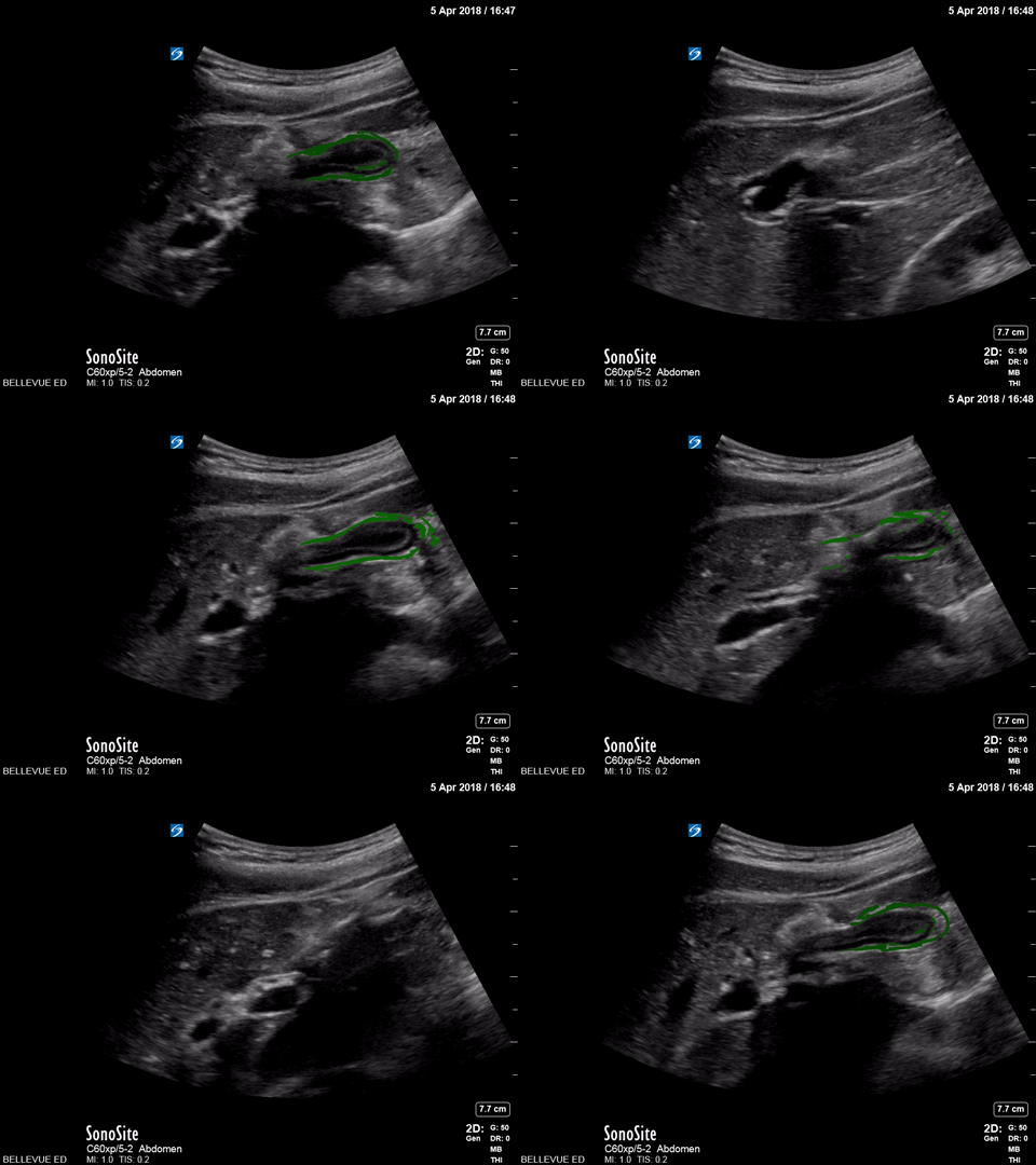
Contracted Gallbladder
Green- Gallbladder
Images: Dr. Lindsay Davis, Dr. Hannah Kopinski. Image Editing: Michael Amador and Dr. Matthew Riscinti
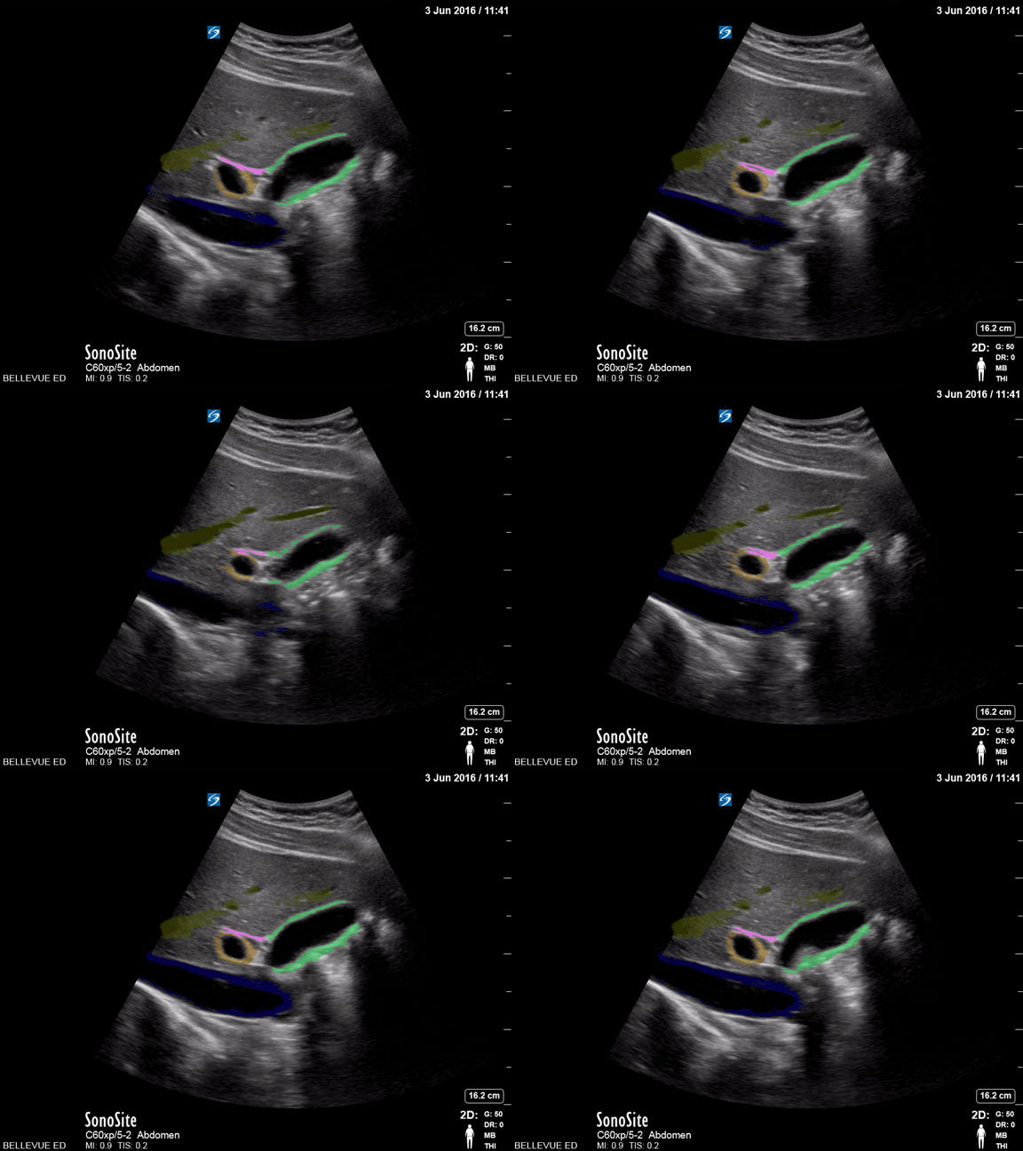
Gallbladder and Common Bile Duct
Green: Gallbladder, Blue: IVC, Orange: Portal vein, Pink: Common bile duct, Yellow: Lumen of hepatic vein
Images: Dr. Lindsay Davis, Dr. Hannah Kopinski. Image Editing: Michael Amador and Dr. Matthew Riscinti
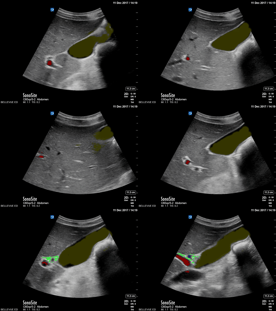
Normal Gallbladder
Yellow: Lumen of gallbladder, Red: Portal vein, Green: Common bile duct
Images: Dr. Lindsay Davis, Dr. Hannah Kopinski. Image Editing: Michael Amador and Dr. Matthew Riscinti
Cholelithiasis
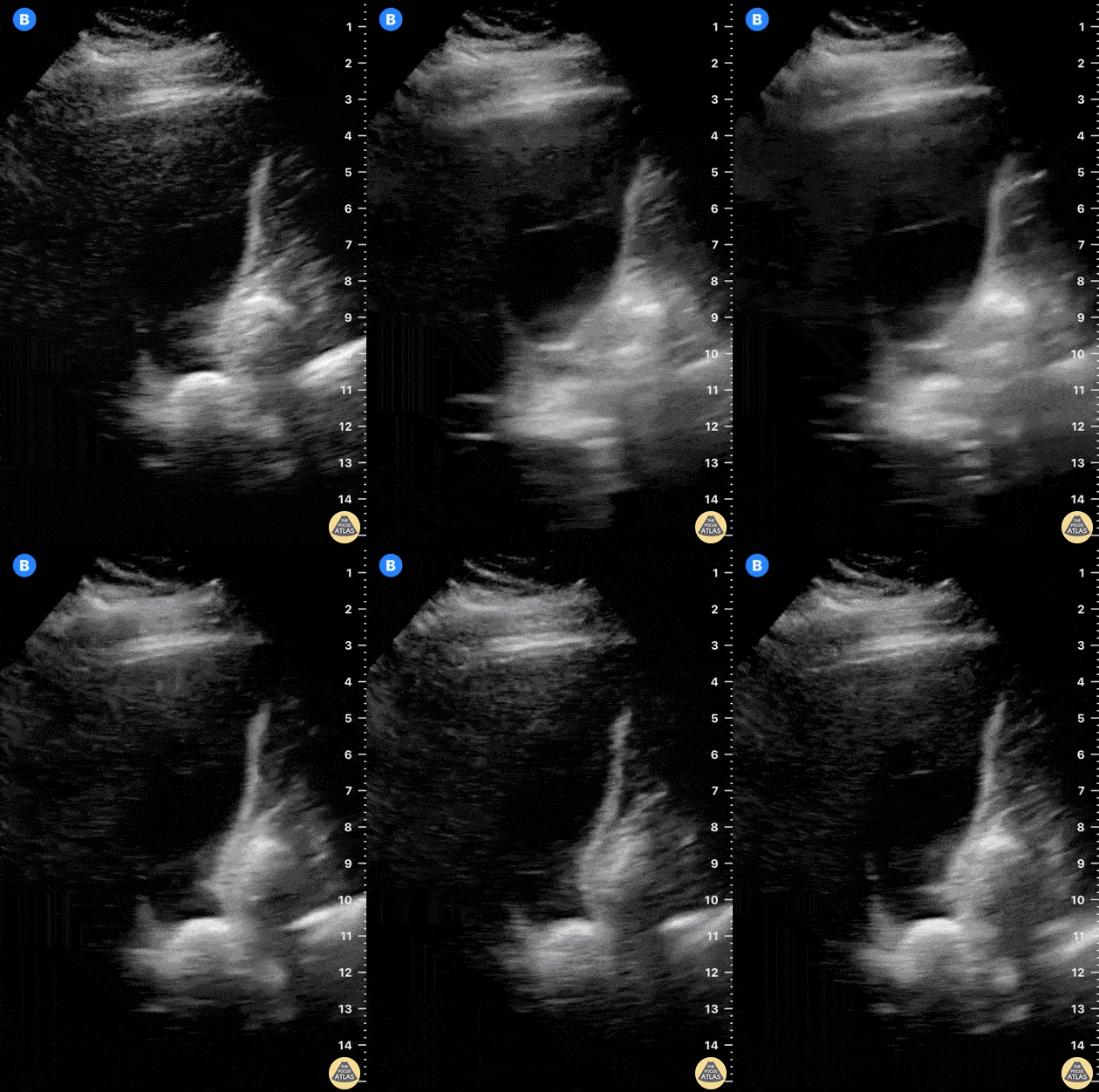
It’s fundamental to study the gallbladder in its whole extension. In this image, there is an impacted stone at the neck.
Dr. Felipe Urriola P.
www.thepocus.com
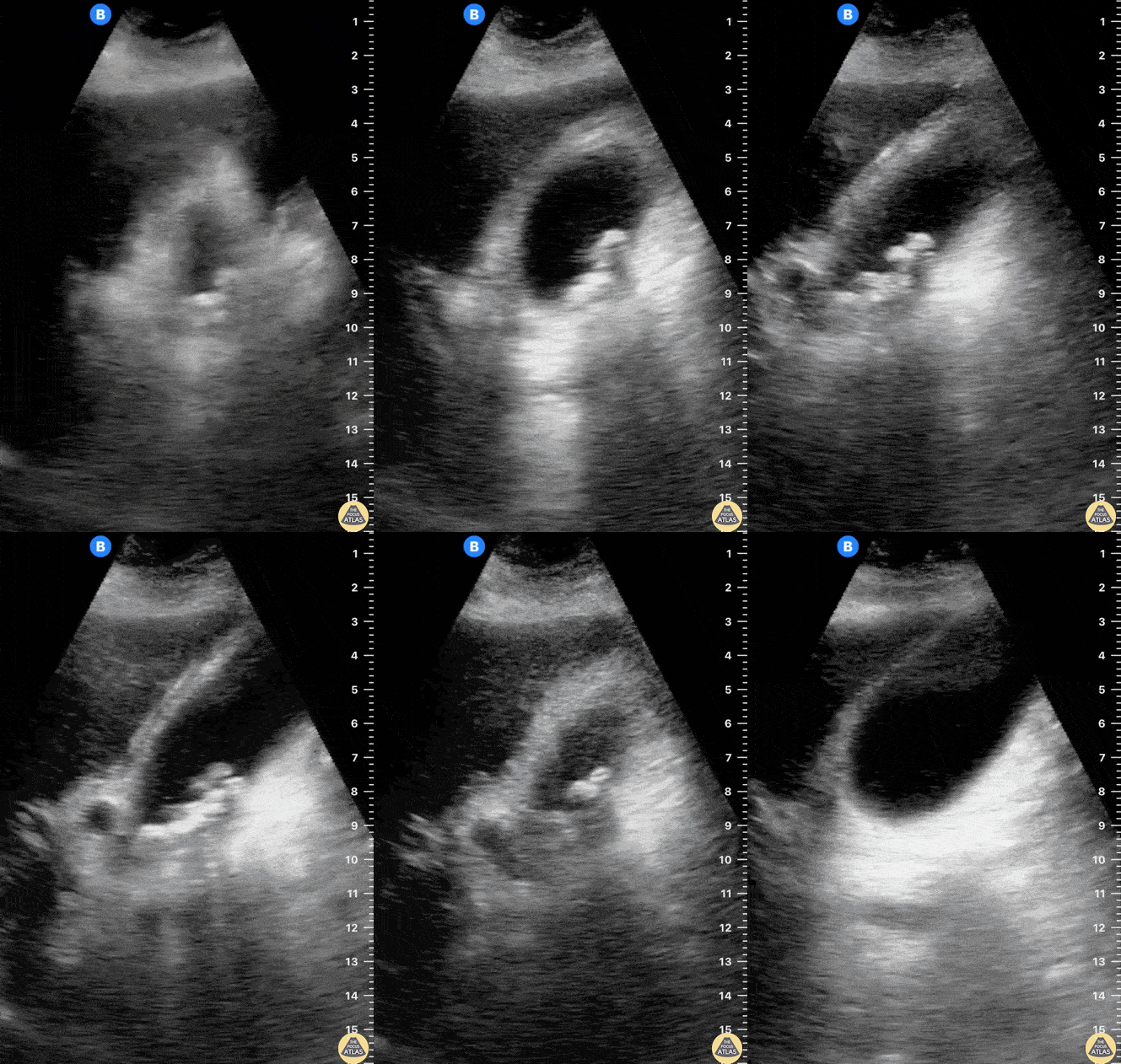
The gallbladder is studied in both planes, revealing multiple small stones generating acoustic shadowing.
Dr. Felipe Urriola P
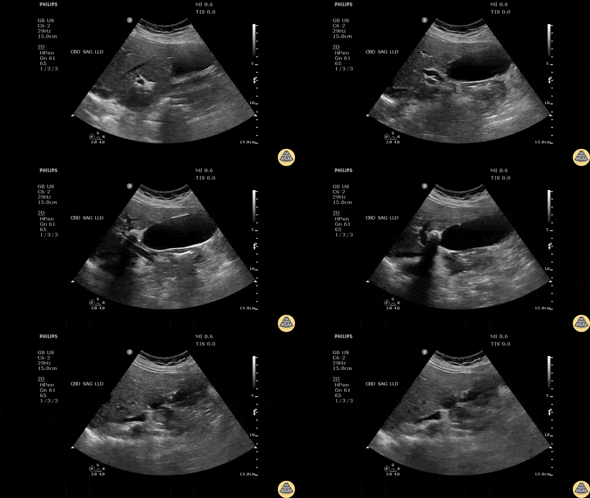
Longitudinal view of the gallbladder revealing two unique findings. A hyperechoic structure with posterior acoustic shadowing indicative of a stone is located in the neck. Two smaller hyperechoic structures can be seen attached to the wall of the GB body, likely adenomyomatosis.
Image courtesy of Robert Jones DO, FACEP @RJonesSonoEM
Director, Emergency Ultrasound; MetroHealth Medical Center; Professor, Case Western Reserve Medical School, Cleveland, OH
View his original post here
Cholecystitis
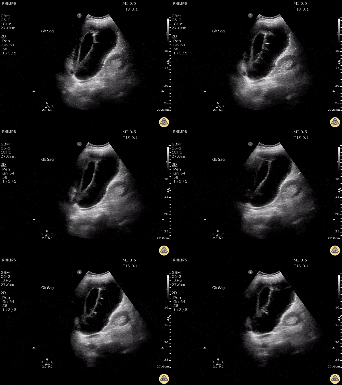
Stone trapped in cystic duct resulted in gallbladder distention as well as sloughing of intraluminal membranes.
Image courtesy of Robert Jones DO, FACEP @RJonesSonoEM
Director, Emergency Ultrasound; MetroHealth Medical Center; Professor, Case Western Reserve Medical School, Cleveland, OH
View his original post here
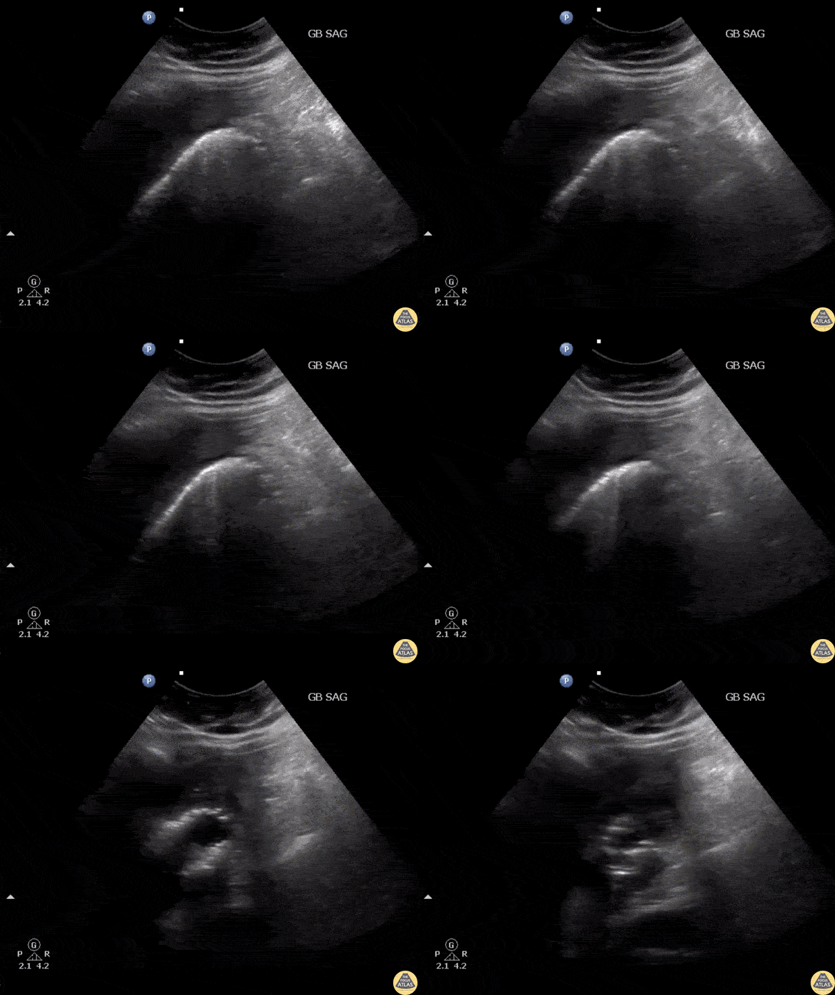
RUQ US of a patient revealed air within the gallbladder consistent with emphysematous cholecystitis, which can easily be confused with bowel gas.
Image courtesy of Robert Jones DO, FACEP @RJonesSonoEM
Director, Emergency Ultrasound; MetroHealth Medical Center; Professor, Case Western Reserve Medical School, Cleveland, OH
View his original post here
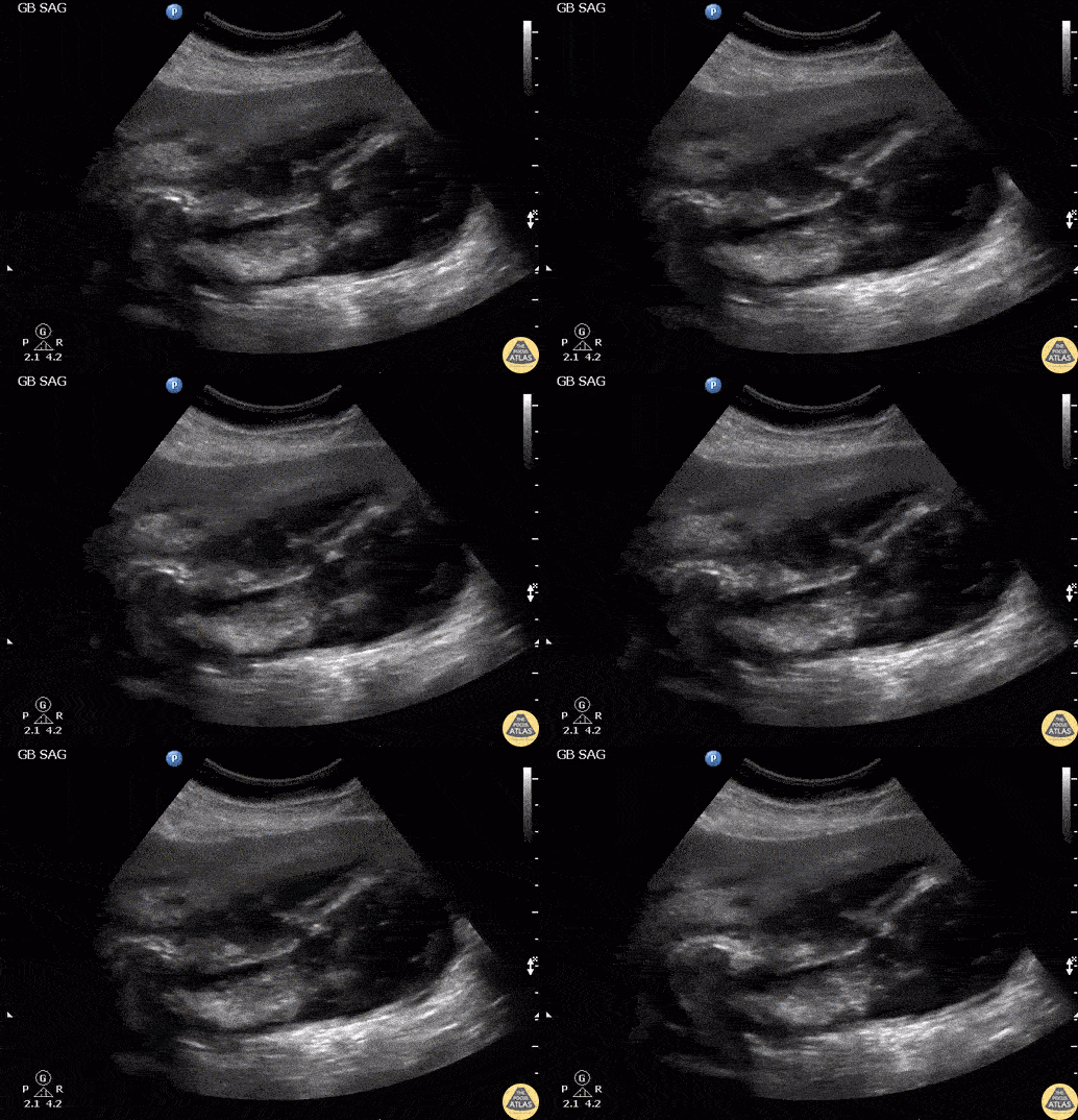
Patient presented with right upper quadrant abdominal pain and fever. RUQ ultrasound showed an impacted stone in the gallbladder neck with gallbladder wall perforation. Note the flow through the perforation and the associated pericholecystic abscess. Labelled image can be found on the original post with the link below.
Image courtesy of Robert Jones DO, FACEP @RJonesSonoEM
Director, Emergency Ultrasound; MetroHealth Medical Center; Professor, Case Western Reserve Medical School, Cleveland, OH
View his original post here
Common Bile Duct Pathology
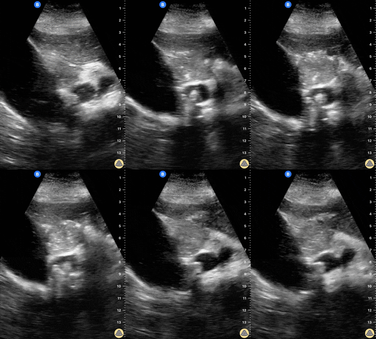
A 65-year-old female with no PMH presented to ED with 6 hour hx RUQ abdominal pain. She reported no associated vomiting or fever.
POCUS seen here reveals the cystic duct emerging from the GB neck to then reach a severely dilated CBD. A hyperechoic round structure with posterior shadowing lies inside the CBD, consistent with a diagnosis of choledocholithiasis.
Dr. Felipe Urriola
Emergency Unit. Puerto Aysen Hospital, Chilean Patagonia.
A patient presented to the ED with RUQ pain. Sagittal view of the common bile duct revealed dilation and stones indicative of choledocolithiasis.
Image courtesy of Robert Jones DO, FACEP @RJonesSonoEM
Director, Emergency Ultrasound; MetroHealth Medical Center; Professor, Case Western Reserve Medical School, Cleveland, OH
View his original post here
A patient presented with RUQ abdominal pain.
Ultrasound showed cholelithiasis and choledocholithiasis. Left image shows a portion of the common bile duct containing a small hyperechoic shadowing mobile stone. Right image shows the same with color Doppler added demonstrating twinkling artifact.
Learning point: Twinkling artifact is most often associated with urinary stones but also can be caused by gallstones (depending on composition), bowel gas, foley balloons, metallic objects, and many others. Twinkling artifact may help identify small stones that do not cause prominent acoustic shadowing.
Michael Cover, MD
@michaelc0ver
Other Pathology
Drainage tube within a located liver fluid collection.
Contributors: Dimitri Livshits, DO; Jane Belyavskaya, MD; Chris Hanuscin, MD
Kings County/SUNY Downstate
Located collection within the liver consistent with likely liver abscess.
Contributors: Dimitri Livshits, DO; Jane Belyavskaya, MD; Chris Hanuscin, MD
Kings County/SUNY Downstate
A 28 year old female comes to the ER with fever, myalgia, hypotension, and abdominal pain.
This case illustrates one of the ultrasound findings in Dengue Fever in the Leakage Syndrome Stage. We can see a large, thick-walled gallbladder and the presence of pericholecystic fluid. This clip demonstrates the gallbladder in short-axis view.
Contributor: Renato Tambelli (@R_Tambelli @JediPocus)