Pulmonary Jr
Pulmonary Jr
Note: These search bar results are only for the Pulmonary Jr image archive. To search the entire site, click here.

Recently Added
Pediatric patient presenting with shortness of breath. Found to have a large left sided pneumothorax. M-mode tracing shows lack of lung sliding
Contributor: Kathryn Pade, MD, Rady Children's Hospital San Diego
11 year old female presented to the emergency department with a puncture wound to her left chest wall. FAST exam did not reveal any intraabdominal free fluid, and there was lung sliding bilaterally, suggesting no pneumothorax. The patient had her wound explored, repaired, and was discharged home.
Contributed by: Zach Boivin, MD, @ZachBoivinMD
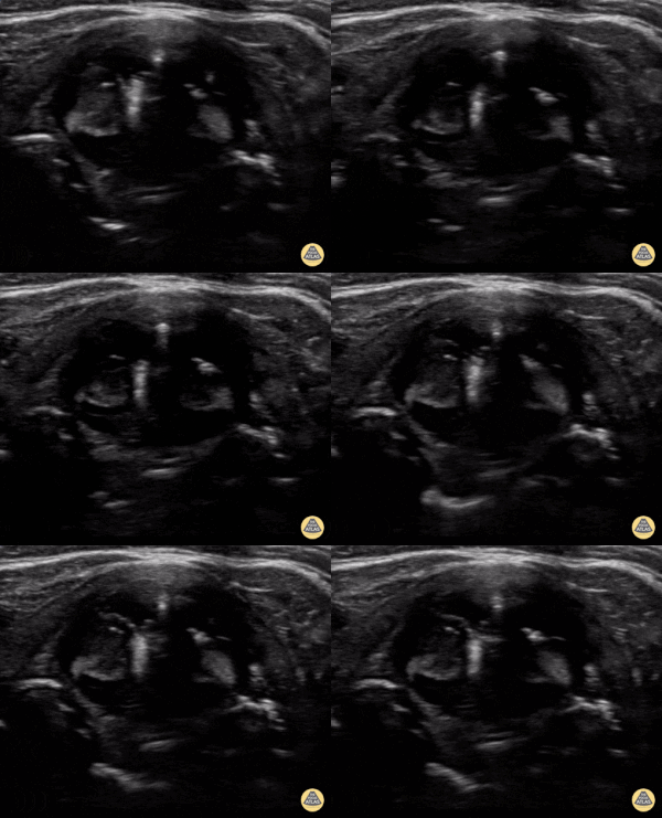
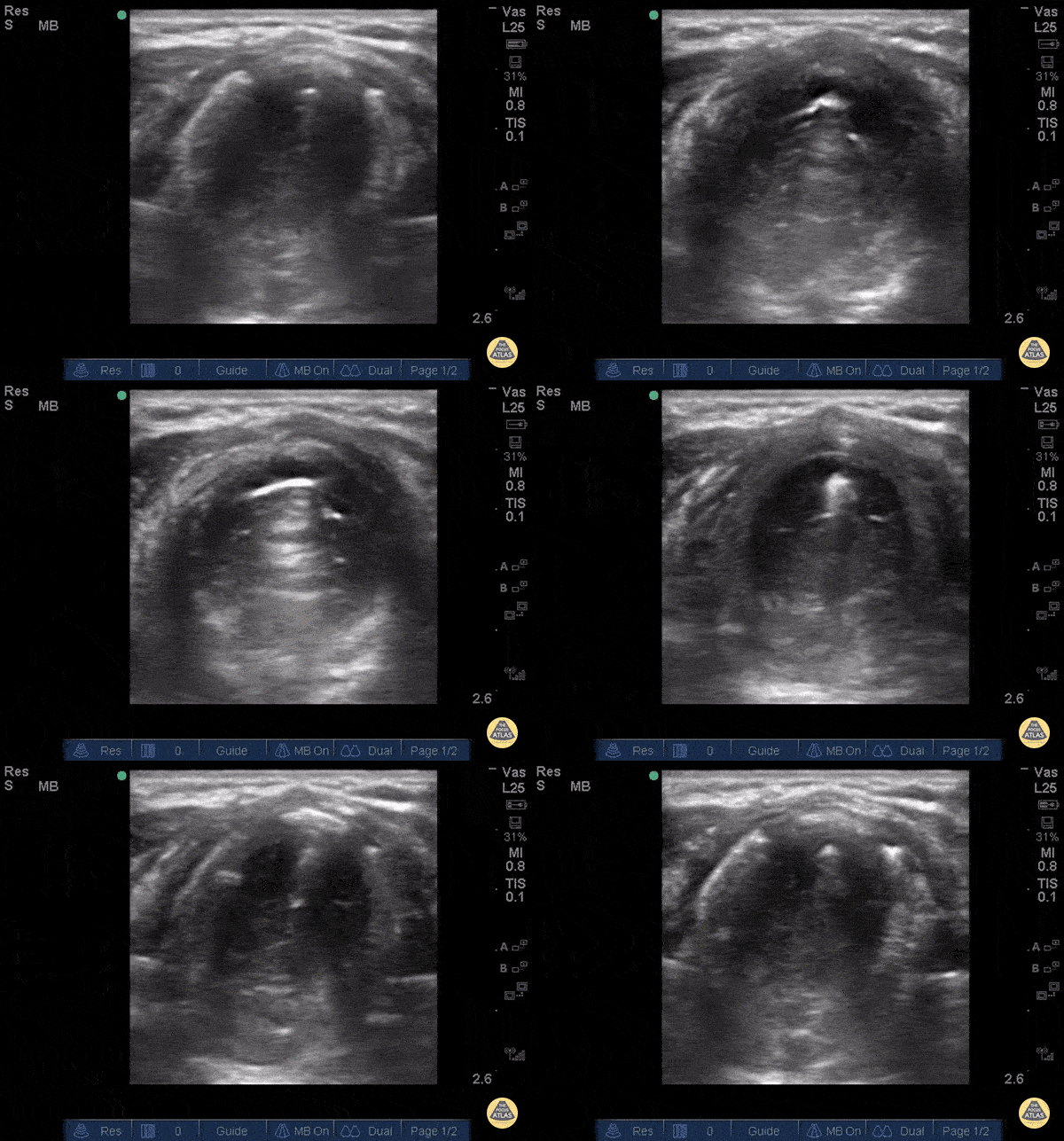
Intubation attempt into the esophagus of an 8 year old male with respiratory distress, notice the soft tissue posterior to the trachea moving with the provider attempting the pass the tube.
Image contributed by: Zach Boivin, MD, @ZachBoivinMD
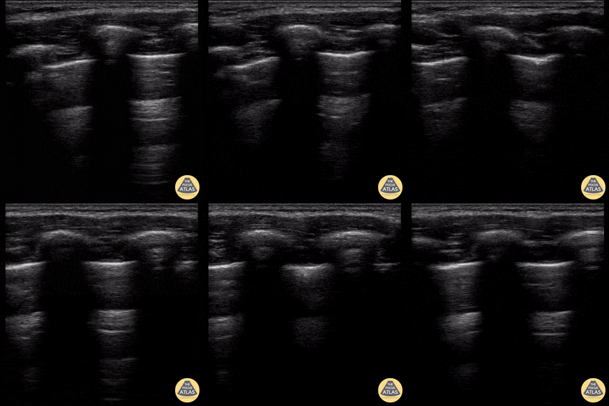
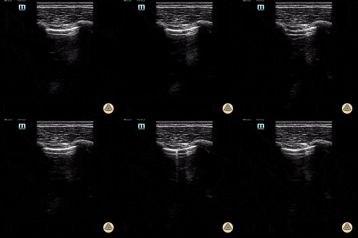
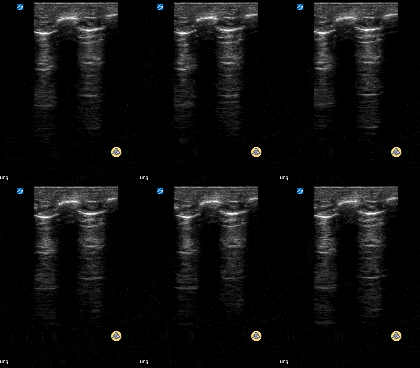
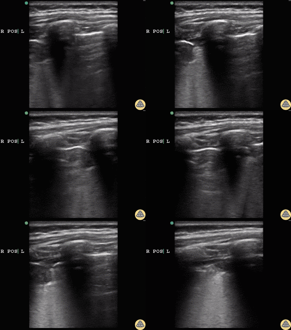
Normal lung sliding using linear probe
Contributor: Peter Gutierrez, MD, FAAP, Emory University School of Medicine/Children's Healthcare of Atlanta, @pocuspete
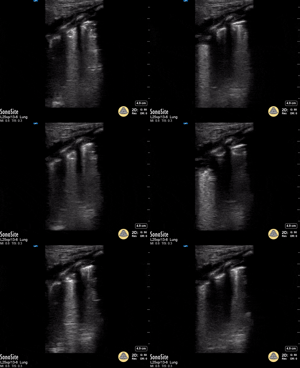
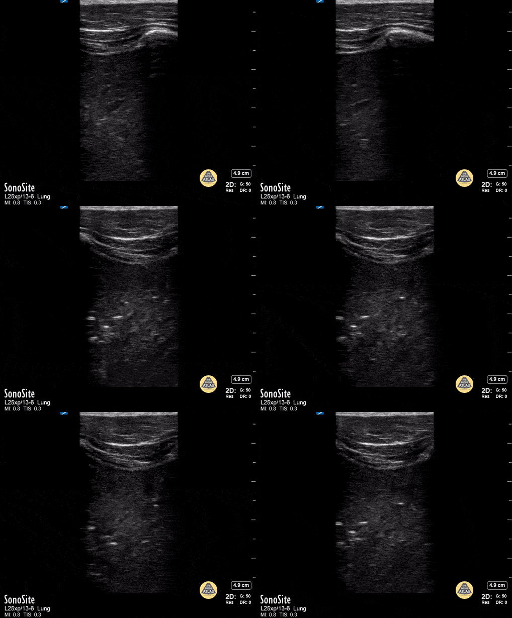
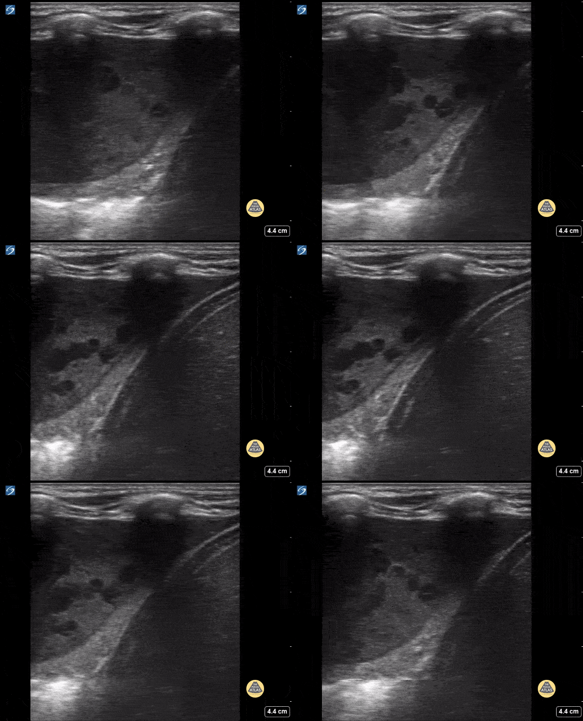
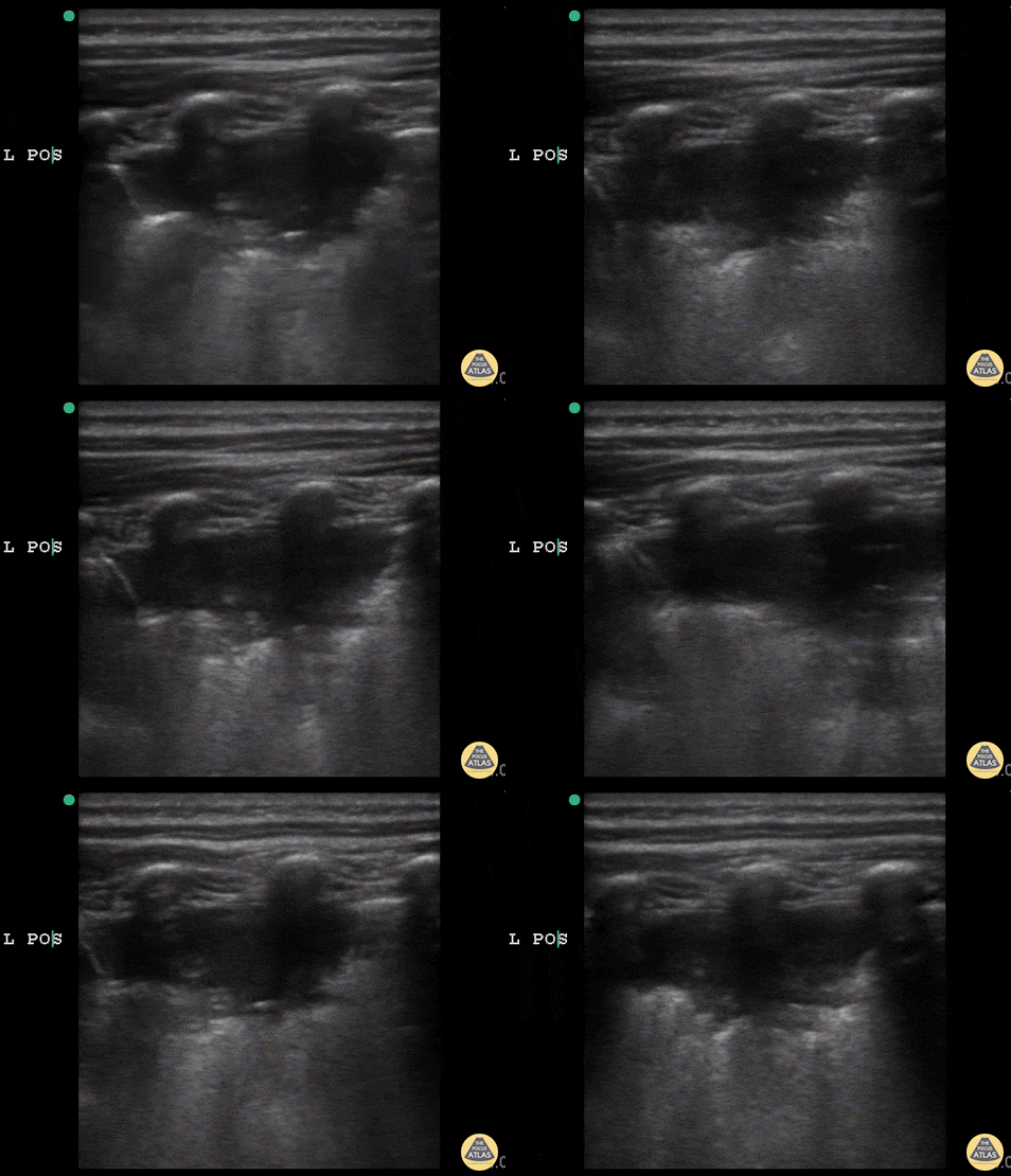
Toddler with pneumonia; multiple B-lines noted with adjacent area of consolidation.
Contributor: Peter Gutierrez, MD, FAAP, Emory University School of Medicine/Children's Healthcare of Atlanta, @pocuspete
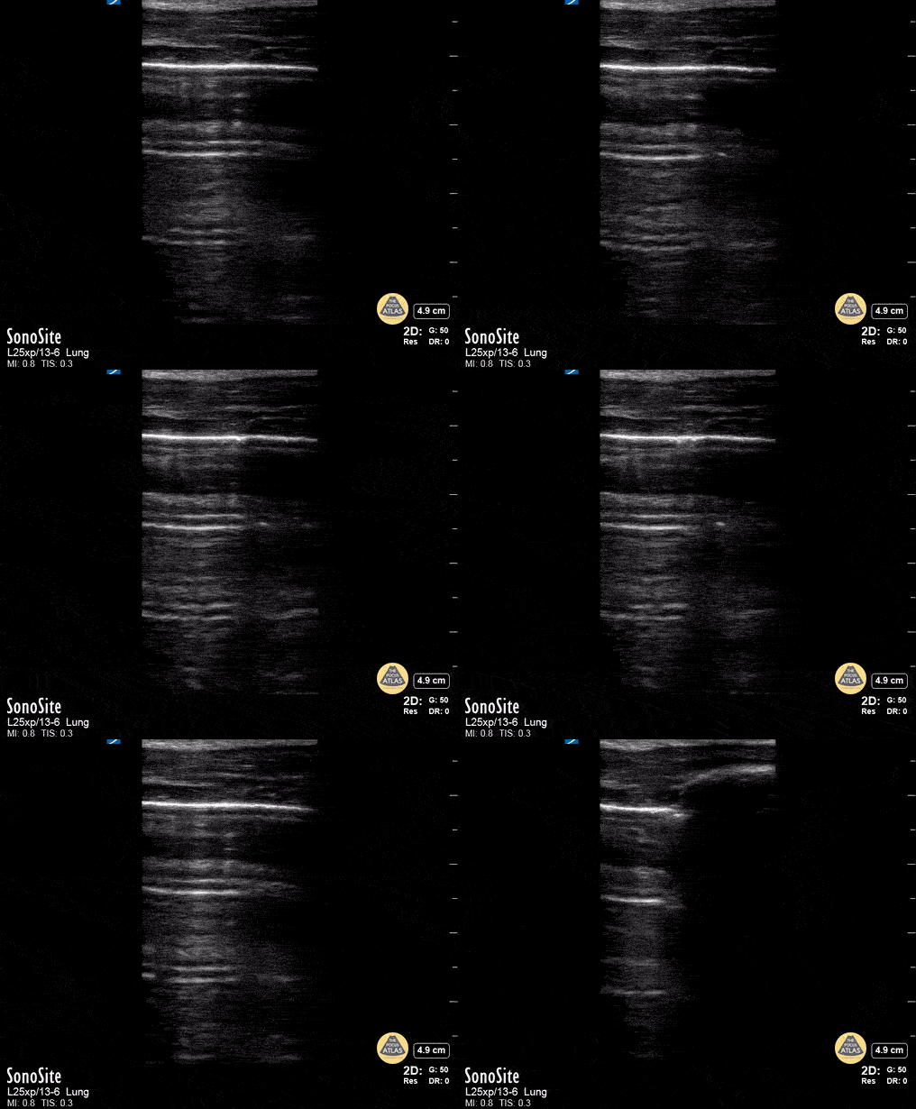
Lung point indicative of pneumothorax.
Contributor: Peter Gutierrez, MD, FAAP, Emory University School of Medicine/Children's Healthcare of Atlanta, @pocuspete
Normal Anatomy
Normal lung sliding using linear probe
Contributor: Peter Gutierrez, MD, FAAP, Emory University School of Medicine/Children's Healthcare of Atlanta, @pocuspete
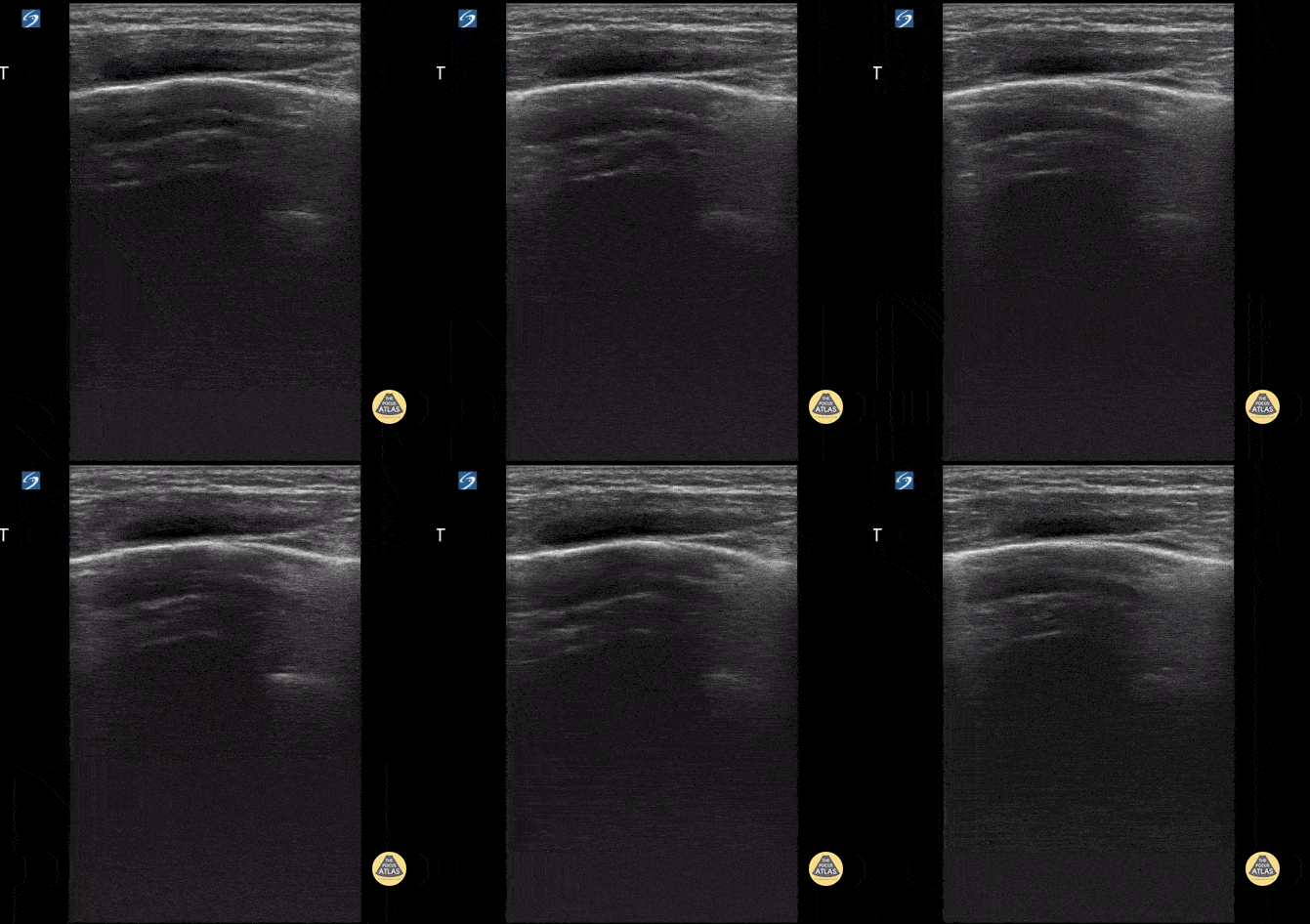
4 y/o with asthma who presents in respiratory distress. Using the linear probe, normal lung sliding is seen with comet tails.
Contributor: Kathryn Pade, MD, Rady Children's Hospital San Diego
Pneumonia & Subpleural Consolidation
Toddler with pneumonia; multiple B-lines noted with adjacent area of consolidation.
Contributor: Peter Gutierrez, MD, FAAP, Emory University School of Medicine/Children's Healthcare of Atlanta, @pocuspete
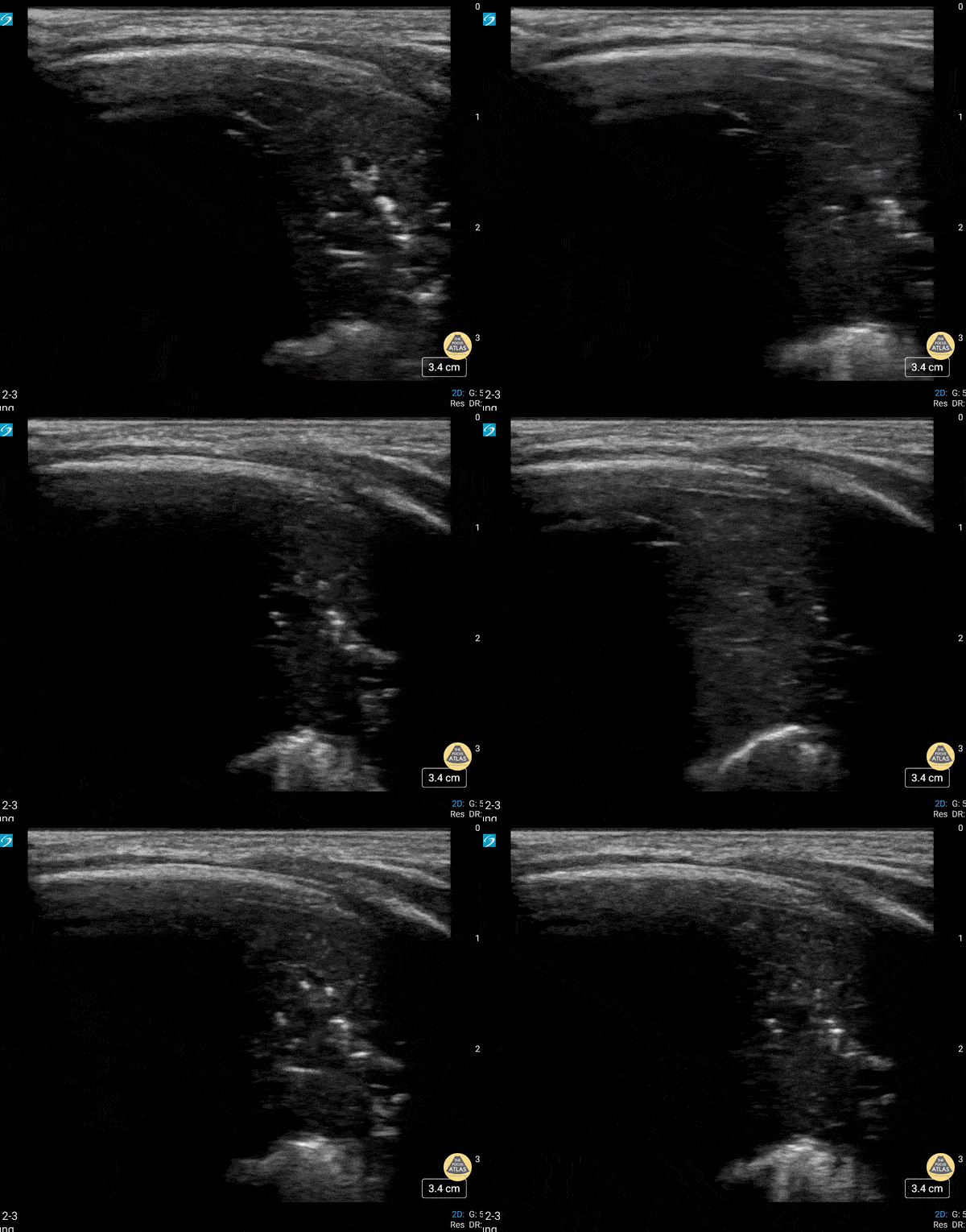
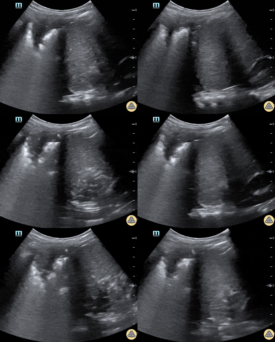
Lung consolidation in a child with fever and cough; faint dynamic air bronchogram seen
Contributor: Peter Gutierrez, MD, FAAP, Emory University School of Medicine/Children's Healthcare of Atlanta, @pocuspete
Neonate in respiratory failure status post intubation. The presence of dynamic air bronchograms helps point away from atelectasis.
Contributor: Peter Gutierrez, MD, FAAP | Emory University School of Medicine/Children's Healthcare of Atlanta | @pocuspete
Pleural Effusion
Large pleural effusion in a teenager
Contributor: Peter Gutierrez, MD, FAAP, Emory University School of Medicine/Children's Healthcare of Atlanta, @pocuspete
16 yo with initial diagnosis of pneumonia. Worsening shortness of breath. Found to have a loculated pleural effusion on POCUS.
Contributor: Kathryn Pade, MD, Rady Children's Hospital San Diego
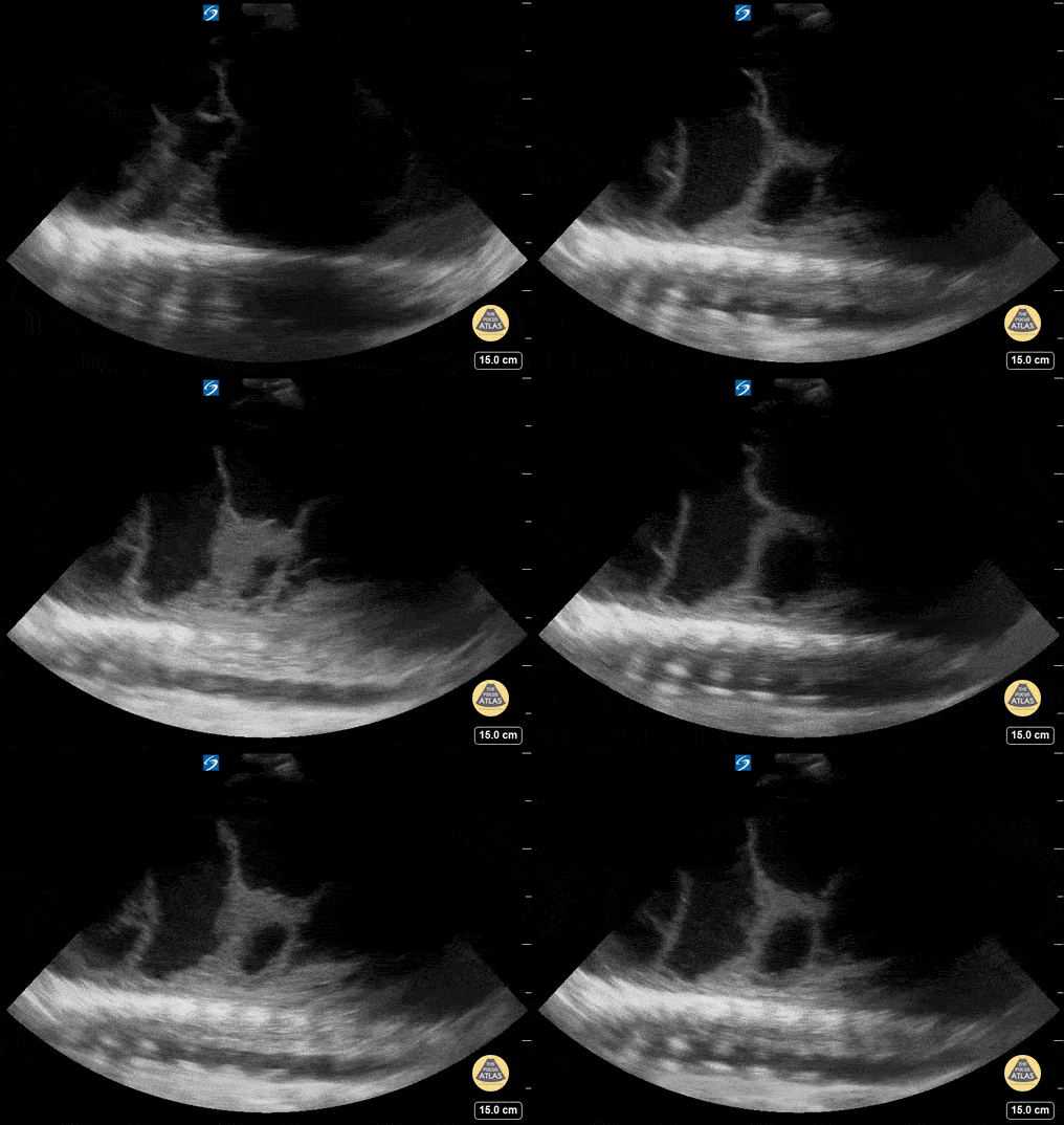
15 year old with complex PMH who presented with respiratory distress. CXR shows a large effusion. Bedside ultrasound showed an empyema.
Pneumothorax
Pediatric patient presenting with shortness of breath. Found to have a large left sided pneumothorax. M-mode tracing shows lack of lung sliding
Contributor: Kathryn Pade, MD, Rady Children's Hospital San Diego
Lung point indicative of pneumothorax.
Contributor: Peter Gutierrez, MD, FAAP, Emory University School of Medicine/Children's Healthcare of Atlanta, @pocuspete
Lack of lung sliding.
Contributor: Peter Gutierrez, MD, FAAP | Emory University School of Medicine/Children's Healthcare of Atlanta | @pocuspete
Other Pathology
Intubation attempt into the esophagus of an 8 year old male with respiratory distress, notice the soft tissue posterior to the trachea moving with the provider attempting the pass the tube.
Image contributed by: Zach Boivin, MD, @ZachBoivinMD
32 weeks premature. 4 weeks of life in the course of weaning from oxygen with respiratory deterioration requiring orotracheal intubation
Evidence of lung collapse with static bronchogram in the right upper lobe.
Contributor: Mg. Andres Silva Horna, Hospital Cayetano Heredia Piura-Peru
The patient, presenting with stridor, is supine and the airway is seen from the anterior neck in transverse orientation. As the probe is fanned, the bright white line is seen to widen. This column of air moves with inspiration. At its narrowest it is only a few millimeters wide. Growth along the lateral tracheal walls has caused significant tracheal stenosis. In this case, US was used to determine the width of the tracheal column and determine that passage of an ETT would not be feasible. The patient was taken to the OR for an emergent surgical airway. Use of US to estimate tracheal diameter is a novel application.
Andrew Liteplo MD, RDMS - Massachusetts General Hospital
Chief, Division of Ultrasound in Emergency Medicine
Director, Emergency Ultrasound Fellowship
5 year old male that drank out of container with gasoline and started coughing and was breathing fast. On exam appeared tachypneic, with air entry bilaterally and subcostal retractions.
POCUS revealed bilateral subpleural consolidations, confirmed with CXR. This imaging finding is similar to findings seen in other consolidative processes. This was suggestive of an aspiration pneumonitis.
Dr. Sathya Subramaniam, Pediatric EM Fellow - Kings County/SUNY Downstate
6 y/o sickle cell (HbSS) coughing with left-sided chest pain and 1 day of fever. Lungs without crackles, good air entry bilaterally.
A consolidative process is seen with a hypoechoic region with posterior enhancement greater than 1 cm in an area where normal A lines should be present. This is highly suggestive of acute chest syndrome given clinical features.
Dr. Sathya Subramaniam, Pediatric EM Fellow - Kings County/SUNY Downstate
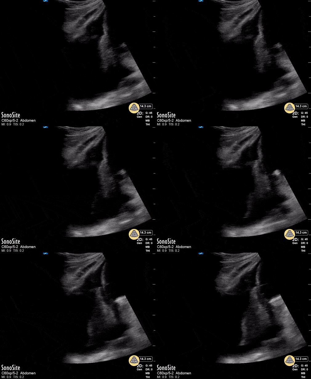
A child with history of sickle cell presented with upper abdominal and back pain and was found to be tachypneic with low grade fever. Ultrasound of the left upper quadrant demonstrated basilar left lung consolidation. Subsequent chest x-ray was interpreted as infiltrate consistent with acute chest syndrome.
Michael Cover, @michaelc0ver