
GI Jr
Gastrointestinal Jr
Note: These search bar results are only for the GI Jr image archive. To search the entire site, click here.
Recently Added
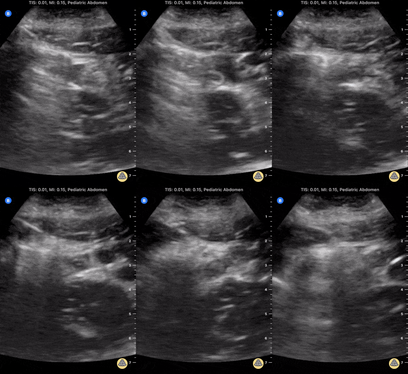
Teenage male presenting with abdominal pain worsening over 24 hours, diagnosed with acute appendicitis via POCUS.
The clip shows a circular structure which measures at 6.5 mm transversely, representing an acutely inflamed appendix with surrounding anechoic free fluid.
POCUS Acute Appendicitis: noncompressible, diameter >6mm, single wall >3mm are direct signs of appendicitis. Use of POCUS for diagnosis of acute appendicits significantly reduces cost and radiation exposure for patients without sacrificing diagnostic accuracy.
Contributor: C. Malcolm Roberson, MD. @ProjectUltraEM
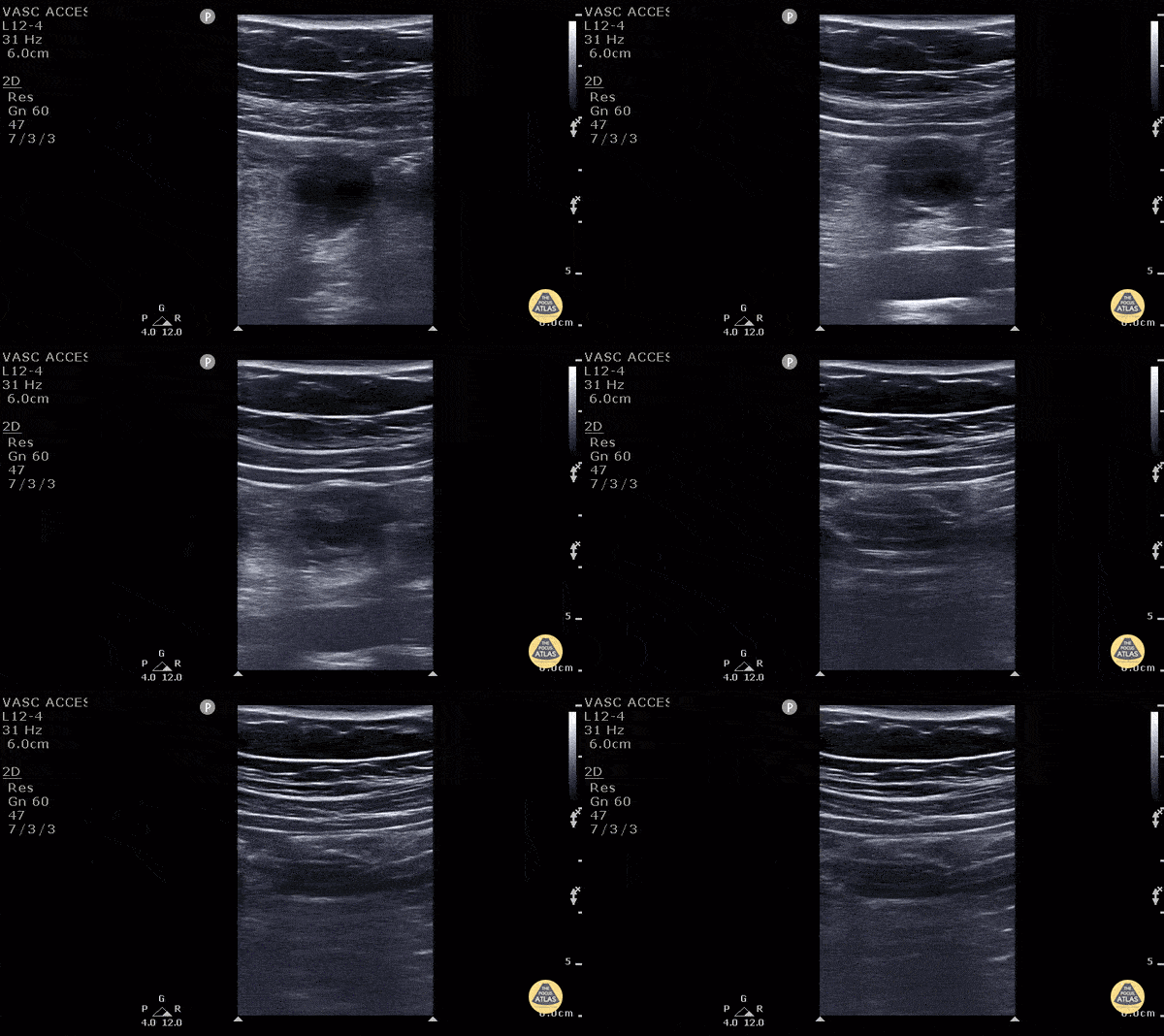
11 y/o M presented with 1 day of periumbilical pain that migrated to the right lower quadrant with nausea, vomiting and anorexia. Ultrasound with high frequency linear probe demonstrates an enlarged appendix with an external diameter of 1.32 cm with trace free fluid posteriorly, as well as a fecalith at the proximal end of the appendix. Surgery was consulted who requested a formal US which was non-diagnostic. Surgery took the patient anyway, and MRI confirmed our findings (including the large diameter).
Diagnostic criteria for appendicitis: a non-compressible, aperistaltic, blind ended structure >6mm diameter. Visualizing free fluid, tenderness in that area, and visualizing a fecalith can also add to the diagnosis.
See the evidence atlas for more info about POCUS diagnosing appendicitis but when visualizing a diagnostic appendicitis, it carries a positive likelihood ratio of 9.24. PMID: 28214369
Jackie Chiou MS4, Dr. Matthew Riscinti and Dr. Tian Liang - Kings County Emergency Medicine
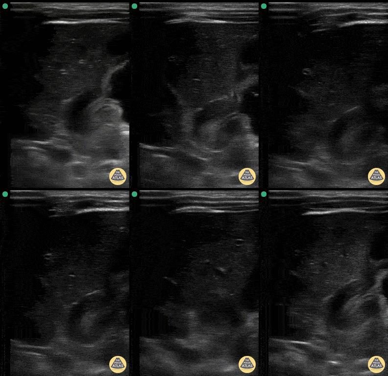
Pyloric Stenosis
4 w/o male with forceful vomiting with each feed. Clip fans through hypertrophic pylorus measuring 14mm in diameter and channel length 24mm. It also shows a fluid filled stomach and the "antral nipple sign" - outpouching of pyloric tissue into antrum.
The "Pi" π = 3.1415 mnemonic for ballpark measurement cut-offs.
>3mm diameter of single muscular wall
>14mm transverse diameter of pylorus
>15mm channel length
Lilly Bellman, MD - PEM /US fellow Harbor-UCLA
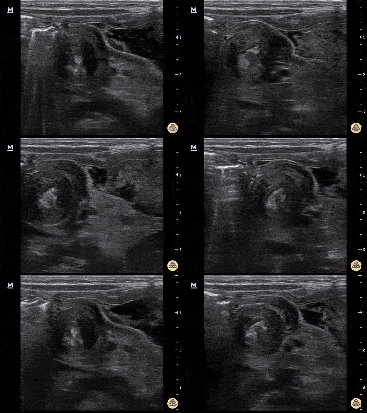
4-week-old, vomiting intermittently for 2 weeks, seen PCP 3 days ago, reassured. Revisit today and PCP concerned for pyloric stenosis, so referred to ED. Exam in ED reassuring for well-appearing neonate.
In ED, POCUS completed revealing hypertrophic pyloric stenosis. Pylorus muscle hypertrophied and thickened in both transverse and logitudinal view. Transverse view demonstrates the classic target sign seen in pyloric stenosis.
Dr. Sathya Subramaniam, Pediatric EM Fellow - Kings County/SUNY Downstate
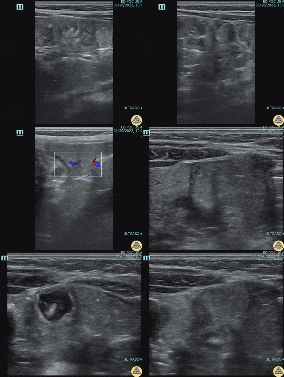
2 year-old male with history of constipation presented with one episode of painless hematochezia that occurred 1 hour prior to arrival. He had a benign abdominal exam. POCUS revealed a focal fluid collection in the RLQ with a bowel wall appearance containing a hyperechoic focus, most suspicious for a Meckel’s diverticulum with fecalith. Surgical resection and pathology confirmed a Meckel's diverticulum.
Dr. Kelly McWilliams, PGY-2, Denver Health Residency in Emergency Medicine
Dr. Anna Abrams, Pediatric Emergency Medicine Fellow, Childrens Hospital Colorado
Dr. Jon Orsborn, Director of Pediatric POCUS, Childrens Hospital Colorado
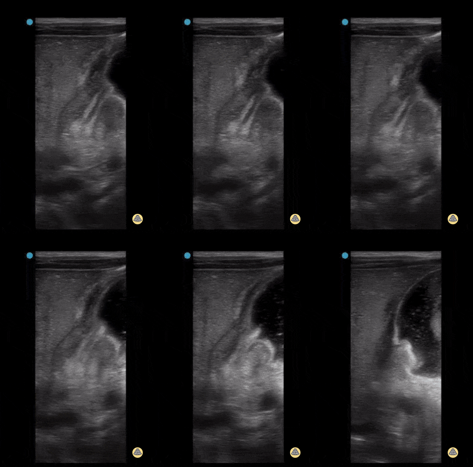
3 week old with projectile vomiting, POCUS showed positive astral nipple sign which is a highly specific finding for pyloric stenosis - the redundant pyloric mucosa protrudes into the gastric antrum. The measurements show increased pyloric muscle thickness (>3mm) and increased pyloric longitudinal measurement (>15 - 17 mm)
Measurements can be remembered using "Pi Rule"
- Pyloric muscle thickness, i.e. diameter of a single muscular wall on a transverse image >3 mm
- Pyloric transverse diameter >14 mm
- Pyloric longitudinal measurement >15 - 17 mm
Contributed by:
Dimitri Livshits DO, Ultrasound Fellow; Jane Belyavskaya MD, Ultrasound Fellow; Chris Hanuscin MD, Ultrasound Division Director (Kings County/SUNY Downstate)
Normal Anatomy
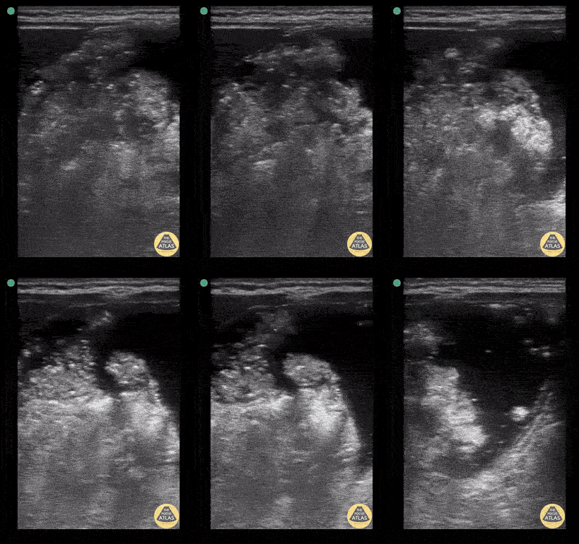
Gastric content eval for patient undergoing procedural sedation. With the patient in the right lateral decubitus position the linear probe is placed in a subxiphoid sagittal axis with the probe marker towards the head. The stomach is seen immediately caudal to the liver. The class 5 layer bowel wall of the stomach can be seen containing large volume, mixed echogenicity content.
Contributor: Matthew Moake, MD PhD
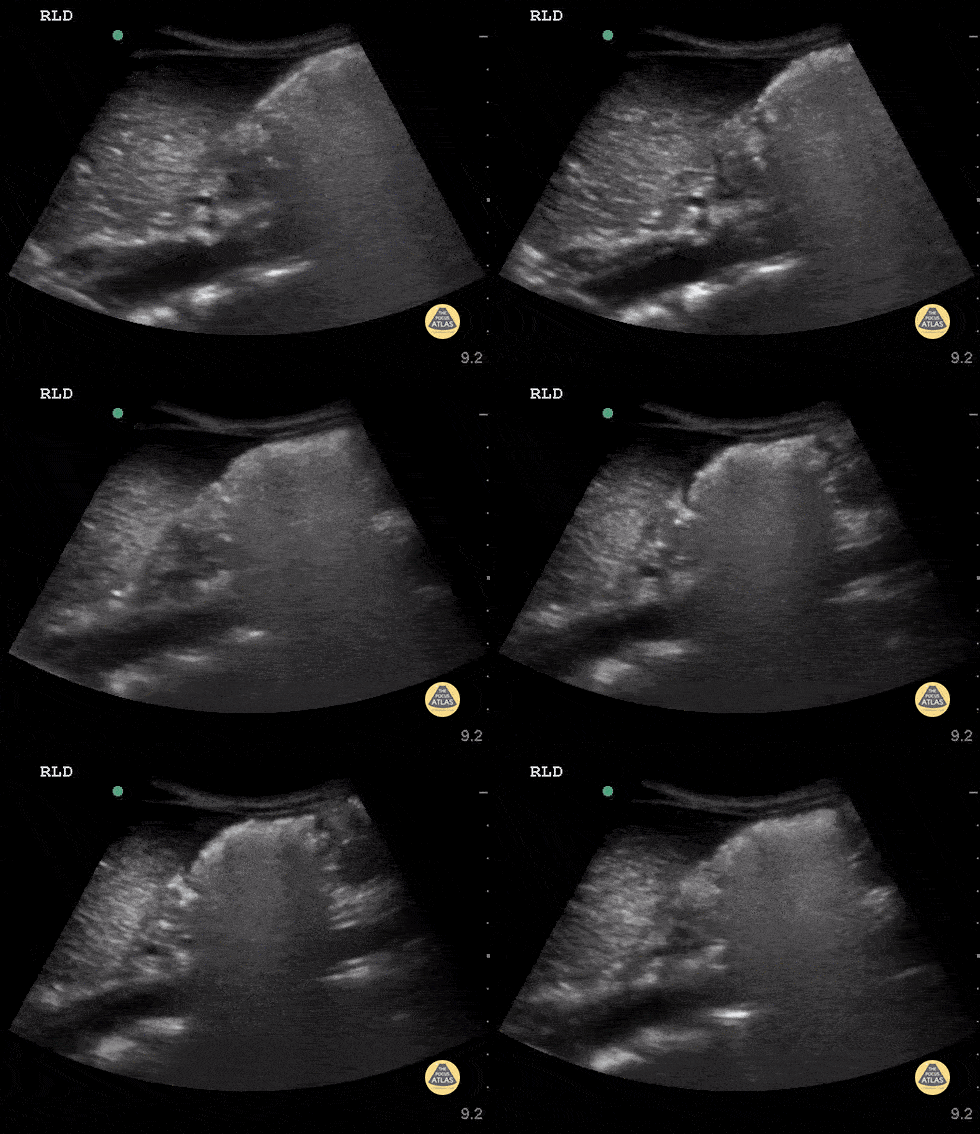
Ultrasound used to assess gastric content prior to procedural sedation. Sagittal view in the epigastric region using the curvilinear probe with the patient in the right lateral decubitus position. Probe indicator cephalad. The gastric antrum is seen in the upper right with heavily air-admixed content with dirty shadow obscuring deeper content. The liver is seen in the upper left and the aorta in the deep field.
Contributor: Matthew Moake, MD PhD
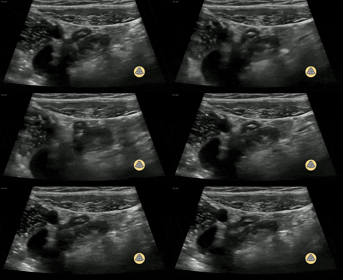
10 y/o with abdominal pain. Normal appendix identified medial to the iliac vessels. Please see other image in series for doppler.
Contributor: Elena Chen, MD
Appendicitis - Check out our Appendicitis Post Here!
Teenage male presenting with abdominal pain worsening over 24 hours, diagnosed with acute appendicitis via POCUS.
The clip shows a circular structure which measures at 6.5 mm transversely, representing an acutely inflamed appendix with surrounding anechoic free fluid.
POCUS Acute Appendicitis: noncompressible, diameter >6mm, single wall >3mm are direct signs of appendicitis. Use of POCUS for diagnosis of acute appendicits significantly reduces cost and radiation exposure for patients without sacrificing diagnostic accuracy.
Contributor: C. Malcolm Roberson, MD. @ProjectUltraEM
11 y/o M presented with 1 day of periumbilical pain that migrated to the right lower quadrant with nausea, vomiting and anorexia. Ultrasound with high frequency linear probe demonstrates an enlarged appendix with an external diameter of 1.32 cm with trace free fluid posteriorly, as well as a fecalith at the proximal end of the appendix. Surgery was consulted who requested a formal US which was non-diagnostic. Surgery took the patient anyway, and MRI confirmed our findings (including the large diameter).
Diagnostic criteria for appendicitis: a non-compressible, aperistaltic, blind ended structure >6mm diameter. Visualizing free fluid, tenderness in that area, and visualizing a fecalith can also add to the diagnosis.
See the evidence atlas for more info about POCUS diagnosing appendicitis but when visualizing a diagnostic appendicitis, it carries a positive likelihood ratio of 9.24. PMID: 28214369
Jackie Chiou MS4, Dr. Matthew Riscinti and Dr. Tian Liang - Kings County Emergency Medicine
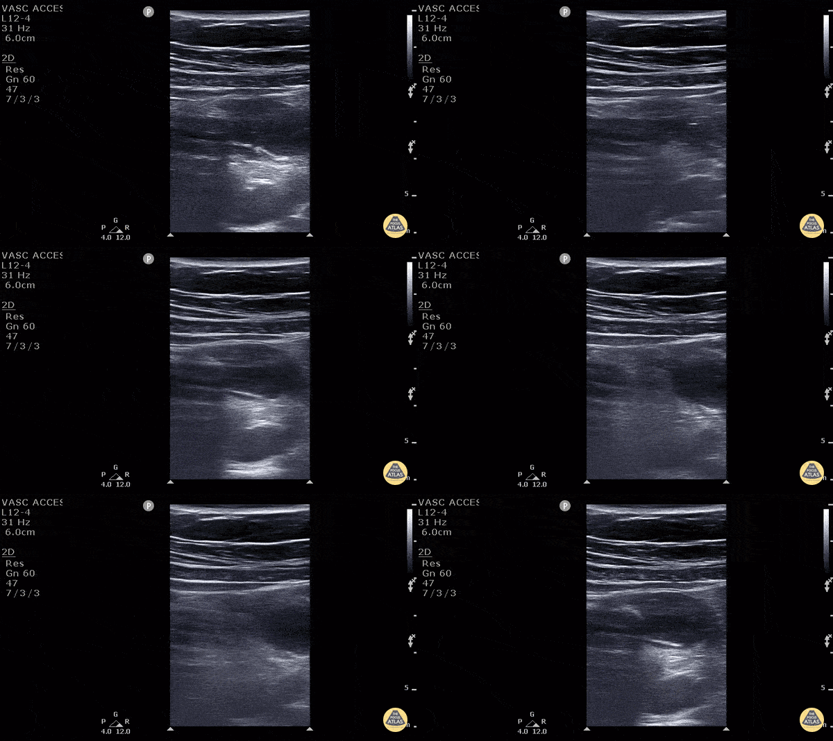
11 y/o M presented with 1 day of periumbilical pain that migrated to the right lower quadrant with nausea, vomiting and anorexia. Ultrasound with high frequency linear probe demonstrates an enlarged appendix with an external diameter of 1.32 cm with trace free fluid posteriorly, as well as a fecalith at the proximal end of the appendix. Surgery was consulted who requested a formal US which was non-diagnostic. Surgery took the patient anyway, and MRI confirmed our findings (including the large diameter).
Diagnostic criteria for appendicitis: a non-compressible, aperistaltic, blind ended structure >6mm diameter. Visualizing free fluid, tenderness in that area, and visualizing a fecalith can also add to the diagnosis.
See the evidence atlas for more info about POCUS diagnosing appendicitis but when visualizing a diagnostic appendicitis, it carries a positive likelihood ratio of 9.24. PMID: 28214369
Jackie Chiou MS4, Dr. Matthew Riscinti and Dr. Tian Liang - Kings County Emergency Medicine
Intussusception
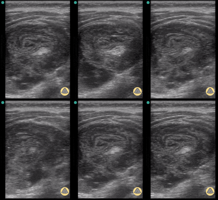
23 month old with ileocolic intussusception. Pseudokidney sign seen here due to oblique orientation of the linear transducer.
Contributor: Antonio Riera, MD
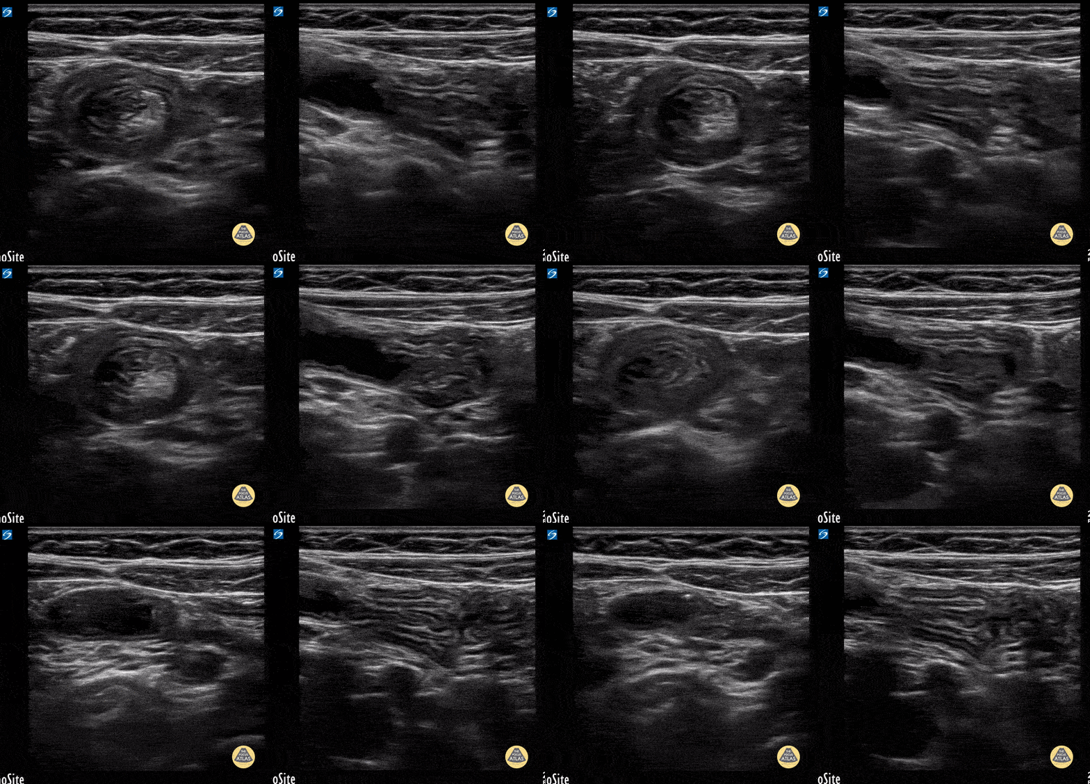
7y female with N/V and tactile fever. Benign abdominal exam. Note the small size of the intuss. In long axis you can easily track the bowel wall as it folds into itself and see it is slowly sliding in and out a small bit.
Contributor: Matthew Moake, MD PhD
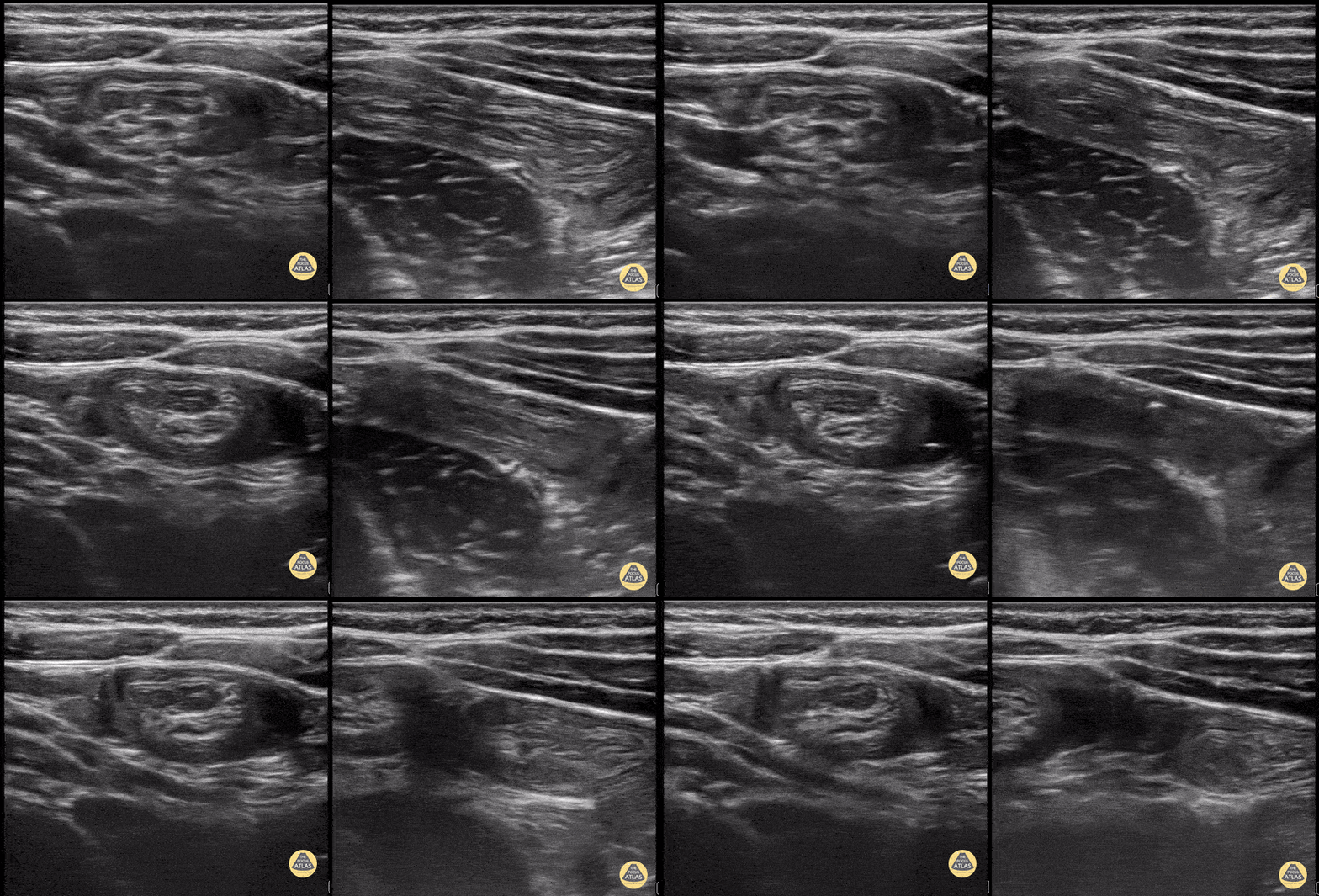
Toddler with colicky abdominal pain. LUQ with ileo-ileal intuss. Note the smaller size and active peristalsis of the intuss.
Contributor: Matthew Moake, MD PhD
Pyloric Stenosis
Pyloric Stenosis
4 w/o male with forceful vomiting with each feed. Clip fans through hypertrophic pylorus measuring 14mm in diameter and channel length 24mm. It also shows a fluid filled stomach and the "antral nipple sign" - outpouching of pyloric tissue into antrum.
The "Pi" π = 3.1415 mnemonic for ballpark measurement cut-offs.
>3mm diameter of single muscular wall
>14mm transverse diameter of pylorus
>15mm channel length
Lilly Bellman, MD - PEM /US fellow Harbor-UCLA
4-week-old, vomiting intermittently for 2 weeks, seen PCP 3 days ago, reassured. Revisit today and PCP concerned for pyloric stenosis, so referred to ED. Exam in ED reassuring for well-appearing neonate.
In ED, POCUS completed revealing hypertrophic pyloric stenosis. Pylorus muscle hypertrophied and thickened in both transverse and logitudinal view. Transverse view demonstrates the classic target sign seen in pyloric stenosis.
Dr. Sathya Subramaniam, Pediatric EM Fellow - Kings County/SUNY Downstate
3 week old with projectile vomiting, POCUS showed positive astral nipple sign which is a highly specific finding for pyloric stenosis - the redundant pyloric mucosa protrudes into the gastric antrum. The measurements show increased pyloric muscle thickness (>3mm) and increased pyloric longitudinal measurement (>15 - 17 mm)
Measurements can be remembered using "Pi Rule"
- Pyloric muscle thickness, i.e. diameter of a single muscular wall on a transverse image >3 mm
- Pyloric transverse diameter >14 mm
- Pyloric longitudinal measurement >15 - 17 mm
Contributed by:
Dimitri Livshits DO, Ultrasound Fellow; Jane Belyavskaya MD, Ultrasound Fellow; Chris Hanuscin MD, Ultrasound Division Director (Kings County/SUNY Downstate)
Bowel Obstruction
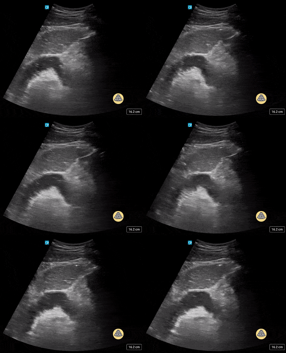
17 yo male with history of abdominal surgeries (g-tube and fundoplication) and chronic constipation who comes in with lower abdominal pain. POCUS shows stool to and fro (tanga sign).
Contributor: Paul Khalil, MD Nicklaus Children's Hospital @khalil3paul
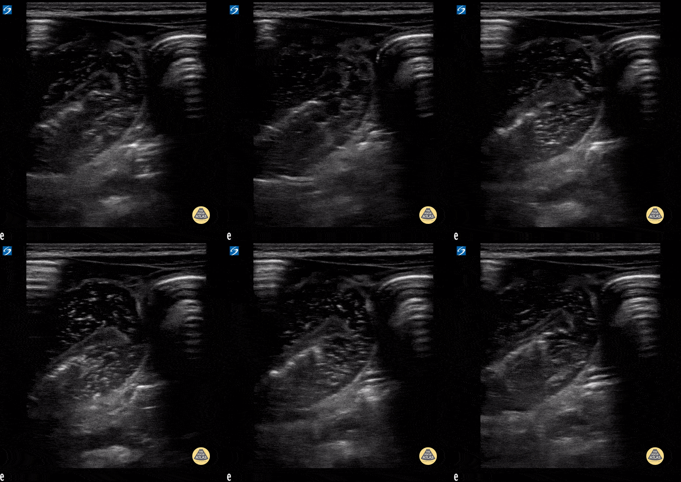
Small bowel obstruction with to and fro peristalsis visualized.
Contributor: Peter Gutierrez, MD FAAP FACEP; Children's Healthcare of Atlanta; @pocuspete
Ascites
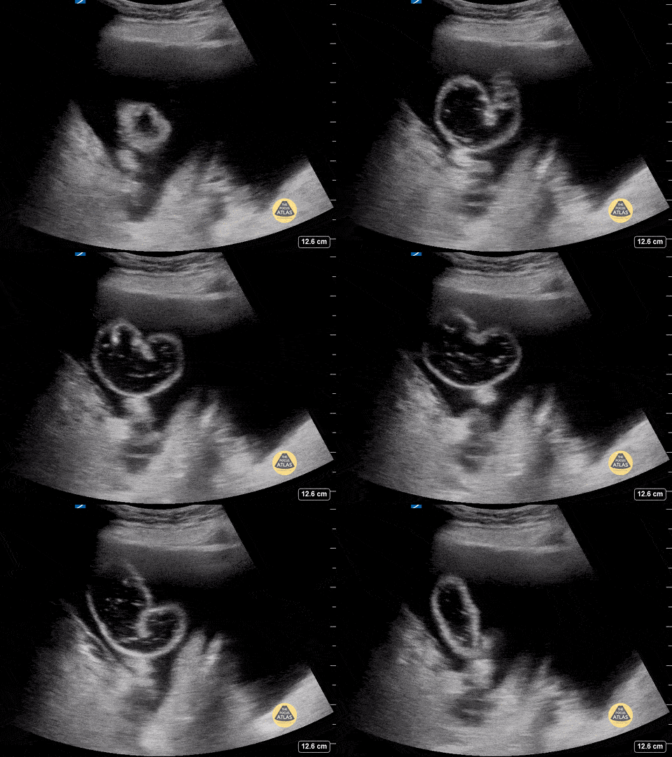
Ascites with floating bowel and questionable to-and-fro peristalsis.
Contributor: Peter Gutierrez, MD, FAAP, Emory University School of Medicine/Children's Healthcare of Atlanta, @pocuspete
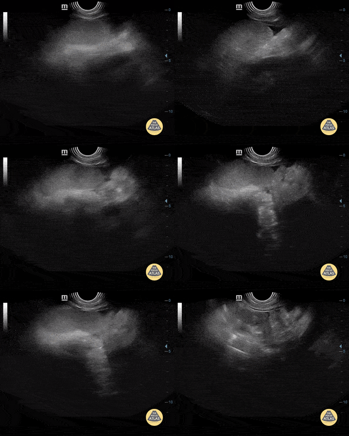
4-year-old boy with severe dengue with free fluid in the abdominal cavity.
Contributor: Mg. Andres Silva Horna, Hospital Cayetano Heredia Piura-Peru
Other Pathology
2 year-old male with history of constipation presented with one episode of painless hematochezia that occurred 1 hour prior to arrival. He had a benign abdominal exam. POCUS revealed a focal fluid collection in the RLQ with a bowel wall appearance containing a hyperechoic focus, most suspicious for a Meckel’s diverticulum with fecalith. Surgical resection and pathology confirmed a Meckel's diverticulum.
Dr. Kelly McWilliams, PGY-2, Denver Health Residency in Emergency Medicine
Dr. Anna Abrams, Pediatric Emergency Medicine Fellow, Childrens Hospital Colorado
Dr. Jon Orsborn, Director of Pediatric POCUS, Childrens Hospital Colorado
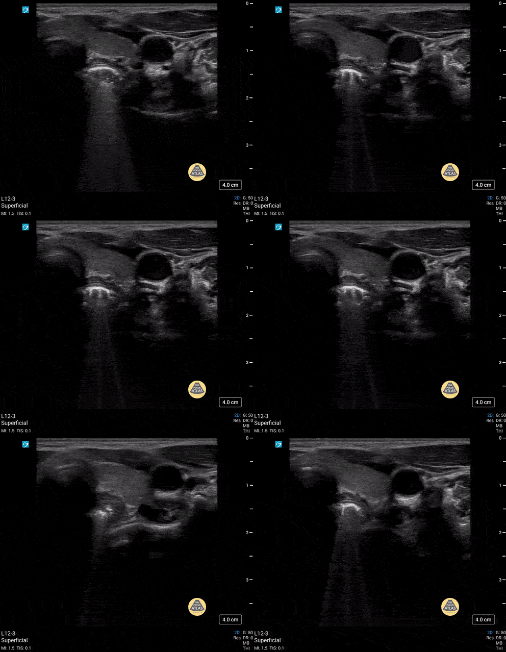
5 yo male presents with swallowed decorative marble. no difficulty breathing. unable to handle secretions. POCUS shows an esophageal FB that was later removed by surgery.
Contributor: Paul Khalil, MD Nicklaus Children's Hospital, @khalil3paul
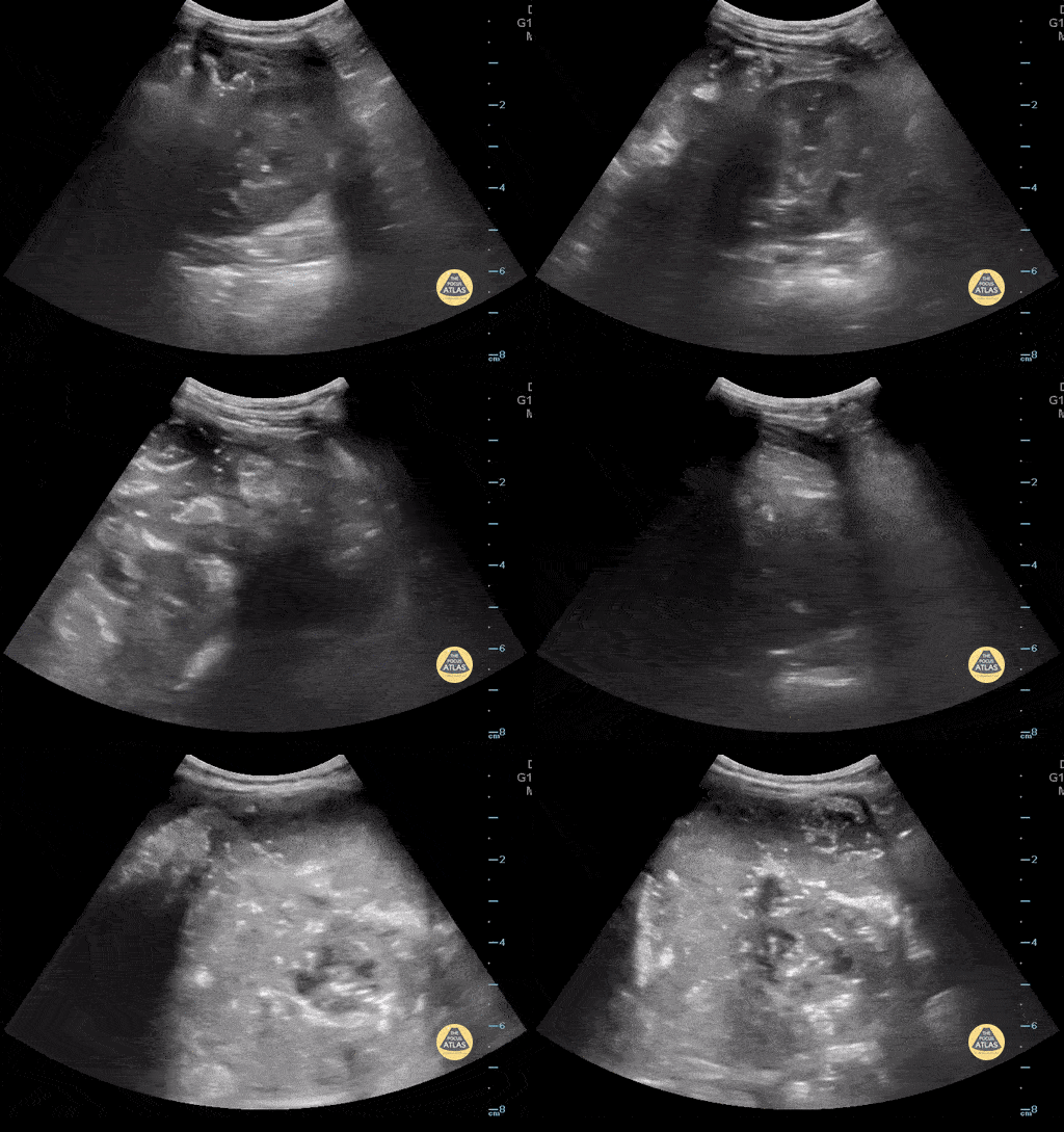
18 mo F sent from PMD for mass in the RUQ. POCUS shows extra renal mass consistent with neuroblastoma that was later confirmed by pathology.
Contributor: Paul Khalil, MD Nicklaus Children's Hospital
Gastric content eval for patient undergoing procedural sedation. With the patient in the right lateral decubitus position the linear probe is placed in a subxiphoid sagittal axis with the probe marker towards the head. The stomach is seen immediately caudal to the liver. The class 5 layer bowel wall of the stomach can be seen containing large volume, mixed echogenicity content.
Contributor: Matthew Moake, MD PhD
Ultrasound used to assess gastric content prior to procedural sedation. Sagittal view in the epigastric region using the curvilinear probe with the patient in the right lateral decubitus position. Probe indicator cephalad. The gastric antrum is seen in the upper right with heavily air-admixed content with dirty shadow obscuring deeper content. The liver is seen in the upper left and the aorta in the deep field.
Contributor: Matthew Moake, MD PhD

3 yo presents with enlarging abdomen and mass palpated. A large abdominal mass was seen on POCUS. The kidney is seen directly below the mass in the image.
Contributor: Kathryn Pade, MD, Rady Children's Hospital San Diego