
Musculoskeletal
Musculoskeletal
Looking for Pediatric MSK Ultrasound Clips? Check out our MSK Junior Page (Beta)!
Note: These search bar results are only for the Musculoskeletal image archive. To search the entire site, click here.
Recently Added
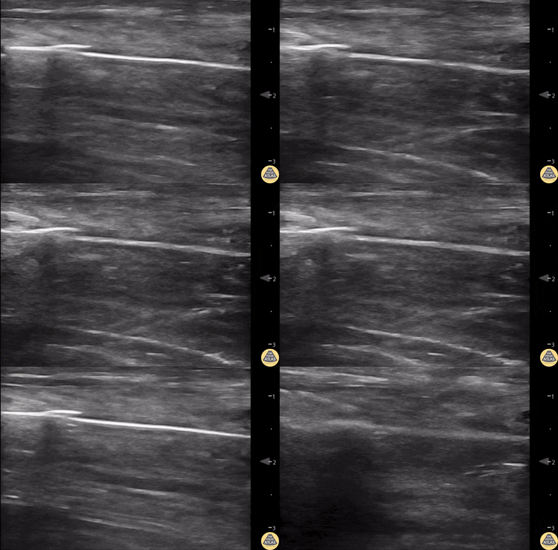
55 yo F s/p RT TKA w/ revision presents after a fall. POCUS was w/o tendinous/bony abnormality. RT knee x-ray was w/o definite acute fracture.
The American College of Radiology (ACR) promotes US evaluation of “pain after...(TKA with) suspect(ed) periprosthetic soft-tissue abnormality unrelated to infection” after radiographic evaluation as “usually appropriate”.
Although ACEP US Guidelines boast 96.8% sensitivity and 99.7% specificity for diagnosing fractures, POCUS evaluation of traumatic arthroplasty complications does not exist. This creates an area of interest within POCUS.
Contributors: Lauren Lowes, DO; Justin Morin, DO; Garrett Richardson, MS4; Nava Kendall, MD
Central Michigan University Residency of Emergency Medicine
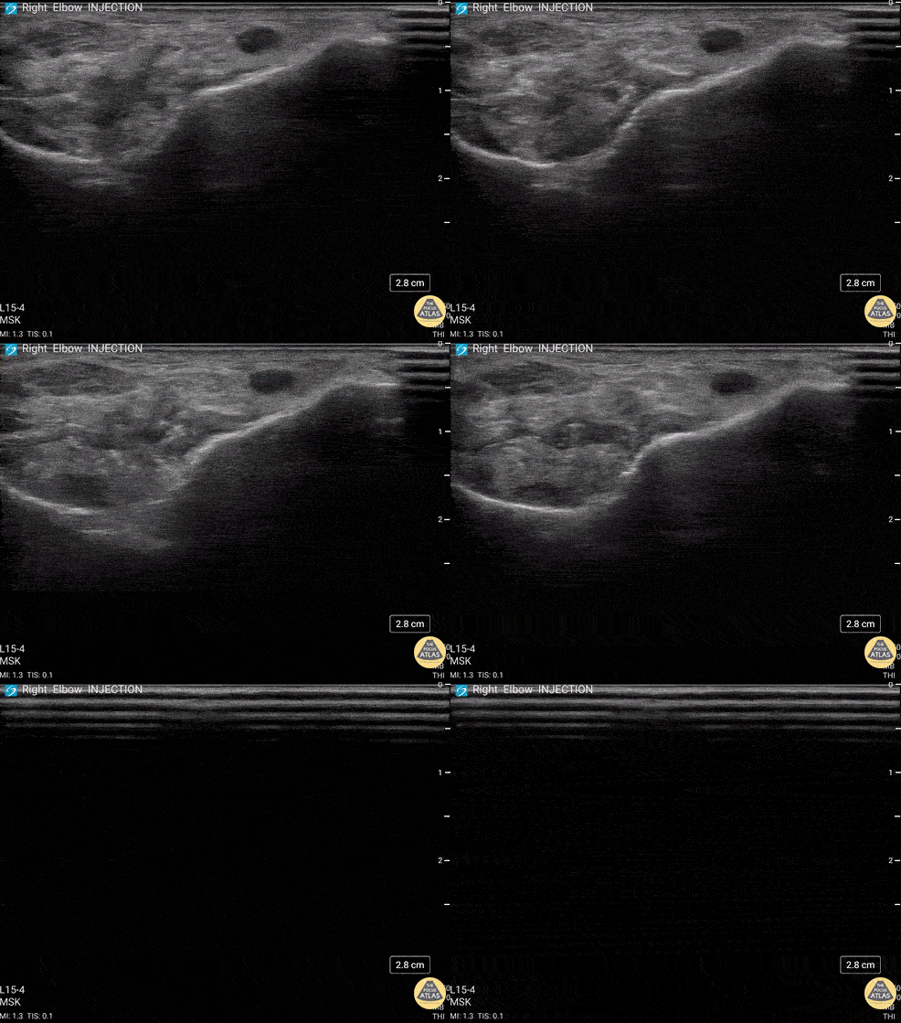
92 y/o M presented with moderate to severe osteoarthritis on elbow x-ray who was treated with ultrasound-guided corticosteroid joint injection.
Video shows transverse view of the posterior elbow as corticosteroid is injected with a posterolateral approach deep to the posterior elbow fat pad located in the olecranon fossa. This area is continuous with the elbow joint capsule. Posterolateral approach is advantageous due to decreased risk of disrupting neurovascular structures or the articular cartilage from other approaches to the joint space. Posterior approach is also advantageous compared to lateral approach in moderate to severe arthritis due to limited access to the radiocapitellar joint space.
Eben Alexander, DO
Devesh Patel, MD
Eastern Virginia Medical School
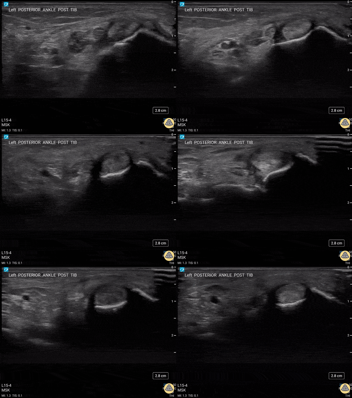
40 y/o F presented with 9 month history insidious onset L medial ankle pain and was found to have tibialis posterior tendon partial tear.
Video shows transverse view scanning proximal to distal at the posteromedial ankle (left is posterior). There is thickening of the posterior tibialis tendon with an anechoic tendon sheath effusion. At the level of the medial malleolus there are two moderate interstitial tears extending distally. Flexor digitorum and hallicus longus appear unaffected.
Eben Alexander, DO
Devesh Patel, MD
Eastern Virginia Medical School
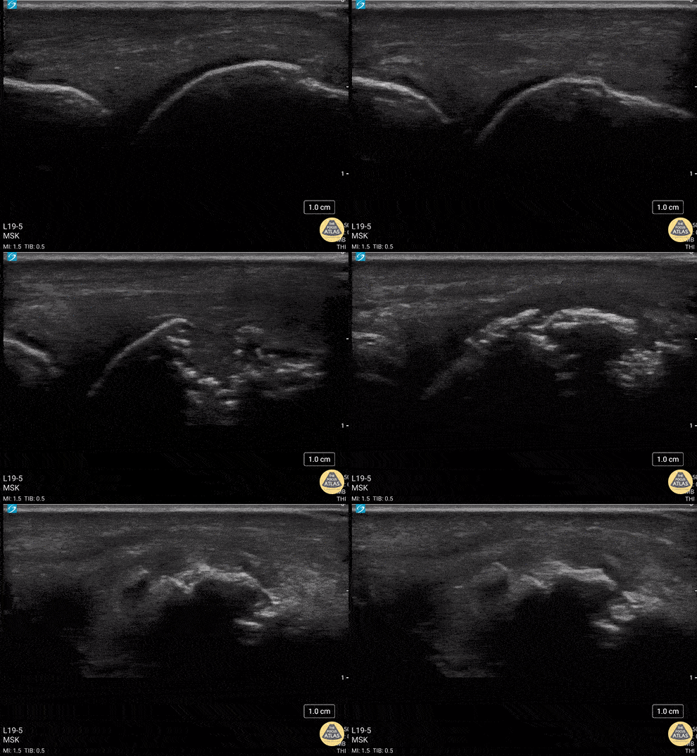
34 y/o M presented with fall while skiing injuring closed fist against ice and was found to have a displaced/impact fracture of his third metacarpal head.
Video shows sagittal view scanning in the ulnar to radial direction at the third metacarpophalangeal joint (right is proximal). There is diffuse disruption of the cortex of the radial aspect of the metacarpal head and displaced/impaction fracture of the metacarpal head. Usually this surface should appear smooth. Articular cartilage can be seen as anechoic at the interface between the two bones. This defect was not visualized on initial or 2 week post x-ray, so ultrasound was able to guide the clinical team to appropriately splint and manage as a fracture.
Eben Alexander, DO
Devesh Patel, MD
Eastern Virginia Medical School
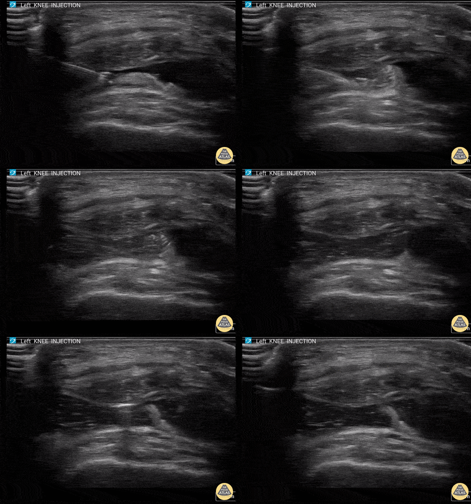
60 y/o F with knee arthritis getting corticosteroid joint injection in left suprapatellar recess.
Ultrasound probe in transverse axis just superior to the patella. Needle approach lateral to medial. Infiltrate seen injected into anechoic suprapatellar recess. The suprapatellar recess is continuous with the tibiofemoral joint. Deep to suprapellar recess in this video is the prefemoral fat pad. Just superior to the suprapatellar recess in this image is the quadriceps tendon in short axis appearing as a fibrillar structure.
Contributors:
Eben Alexander, DO
Devesh Patel, MD
Eastern Virginia Medical School
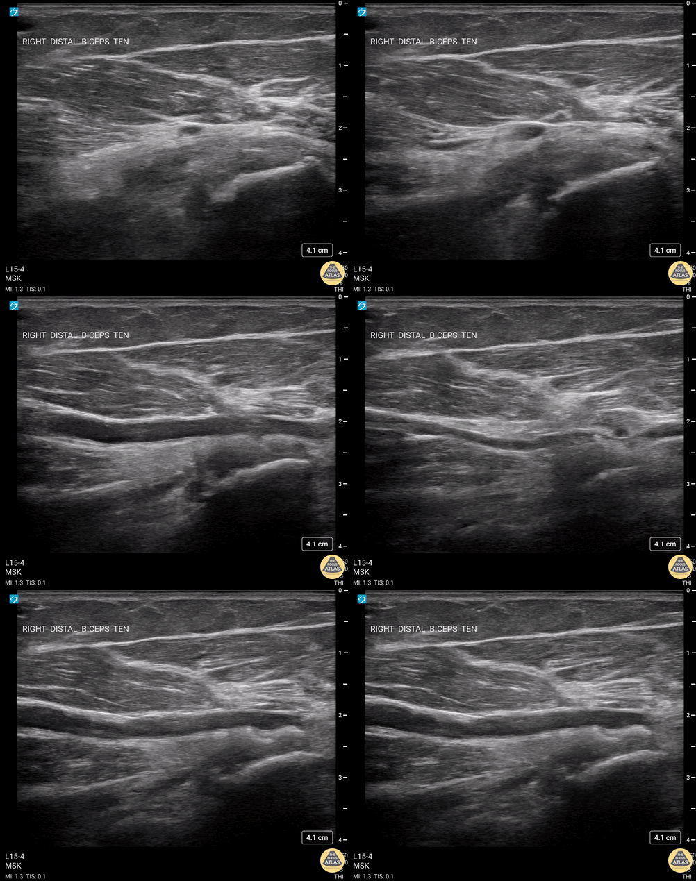
60 y/o F w/R elbow pain for 8 months found to have distal biceps tendinosis.
L=proximal; R=distal. Medial long-axis view of distal biceps tendon. Radial artery seen pulsating directly superficial to biceps tendon. Distal biceps tendon diffusely thickened with decreased echogenicity at its attachment site.
Contributor:
Eben Alexander, DO
Devesh Patel, MD
Eastern Virginia Medical School
Normal Anatomy
55 yo F s/p RT TKA w/ revision presents after a fall. POCUS was w/o tendinous/bony abnormality. RT knee x-ray was w/o definite acute fracture.
The American College of Radiology (ACR) promotes US evaluation of “pain after...(TKA with) suspect(ed) periprosthetic soft-tissue abnormality unrelated to infection” after radiographic evaluation as “usually appropriate”.
Although ACEP US Guidelines boast 96.8% sensitivity and 99.7% specificity for diagnosing fractures, POCUS evaluation of traumatic arthroplasty complications does not exist. This creates an area of interest within POCUS.
Contributors: Lauren Lowes, DO; Justin Morin, DO; Garrett Richardson, MS4; Nava Kendall, MD
Central Michigan University Residency of Emergency Medicine
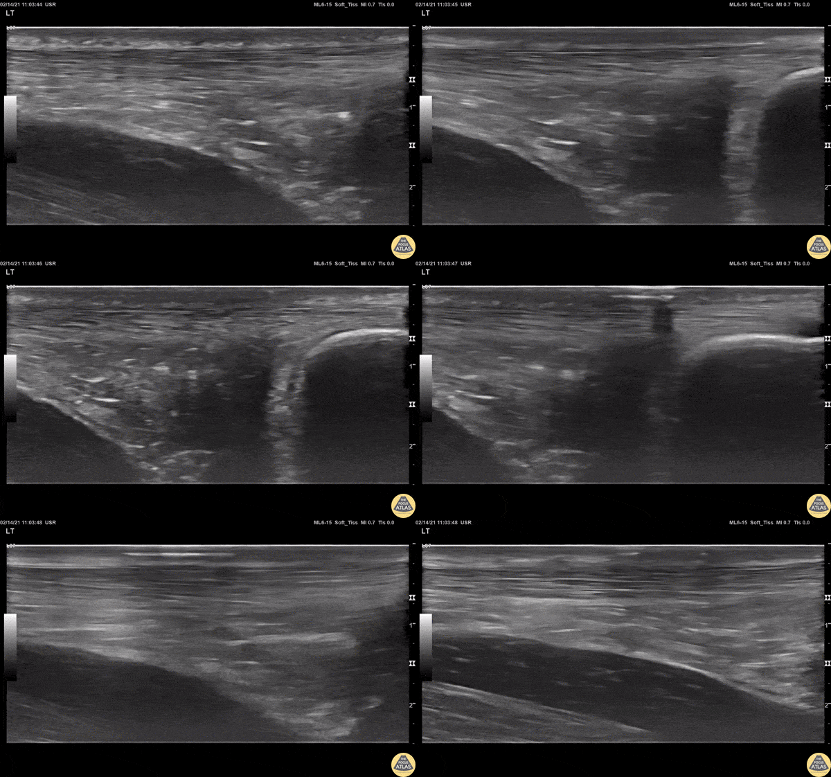
View of a normal Achilles tendon as seen in long axis, using a linear transducer. The relatively hyperechoic, linear fibers of the tendon are seen running from the calcaneus (right of screen, or inferior) to the gastrocnemius muscle (off screen to the left, or superior).
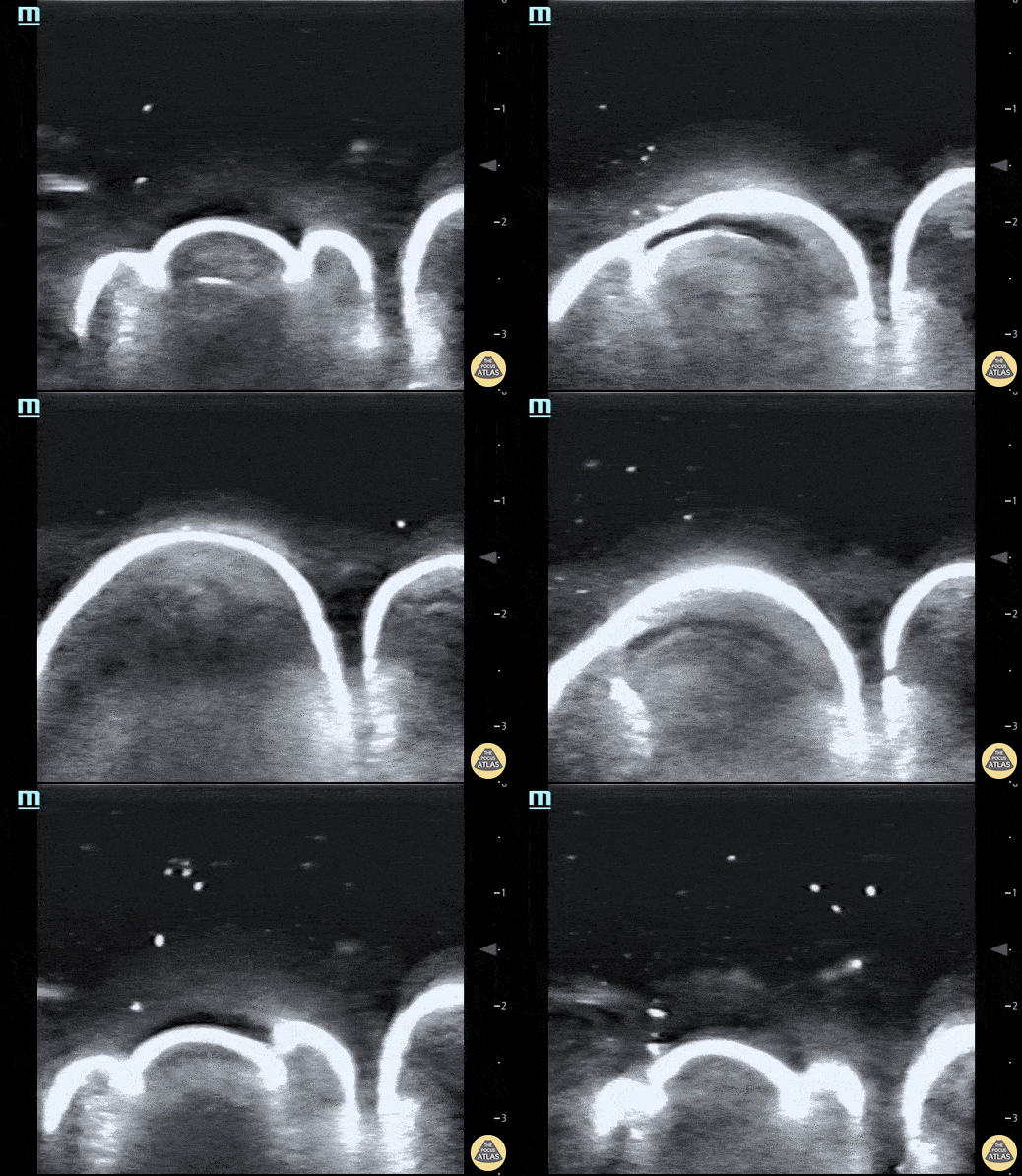
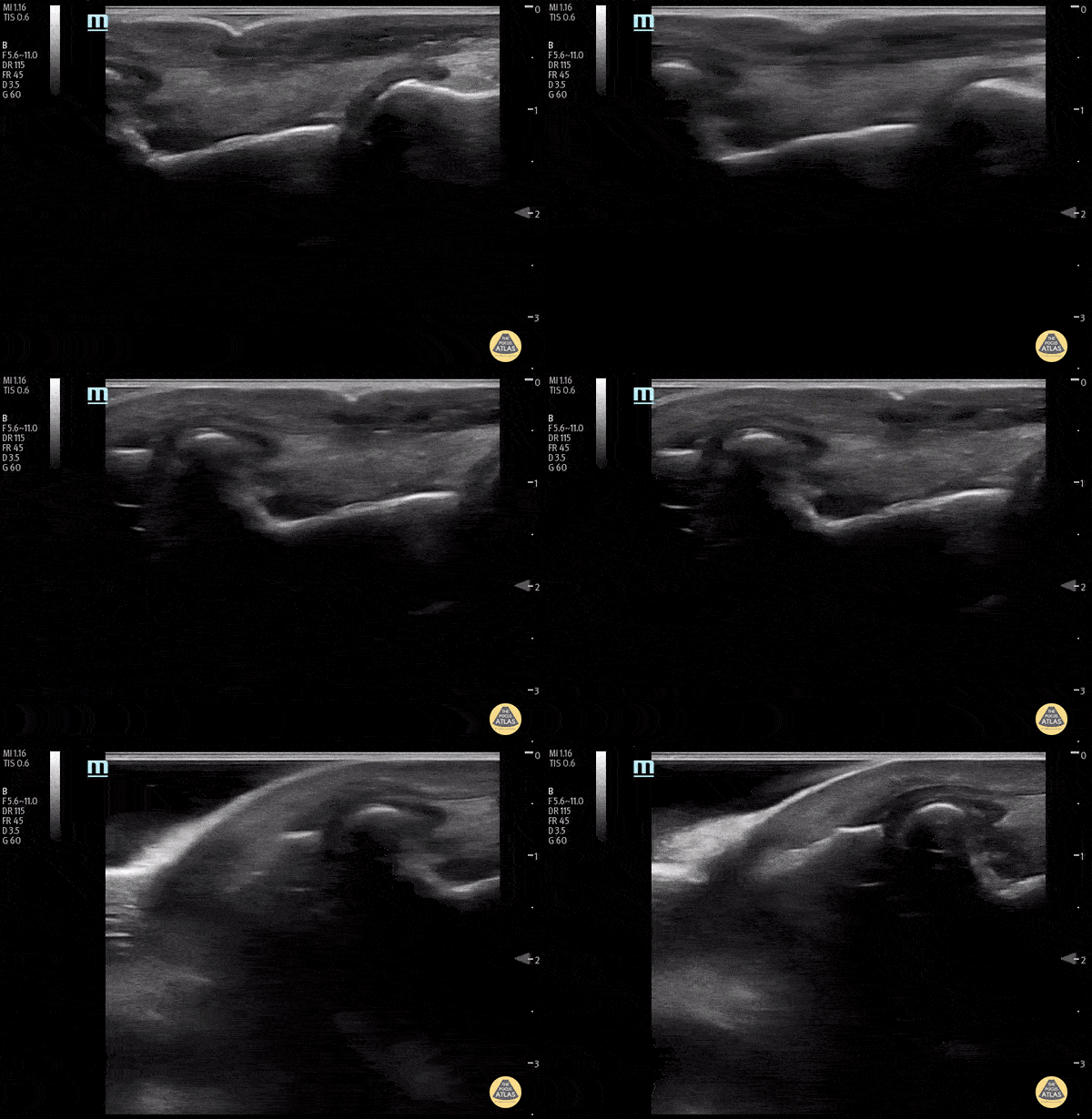
Pictured here is a water bath scan using a high frequency linear transducer; specifically this image visualizes the distal right great toe of a healthy young adult. Water bath scanning is a technique that augments the clarity and distinctness of structures that are otherwise challenging to visualize with POCUS including distal digital examinations. In addition to yielding higher quality images, the use of a water bath circumvents the need for direct contact with the area of interest, often resulting in less patient discomfort.
Moudi Hubeishy, MD. Rural Medicine PGY1. California, USA
@Hubeishy_MD
Colorized Normal Anatomy
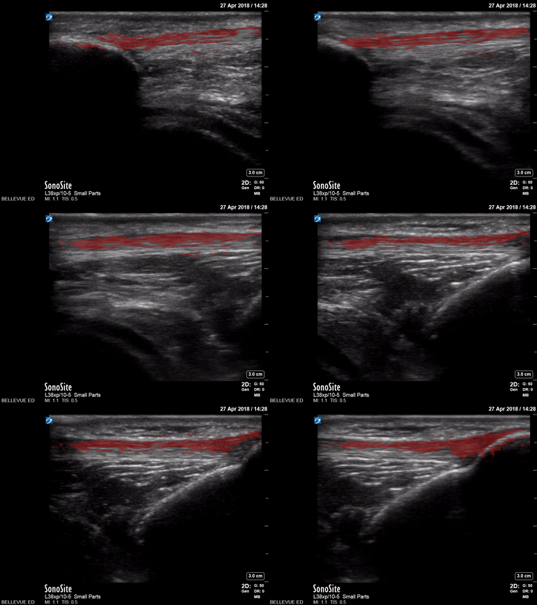
Patellar Tendon
Red: Patellar tendon
Images: Dr. Lindsay Davis, Dr. Hannah Kopinski. Image Editing: Michael Amador and Dr. Matthew Riscinti
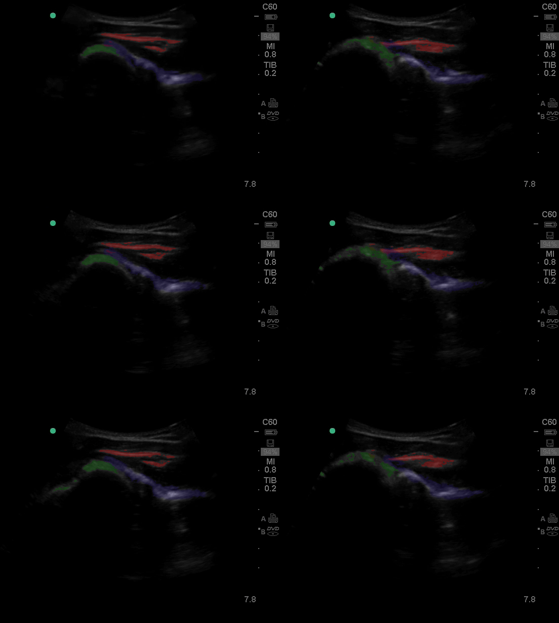
Shoulder
Green: Humeral head, Blue: Infraspinatus, Red: Rotator Cuff
Images: Dr. Lindsay Davis, Dr. Hannah Kopinski. Image Editing: Michael Amador and Dr. Matthew Riscinti
Fractures
34 y/o M presented with fall while skiing injuring closed fist against ice and was found to have a displaced/impact fracture of his third metacarpal head.
Video shows sagittal view scanning in the ulnar to radial direction at the third metacarpophalangeal joint (right is proximal). There is diffuse disruption of the cortex of the radial aspect of the metacarpal head and displaced/impaction fracture of the metacarpal head. Usually this surface should appear smooth. Articular cartilage can be seen as anechoic at the interface between the two bones. This defect was not visualized on initial or 2 week post x-ray, so ultrasound was able to guide the clinical team to appropriately splint and manage as a fracture.
Eben Alexander, DO
Devesh Patel, MD
Eastern Virginia Medical School
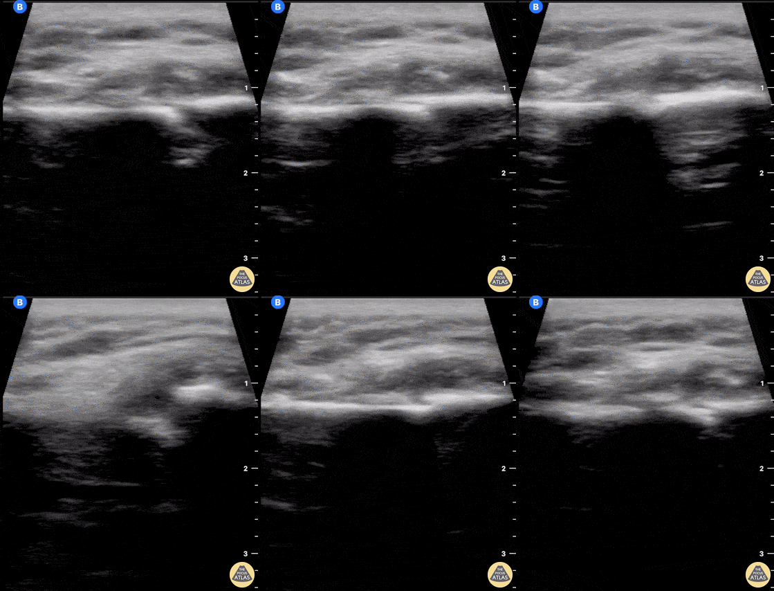
60 year old female with a subacute left clavicular fracture (occurred 2 weeks ago) presented with worsening pain at fracture site of onset while working with occupational therapy. Seen here is the left clavicle (hyperechoic structure) with noted fracture and mild heterogeneous (concern for bloody accumulation) edema around fracture site as observed in long axis view.
Kwasi Ampomah, DO, Eben Alexander IV, DO, Tariq Niazi, MD
EVMS PM&R
Dislocations
A 30-year-old male presented to the emergency department with an inferior shoulder dislocation (luxatio erecta). Studies have shown that ultrasound has a very high sensitivity and specificity nearing 100% each (Secko 2020) in identification of the same. On ultrasound, the humeral head is shown dislocated anterior to the glenoid fossa, similar to typical anterior shoulder dislocations. This patient's shoulder was reduced with simple traction/countertraction under procedural sedation without any complications.
Mark Zhang
Secko MA, Reardon L, Gottlieb M, et al. Musculoskeletal ultrasonography to diagnose dislocated shoulders: a prospective cohort. Annals of Emergency Medicine. 2020;76(2):119-128.
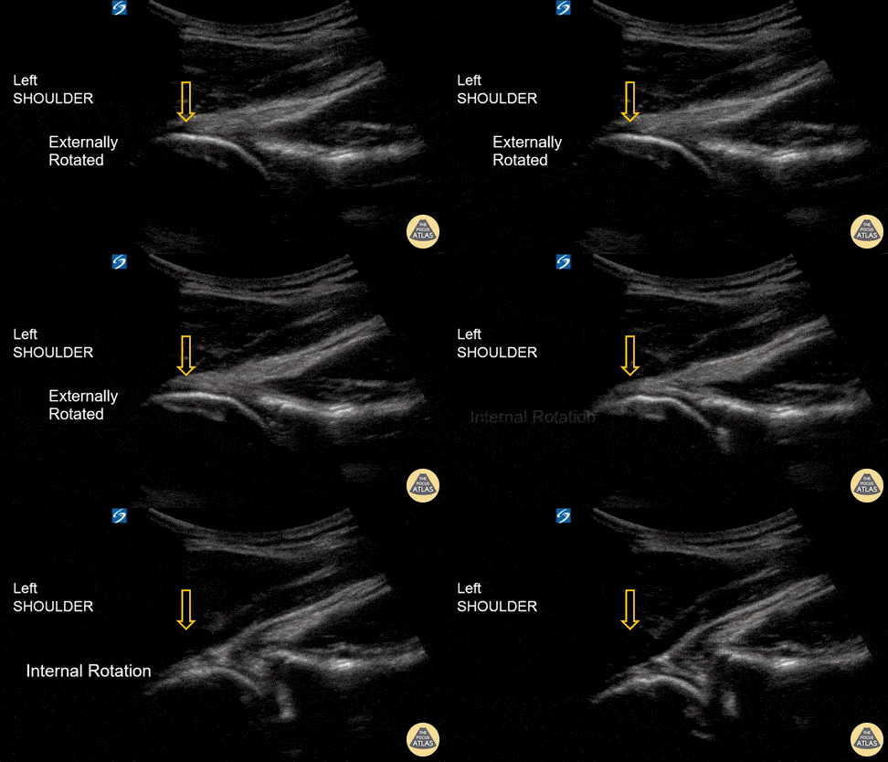
An 18-year-old male with acute left shoulder pain after a physical altercation in which his left arm was pulled. Video clip reveals the left humeral head sliding in and out of the glenoid fossa with internal and external rotation, confirming the diagnosis of shoulder subluxation.
Read more about the case and shoulder POCUS on: https://www.aliem.com/ultrasound-for-win-acute-shoulder-injury-us4tw/
Mark Rivera-Morales, MD, PGY-3
Fernando Rivera, MD, PGY-3
Javier Rosario, MD, FACEP @javimedsimus
UCF/HCA Emergency Medicine Residency Program of Greater Orlando. Osceola Regional Medical Center @UCFEMOrlando
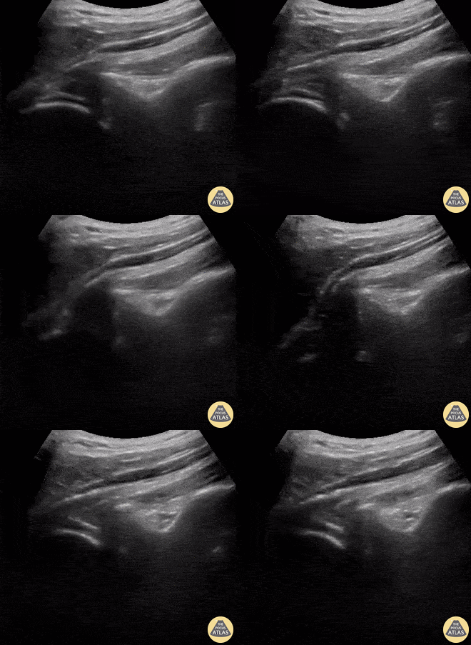
A 9-year-old female presented with left shoulder pain. She has a history of multiple dislocations and, as seen here on POCUS, is able to reduce the dislocation herself.
Julie Klensch, PEM Fellow & Paul Khalil, MD
University of Louisville/Norton Children's Hospital
Tendon/Ligamentous Pathology
40 y/o F presented with 9 month history insidious onset L medial ankle pain and was found to have tibialis posterior tendon partial tear.
Video shows transverse view scanning proximal to distal at the posteromedial ankle (left is posterior). There is thickening of the posterior tibialis tendon with an anechoic tendon sheath effusion. At the level of the medial malleolus there are two moderate interstitial tears extending distally. Flexor digitorum and hallicus longus appear unaffected.
Eben Alexander, DO
Devesh Patel, MD
Eastern Virginia Medical School
60 y/o F w/R elbow pain for 8 months found to have distal biceps tendinosis.
L=proximal; R=distal. Medial long-axis view of distal biceps tendon. Radial artery seen pulsating directly superficial to biceps tendon. Distal biceps tendon diffusely thickened with decreased echogenicity at its attachment site.
Contributor:
Eben Alexander, DO
Devesh Patel, MD
Eastern Virginia Medical School
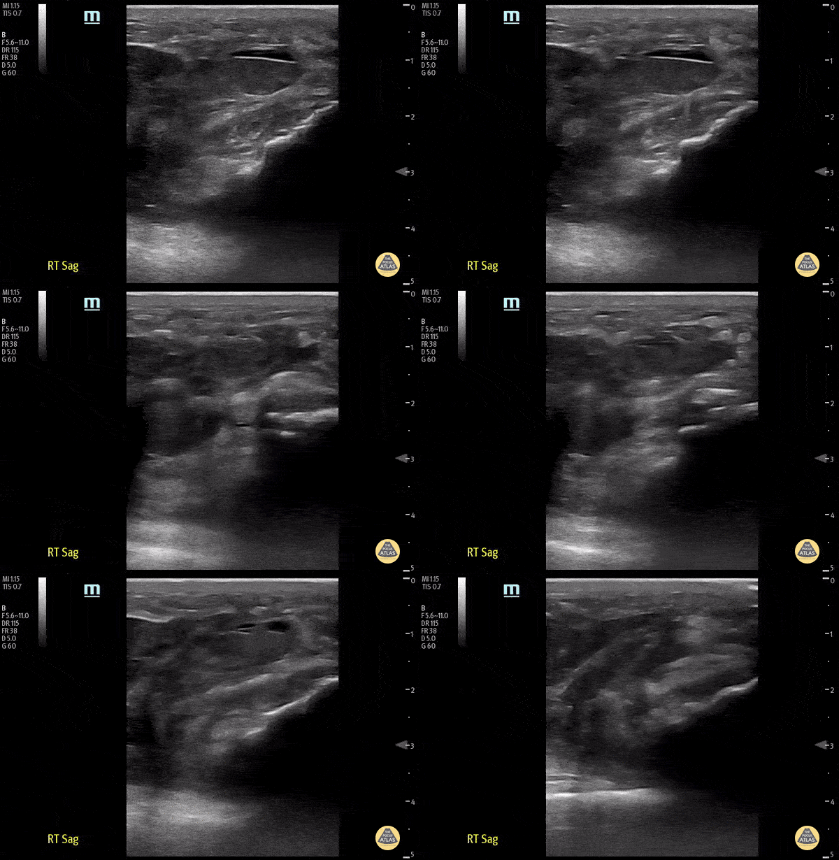
70 y/o male fell onto his right knee presented with visible deformity of the distal quadriceps muscle. Physical exam demonstrated weakness to active extension of the right knee. POCUS with a linear transducer demonstrated complete loss of tendon architecture of the right knee, including a hypoechoic defect between the tendon fibers. When compared to normal knee radiographs, the patient has significant displacement of the patella distally. Radiologists interpreted this as possible quadriceps tendon rupture. During operative repair, a complete rupture of the quadriceps was discovered and repaired.
Justin Morin, DO PGY-1 @justinjmorin; Alex Schlangen, DO PGY-1; Lauren Lowes, DO PGY-3; EM Residents at Central Michigan University
Infectious Disease
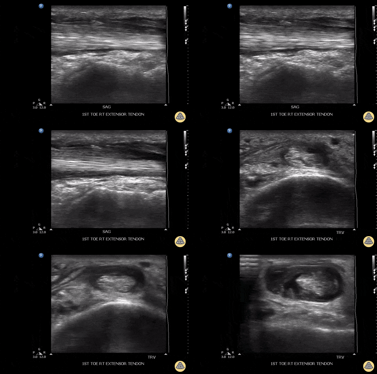
Patient initially suffered a puncture wound 8 days ago to the top of the foot. He presented a week later with pain in the toe, especially with flexion. US imaging showed extensive fluid surrounding the extensor hallucis longus tendon consistent with infectious tenosynovitis. The patient was taken to OR for debridement with good outcome.
Image courtesy of Robert Jones DO, FACEP @RJonesSonoEM
Director, Emergency Ultrasound; MetroHealth Medical Center; Professor, Case Western Reserve Medical School, Cleveland, OH
View his original post here
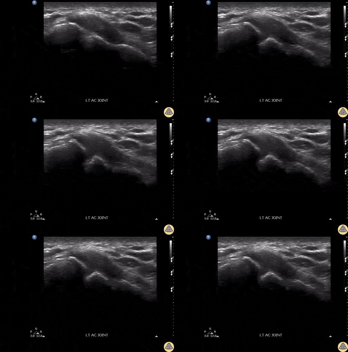
Patient presenting with shoulder pain and AC joint tenderness. Febrile to 102. POCUS revealed an AC joint effusion.
Image courtesy of Robert Jones DO, FACEP @RJonesSonoEM
Director, Emergency Ultrasound; MetroHealth Medical Center; Professor, Case Western Reserve Medical School, Cleveland, OH
View his original post here
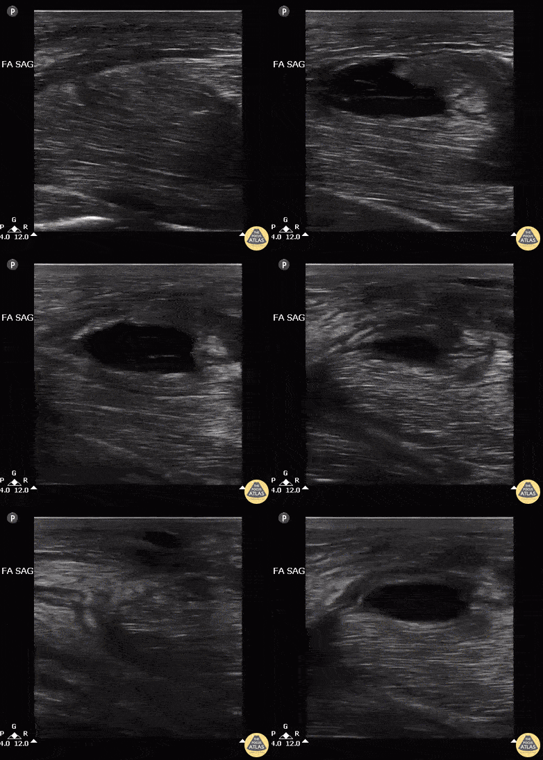
Male with LUE swelling after attempted IV drug use. Ultrasound of bicep reveals intramuscular abscess created from missed injection. Abscess was drained, and pt was admitted and given IV Abx.
Image courtesy of Robert Jones DO, FACEP @RJonesSonoEM
Director, Emergency Ultrasound; MetroHealth Medical Center; Professor, Case Western Reserve Medical School, Cleveland, OH
View his original post here
Joint Effusion
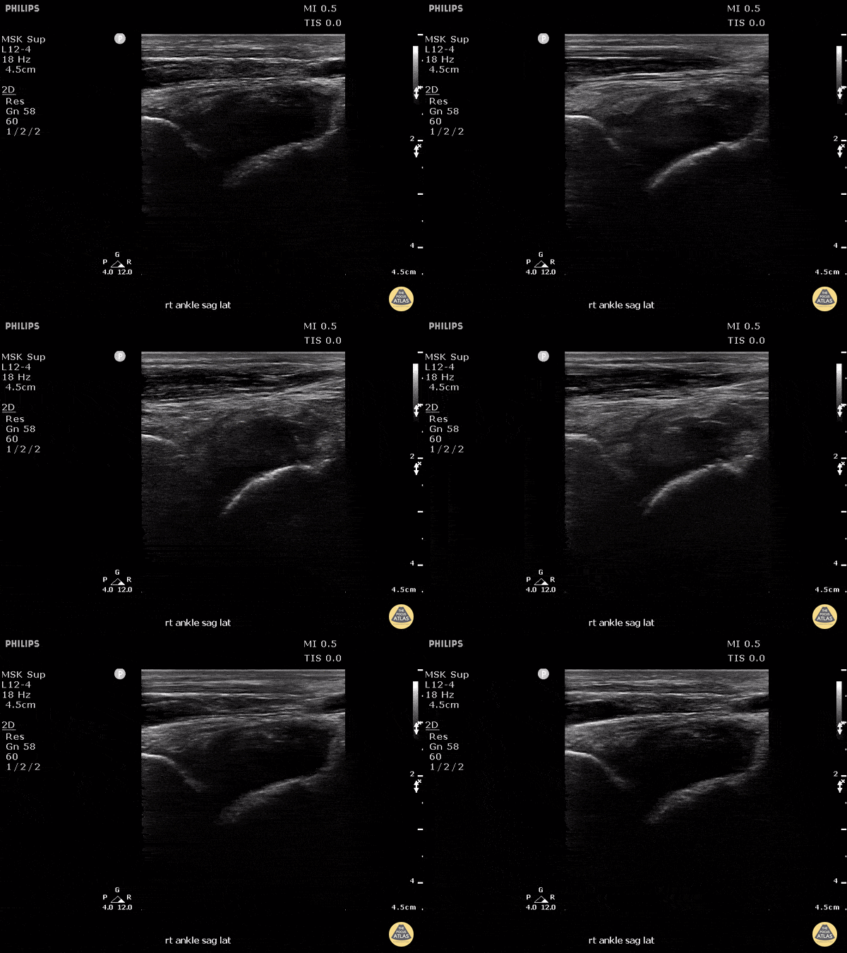
This patient presented to the emergency department for several days of ankle pain. Ultrasound of the joint revealed anechoic fluid within the joint, which normally suggests a non-exudative cause. However, this patient’s joint was eventually tapped which revealed thick purulent fluid that grew Fusobacterium, and a rare cause of septic joint.
Image courtesy of Robert Jones DO, FACEP @RJonesSonoEM
Director, Emergency Ultrasound; MetroHealth Medical Center; Professor, Case Western Reserve Medical School, Cleveland, OH
View his original post here
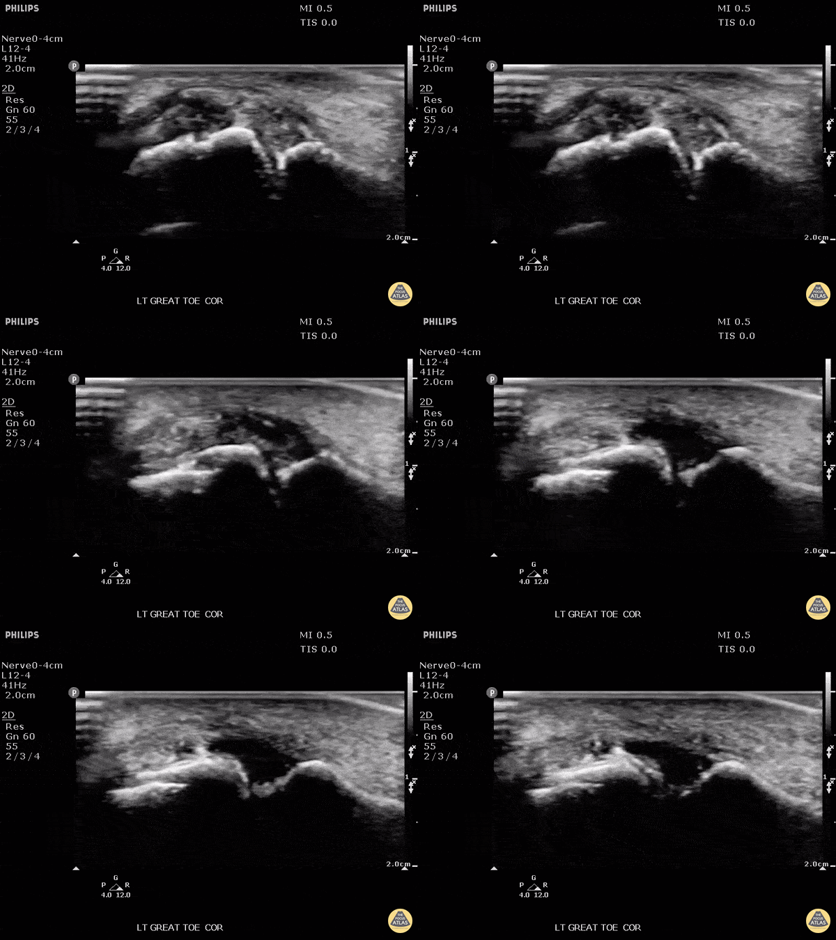
Patient presented with great toe pain. POCUS performed and detected effusion in first metatarsophalangeal joint. Eventually determined to be monosodium urate crystals as seen in gout.
Image courtesy of Robert Jones DO, FACEP @RJonesSonoEM
Director, Emergency Ultrasound; MetroHealth Medical Center; Professor, Case Western Reserve Medical School, Cleveland, OH
View his original post here
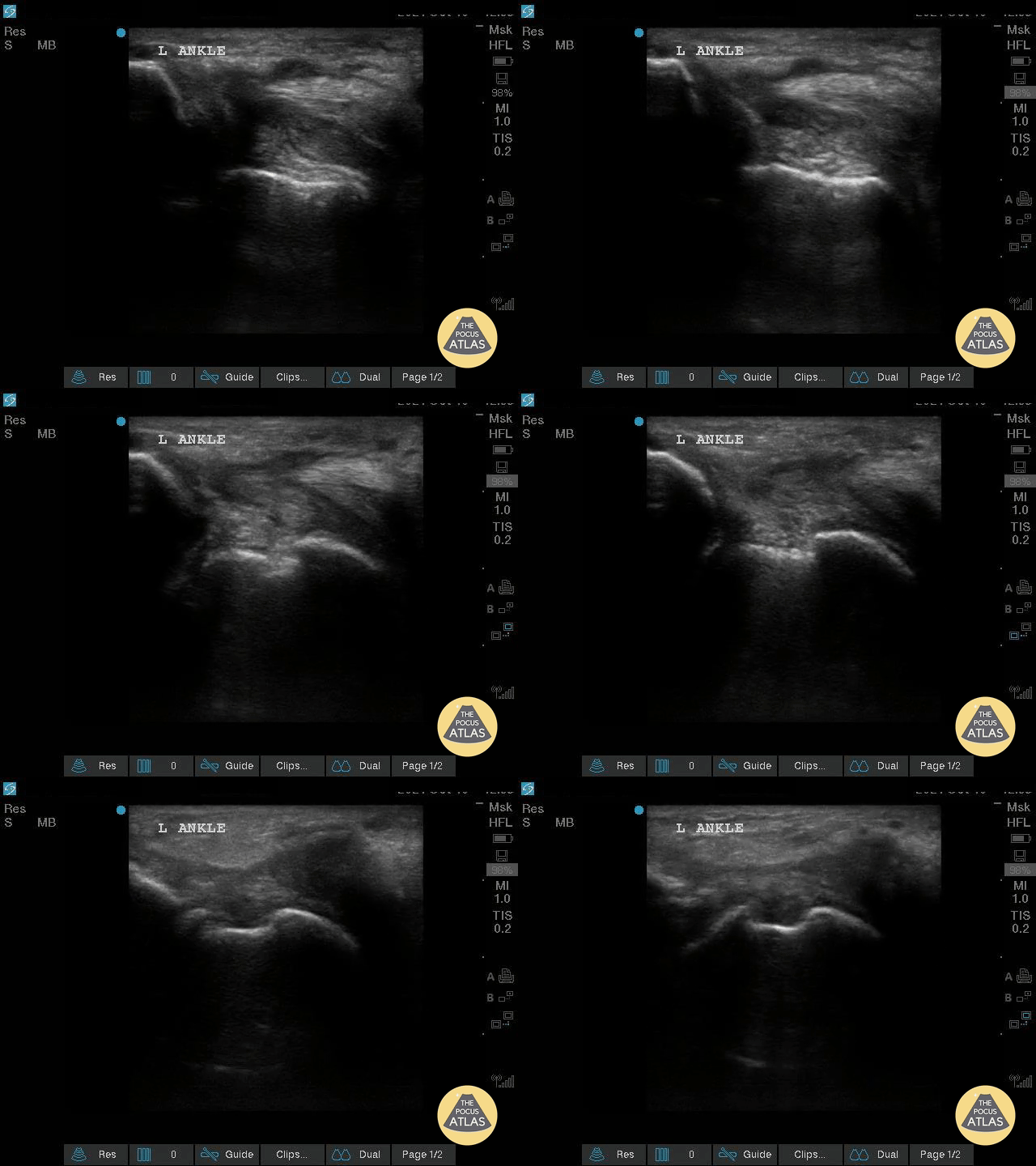
This is an image of a left ankle demonstrating a complex effusion in a patient presenting with ankle pain and swelling. Arthrocentesis was performed confirming septic arthritis.
Michael Macias
Other MSK Pathology
92 y/o M presented with moderate to severe osteoarthritis on elbow x-ray who was treated with ultrasound-guided corticosteroid joint injection.
Video shows transverse view of the posterior elbow as corticosteroid is injected with a posterolateral approach deep to the posterior elbow fat pad located in the olecranon fossa. This area is continuous with the elbow joint capsule. Posterolateral approach is advantageous due to decreased risk of disrupting neurovascular structures or the articular cartilage from other approaches to the joint space. Posterior approach is also advantageous compared to lateral approach in moderate to severe arthritis due to limited access to the radiocapitellar joint space.
Eben Alexander, DO
Devesh Patel, MD
Eastern Virginia Medical School
60 y/o F with knee arthritis getting corticosteroid joint injection in left suprapatellar recess.
Ultrasound probe in transverse axis just superior to the patella. Needle approach lateral to medial. Infiltrate seen injected into anechoic suprapatellar recess. The suprapatellar recess is continuous with the tibiofemoral joint. Deep to suprapellar recess in this video is the prefemoral fat pad. Just superior to the suprapatellar recess in this image is the quadriceps tendon in short axis appearing as a fibrillar structure.
Contributors:
Eben Alexander, DO
Devesh Patel, MD
Eastern Virginia Medical School
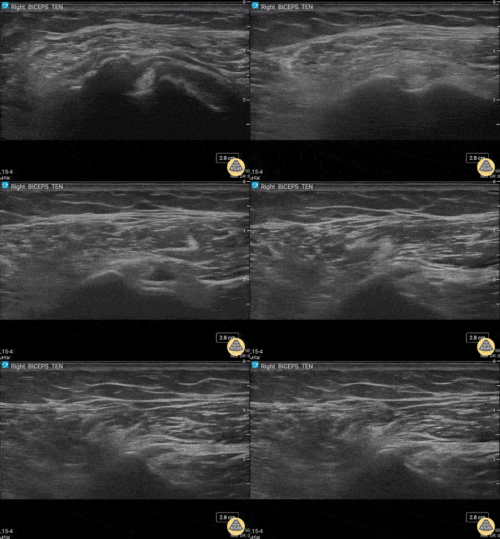
55 y/o F presented 2 months after a FOOSH with adhesive capsulitis and a biceps tendon sheath effusion.
Ultrasound scanning transverse axis proximal to distal along humeral shaft (left screen is lateral). Anechoic space around hyperechoic biceps tendon (center of screen) represents fluid that is continuous with the glenohumeral joint space. Inflammation from the adhesive capsulitis is drawing in fluid into the joint that is extending into biceps tendon sheath.
Contributor:
Eben Alexander, DO
Devesh Patel, MD
Eastern Virginia Medical School