
Pulmonary
Pulmonary
Looking for Pediatric Lung Ultrasound Clips? Check out our Pulmonary Junior Page (Beta)!
Note: These search bar results are only for the Pulmonary image archive. To search the entire site, click here.
Recently Added
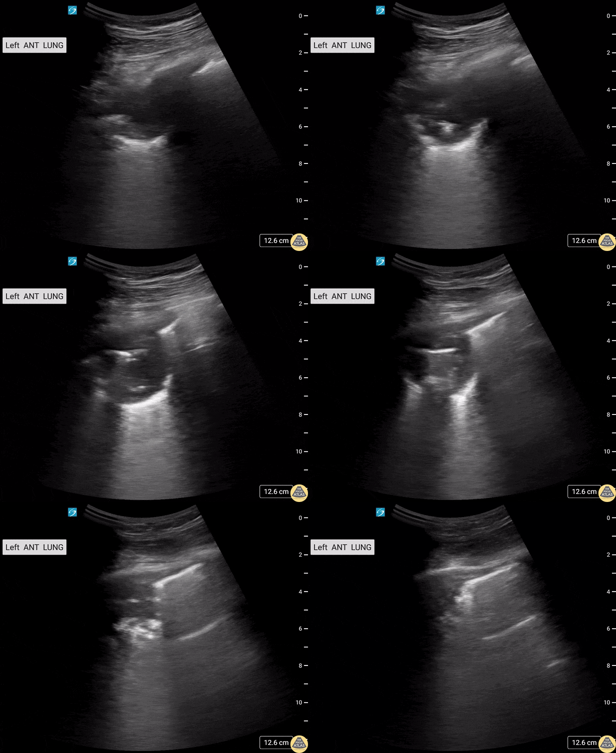
HIV+ male patient presents with cough and shortness of breath. Thoracic US demonstrated a left upper lobe cavitary lesion with calcifications.
Differentials included TB, aspergilloma, lung abscess.
Contributors: Sara Schambach, MD; Garrett Mason, MD
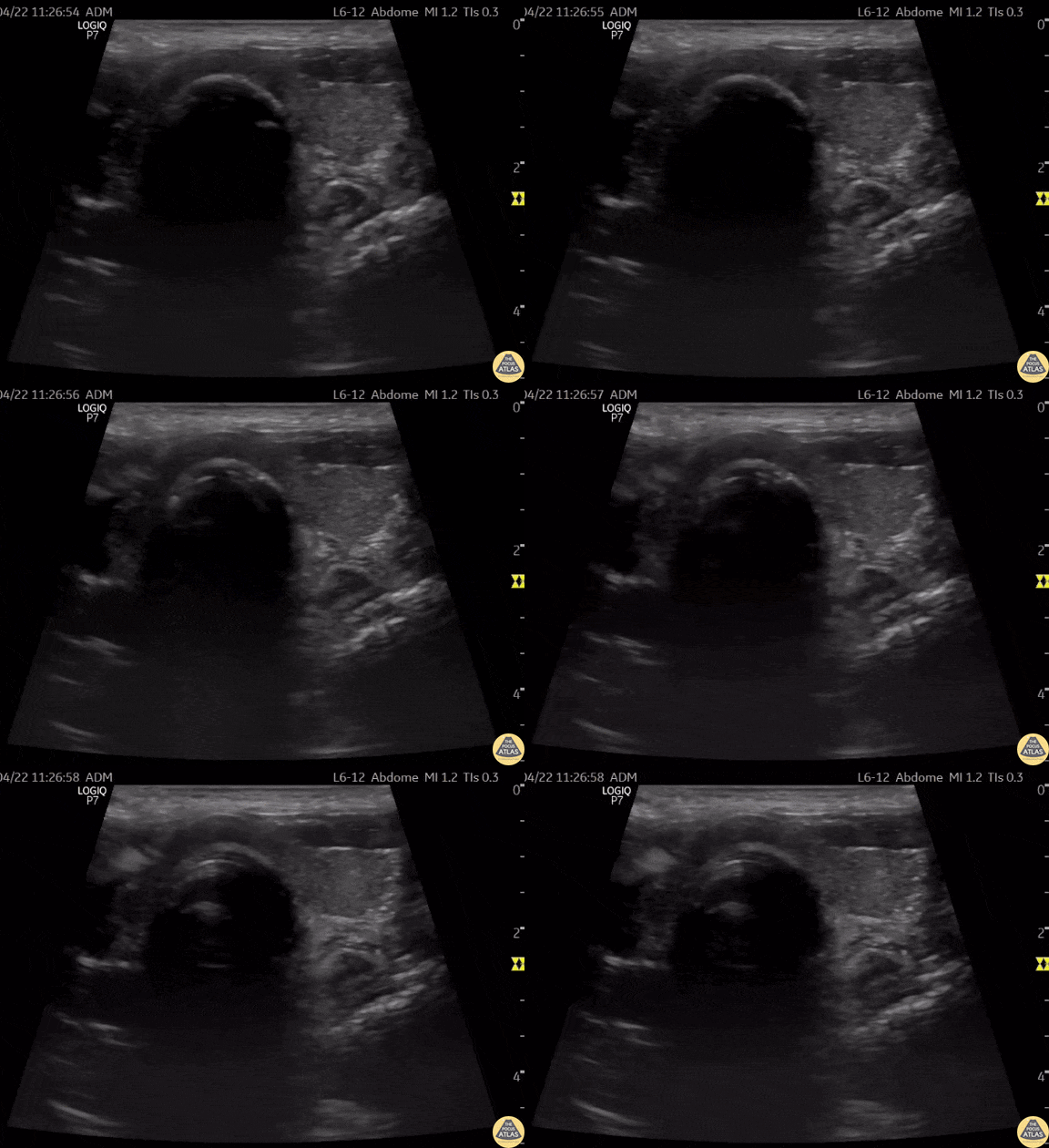
This is a clip of a cervical airway view during intubation attempt.
Some artifact is visible in the trachea when passing the tube demonstrating a "Snowstorm Sign" which can be used to confirm the position of an endotracheal tube. In this view it is possible to see the esophagus next to the trachea. As demonstrated there is no motion detected in esophagus during intubation therefore confirming tracheal intubation.
Contributor: Renato Tambelli (@R_Tambelli @Jedipocus)
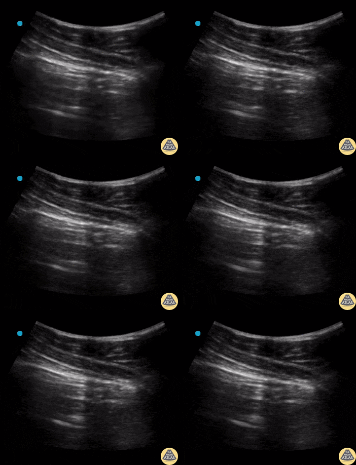
Lung point is a pathognomonic finding on US for pneumothorax. It refers to the junction between healthy lung and collapsed lung.
This is represented in the US recording as lung sliding seen on the left of the pleural line but no lung sliding seen on the right of the pleural line.
Contributors: Dimitri Livshits, DO; Jane Belyavskaya, MD; Chris Hanuscin, MD
Kings County/SUNY Downstate
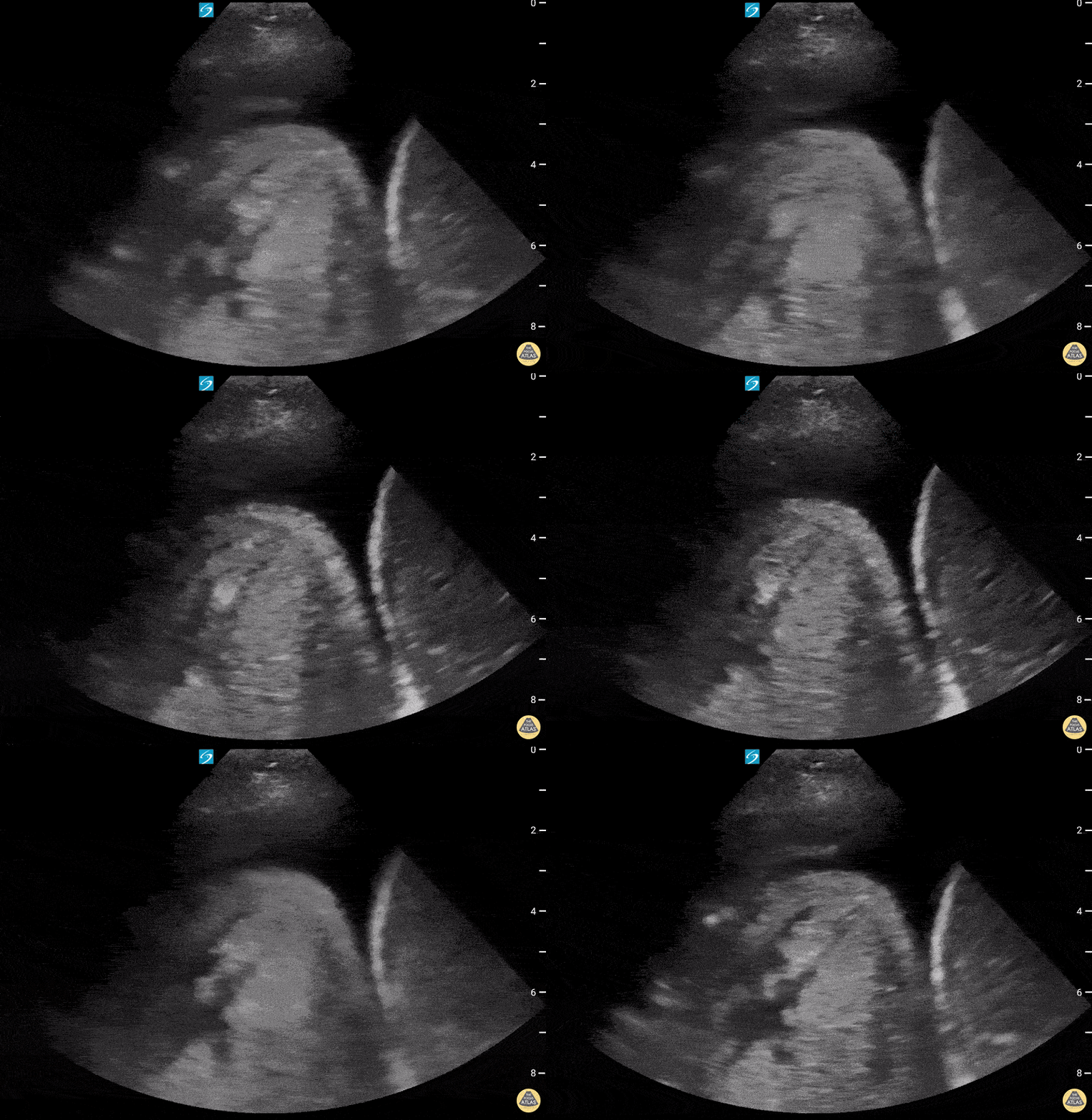
72 year old with past medical history of hiatal hernia presenting to the ED with new onset shortness of breath requiring rescue BPAP. She was diagnosed and treated with CHF exacerbation. Lung ultrasound showed a large pleural effusion with uncertain mass-like object with heterogenous fluid contents on the right (shown here), along with dense B-lines. Correlation with prior CT suggested that the structure on the right chest was a large hiatal hernia.
Contributed by: William McGill, PA-C
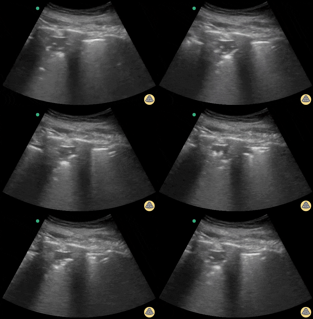
Patient admitted to ICU with sepsis of unknown origin. Complaints of tachypnea led to ultrasound scanning, which showed a subpleural hypoechoic image in the posterolateral region of the left chest compatible with pneumonia.
Contributed by: @intensivanaveia
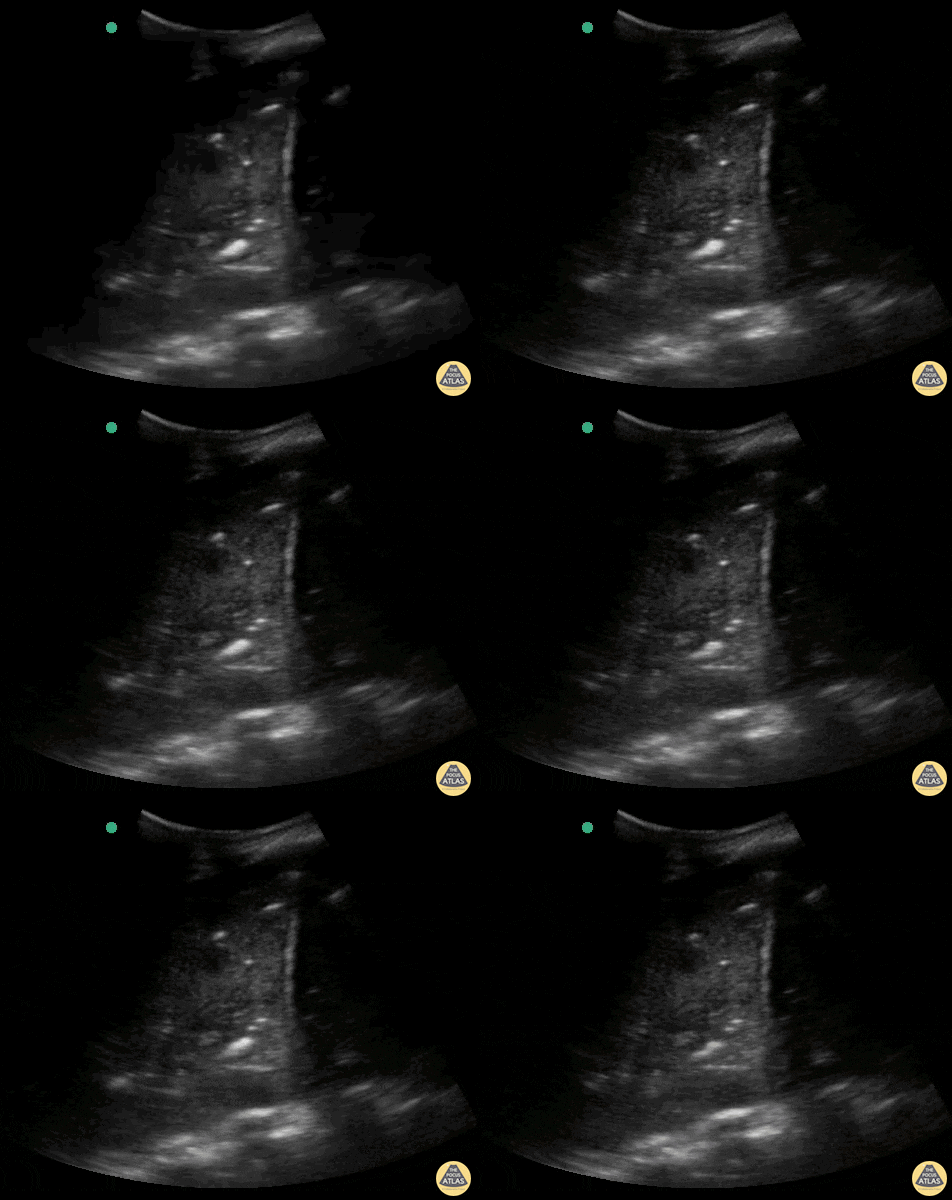
Massive lung consolidation in a patient diagnosed with COVID-19 with bacterial component.
Contributor: Rafael Intensivanaveia - Critical Care Physician at Hospital Israelita Albert Einstein
Normal Anatomy
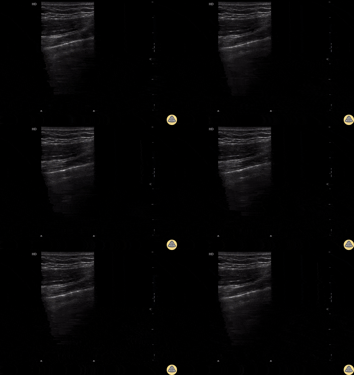
Normal lung sliding with regular appearing pleural lining.
Image courtesy of Robert Jones DO, FACEP @RJonesSonoEM
Director, Emergency Ultrasound; MetroHealth Medical Center; Professor, Case Western Reserve Medical School, Cleveland, OH
View his original post here
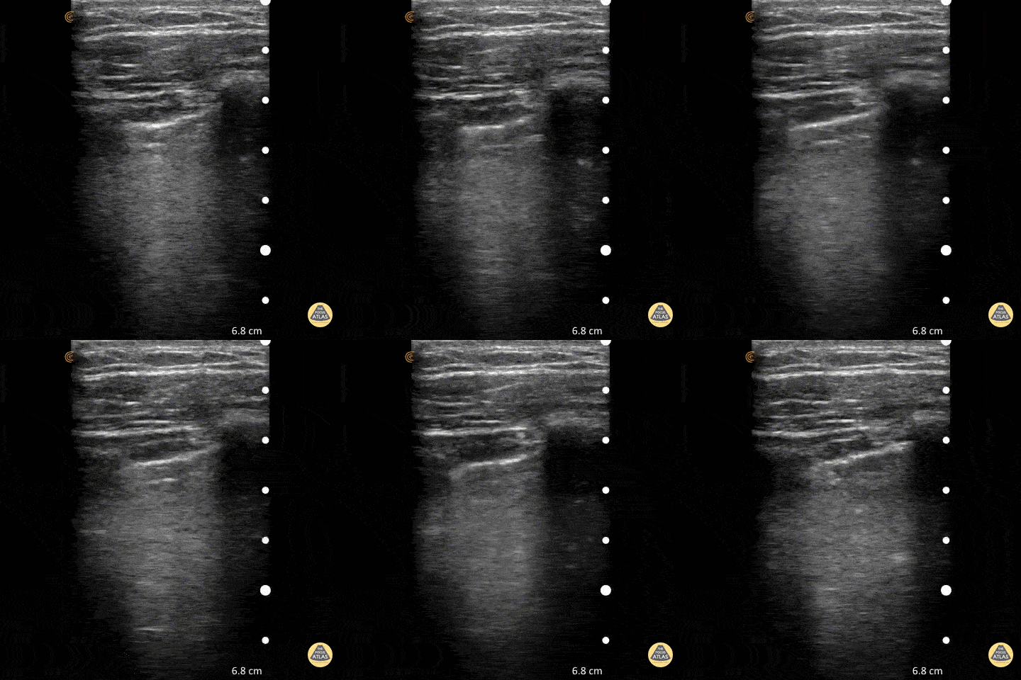
To assess for lung slide, look between two ribs. The two layers of pleura can be seen as the hyperechoic, shimmering line just under the subcutaneous tissue. This represents the sliding of the parietal and visceral pleura.
Normal lung slide should have: Shimmering aka "ants marching"
This may be augmented by m-mode as pictured in another post.
Dr. Matthew Riscinti - Kings County Emergency Medicine
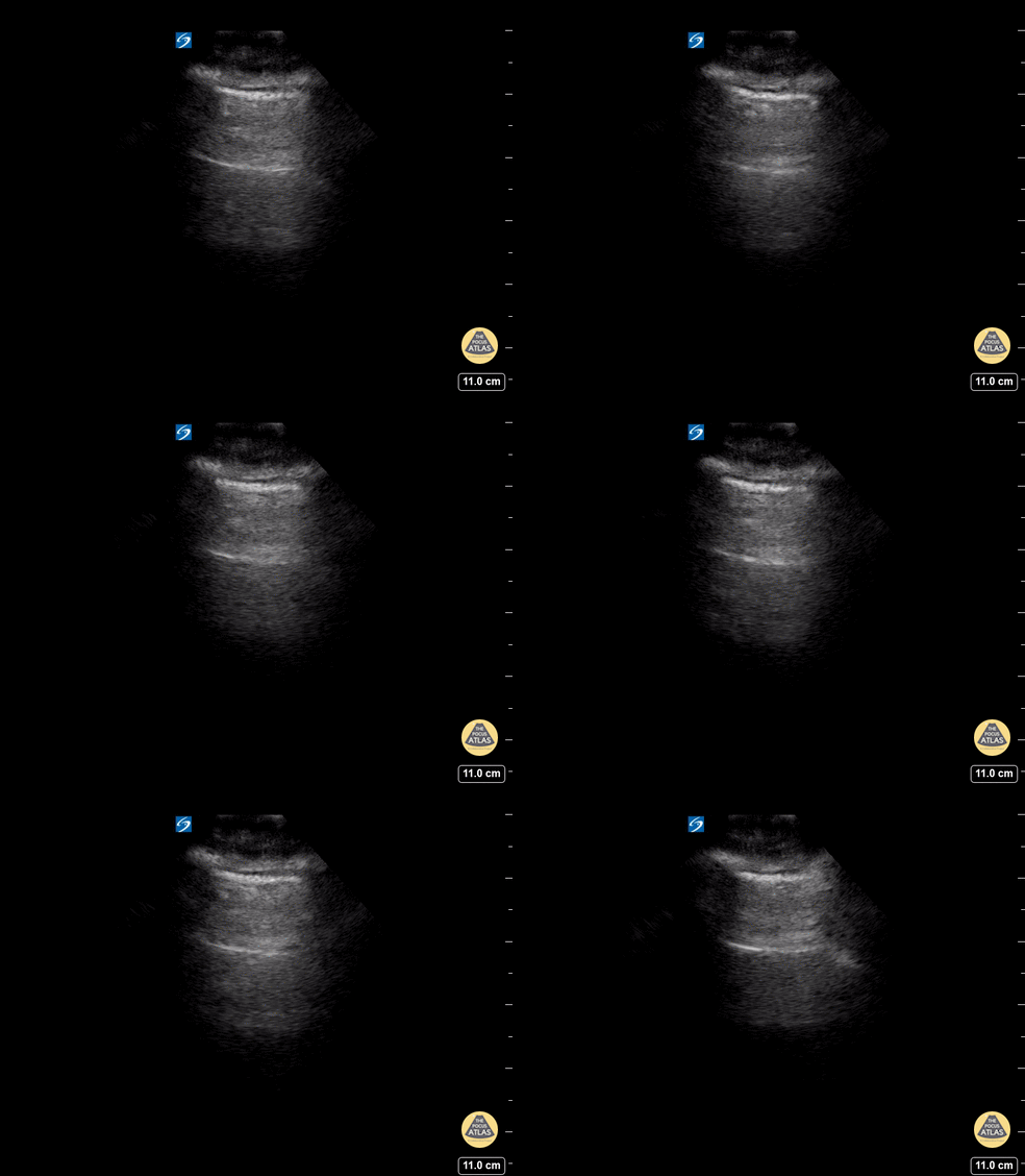
A lines appear as horizontal lines that represent normal aerated lung (dry interlobular septa). They are a reverberation artifact caused by the sound waves bouncing off the highly echogenic pleura and back to the probe, and repeating.
Hannah Kopinski (MS4) and Dr. Lindsay Davis - NYU Emergency Medicine, Matthew Riscinti - Kings County Emergency Medicine
Colorized Normal Anatomy
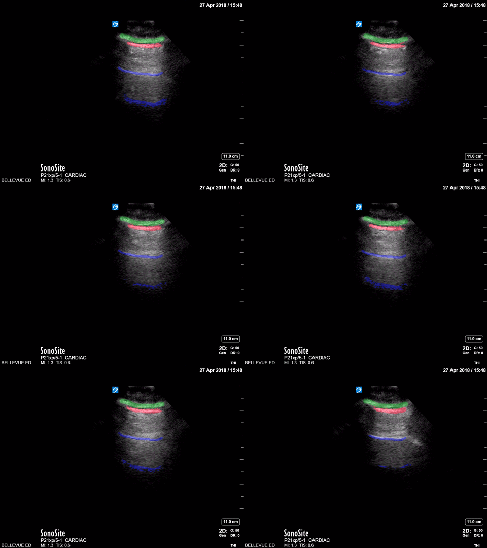
Lung sliding
Green: Subcutaneous tissue, Red: Pleural space, Blue: A lines
Images: Dr. Lindsay Davis, Dr. Hannah Kopinski. Image Editing: Michael Amador and Dr. Matthew Riscinti
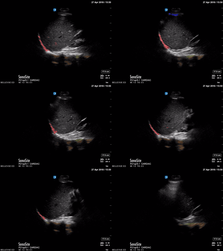
Pleural space
Red: Diaphragm, Blue: Pleural space, Green: A lines, Orange: Spine
Images: Dr. Lindsay Davis, Dr. Hannah Kopinski. Image Editing: Michael Amador and Dr. Matthew Riscinti
Pulmonary Edema
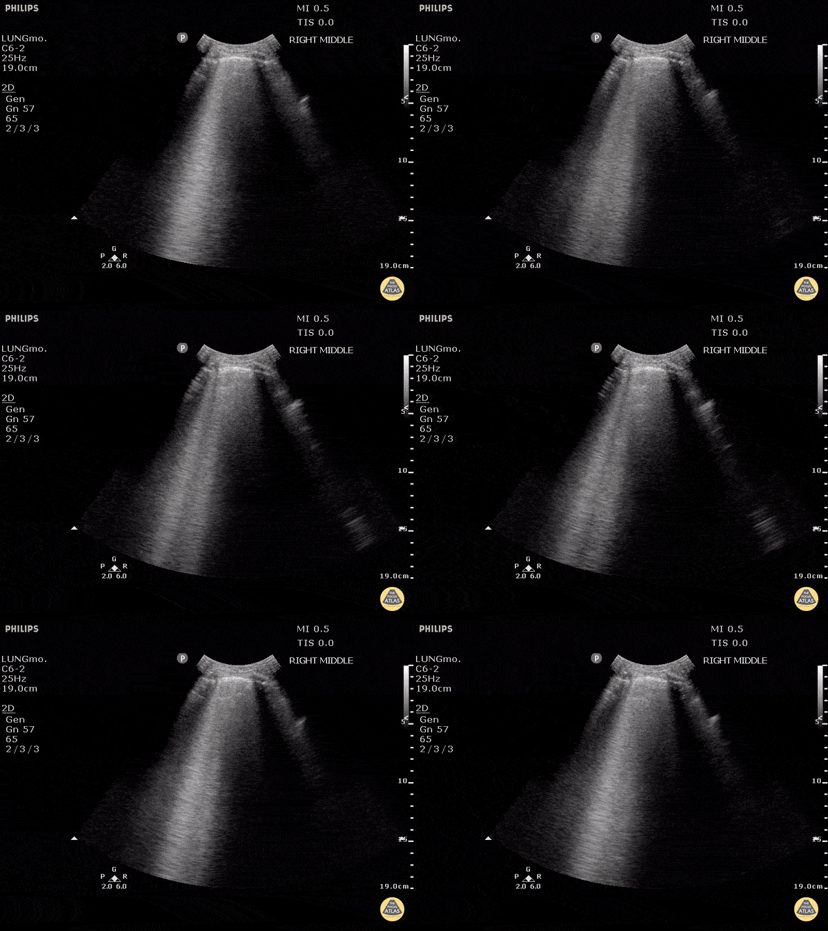
B-lines are vertical artifacts that moves with respiration from the pleural surface. They represent increased water in an area of the lung. In the right clinical context this could represent pulmonary edema. An increase in B-lines correlates with the degree of pulmonary edema.
Keep in mind that in different clinical contexts, they can represent different diagnoses including pulmonary contusions and pneumonia.
Justin Bowra MBBS, FACEM, CCPU Emergency Physician, RNSH et al.
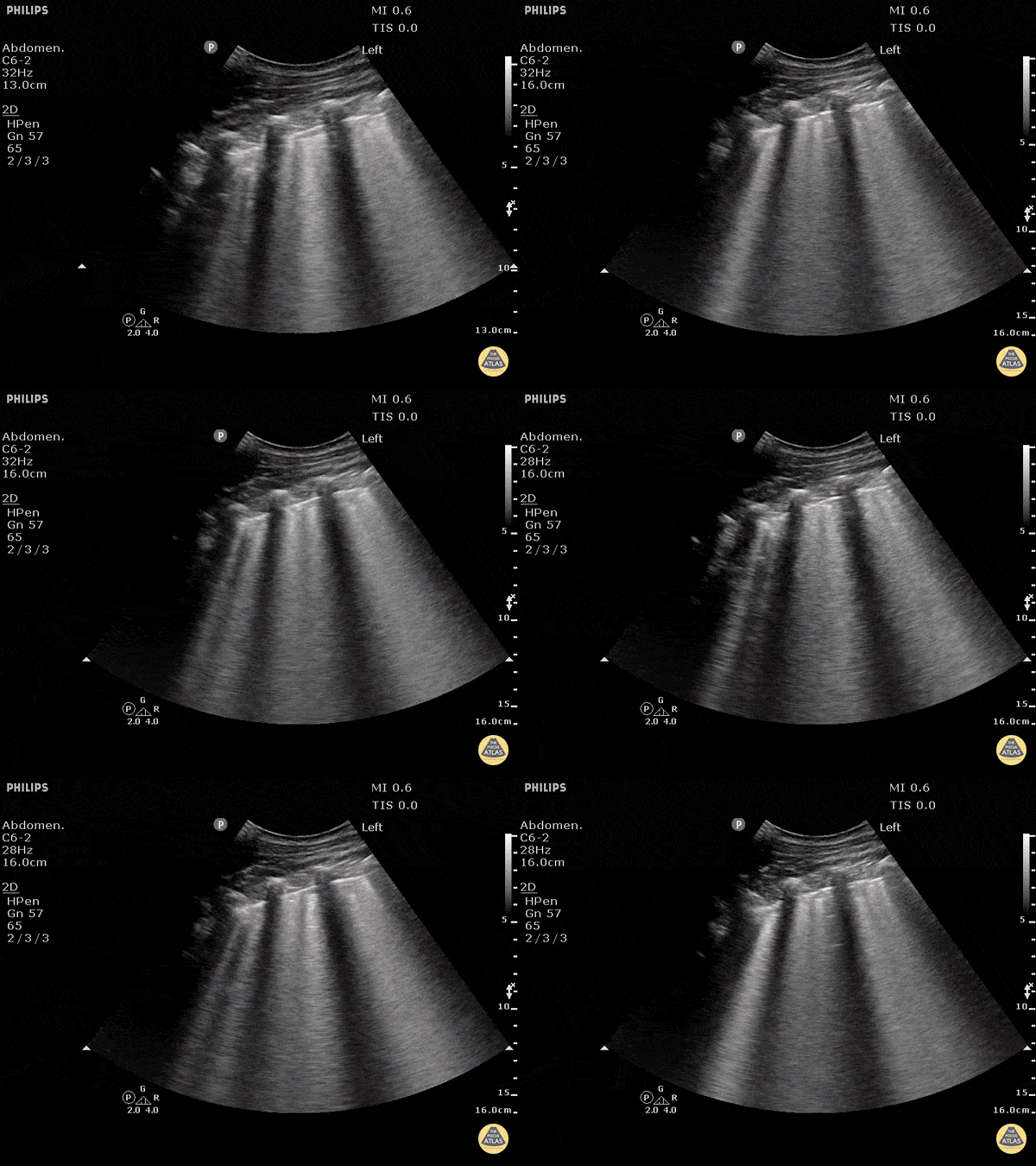
B-lines obtained with curved probe.
B-lines are vertical artifacts that move with respiration from the pleural surface. They represent increased water in an area of the lung. In the right clinical context this could represent pulmonary edema. An increase in B-lines correlates with the degree of pulmonary edema.
3 B-lines in an intercostal space represent a "positive" region of the lung, and if there are two regions of the lung that are positive, you can diagnose pulmonary edema.
Dr. Justin Bowra et al. (Dr. D Browne and Dr. J Knights)
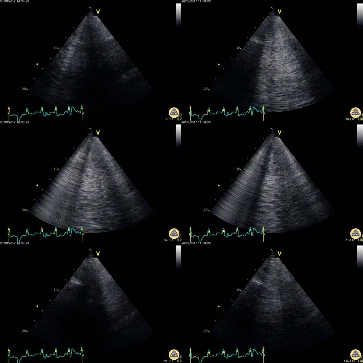
WCUME 2017 Submission for "Best POCUS"
An acutely dyspnoeic patient presents with ventricular tachycardia and has no response to initial chemical cardioversion. Lung POCUS shows widespread bilateral confluent B lines indicating acute pulmonary edema. Unstable tachycardia terminated using synchronized electrical cardioversion.
Dr. Cian McDermott - Dublin, Ireland
Pneumonia/Consolidation
Patient admitted to ICU with sepsis of unknown origin. Complaints of tachypnea led to ultrasound scanning, which showed a subpleural hypoechoic image in the posterolateral region of the left chest compatible with pneumonia.
Contributed by: @intensivanaveia
Massive lung consolidation in a patient diagnosed with COVID-19 with bacterial component.
Contributor: Rafael Intensivanaveia - Critical Care Physician at Hospital Israelita Albert Einstein
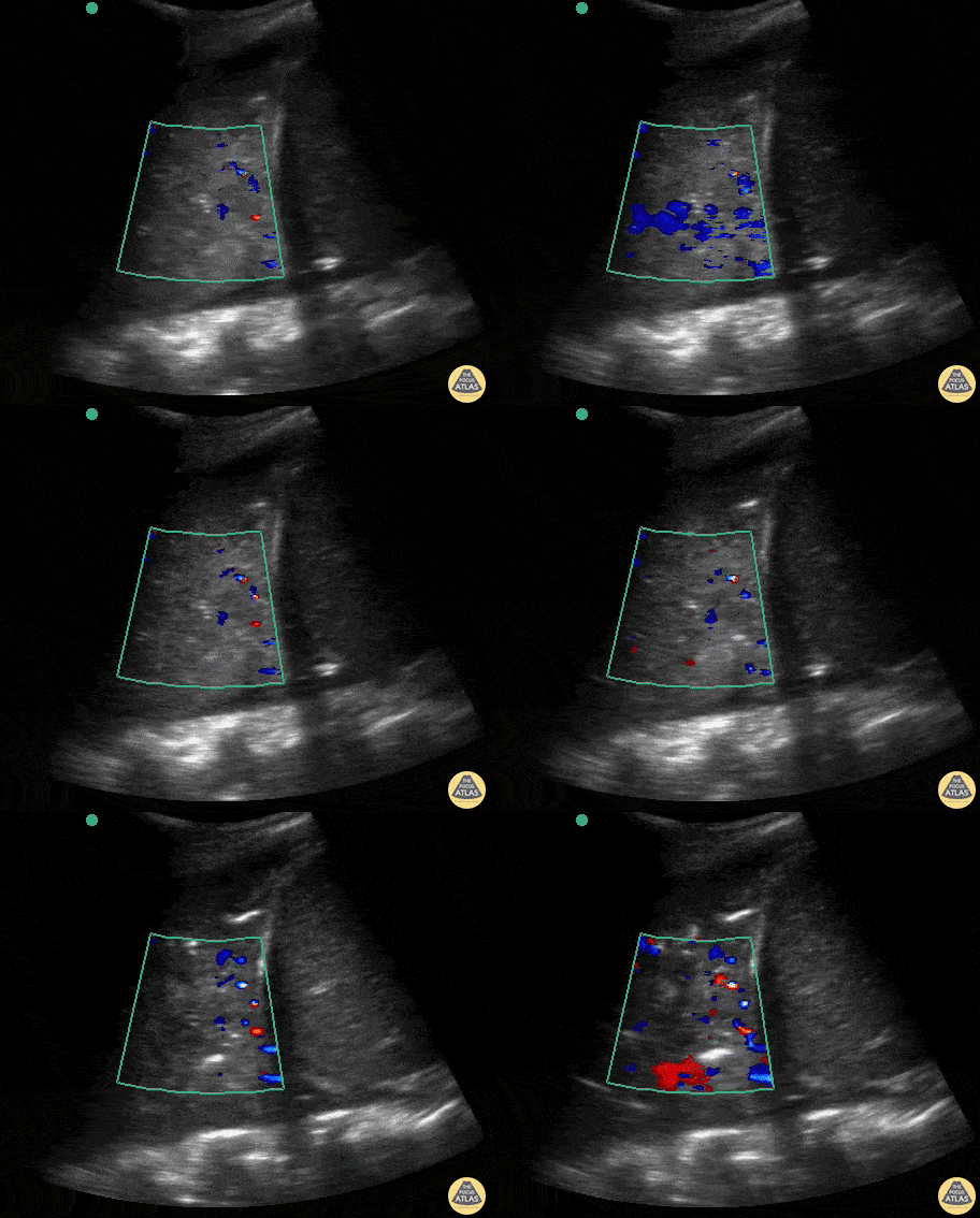
Massive lobar consolidation showing pulsatile flow on color doppler, befitting pneumonia.
Contributor: Rafael Intensivanaveia
COVID-19
Massive lung consolidation in a patient diagnosed with COVID-19 with bacterial component.
Contributor: Rafael Intensivanaveia - Critical Care Physician at Hospital Israelita Albert Einstein
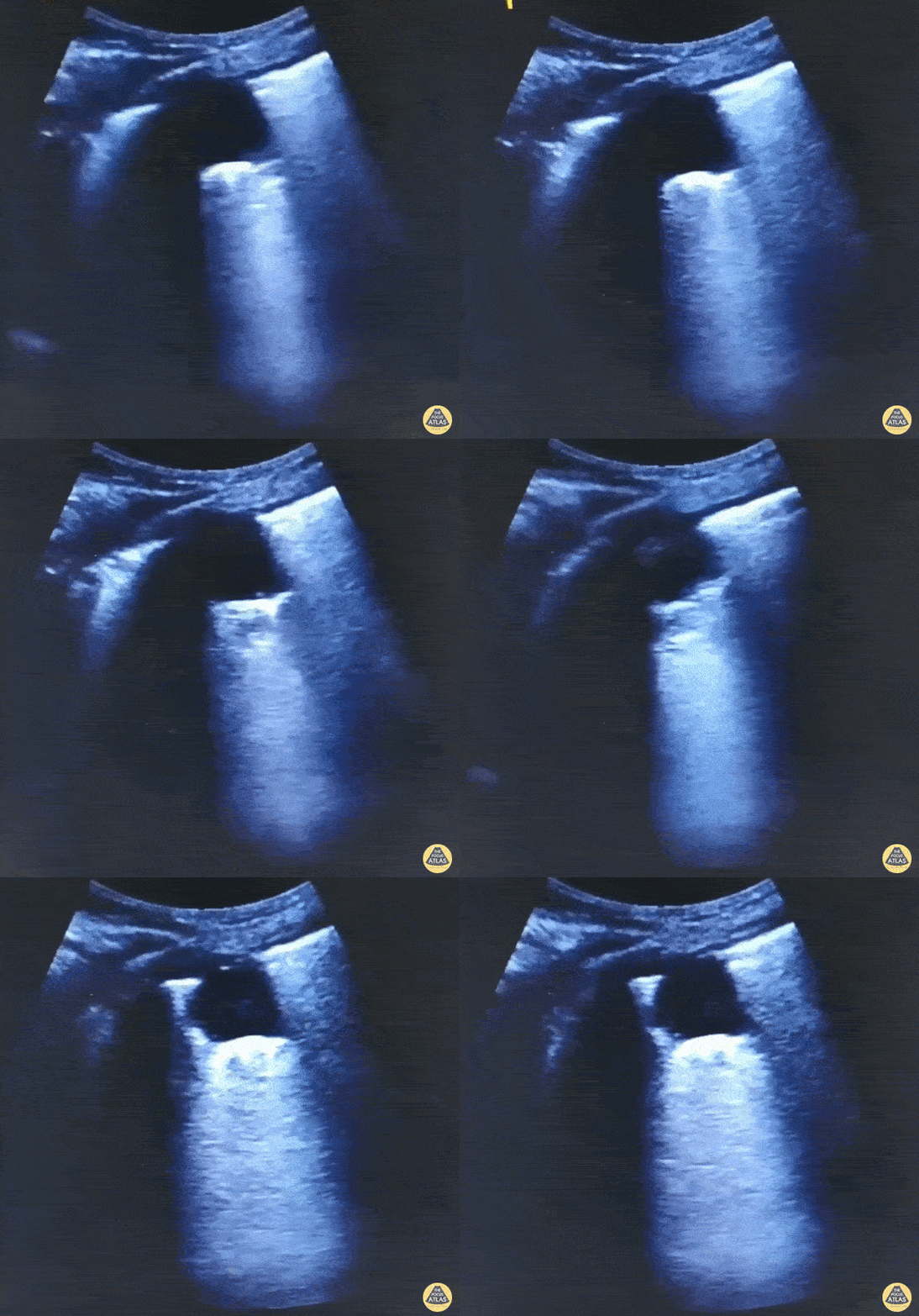
Subpleural consolidation (shred sign) in a COPD patient with Covid 19, admitted to the ICU with acute respiratory failure progressing to intubation and mechanical ventilation.
Contributor: Bruno Souza @BrunoSo03038122
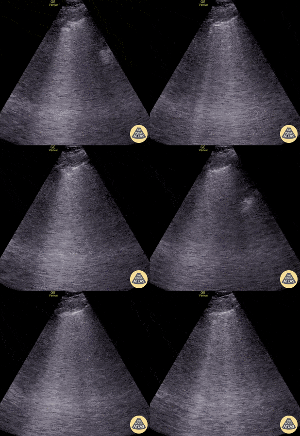
Seen here is an irregular and thickened pleural line with associated focal and confluent B lines in a patient with COVID-19 pneumonia.
Edgar Miranda
Pneumothorax
Lung point is a pathognomonic finding on US for pneumothorax. It refers to the junction between healthy lung and collapsed lung.
This is represented in the US recording as lung sliding seen on the left of the pleural line but no lung sliding seen on the right of the pleural line.
Contributors: Dimitri Livshits, DO; Jane Belyavskaya, MD; Chris Hanuscin, MD
Kings County/SUNY Downstate
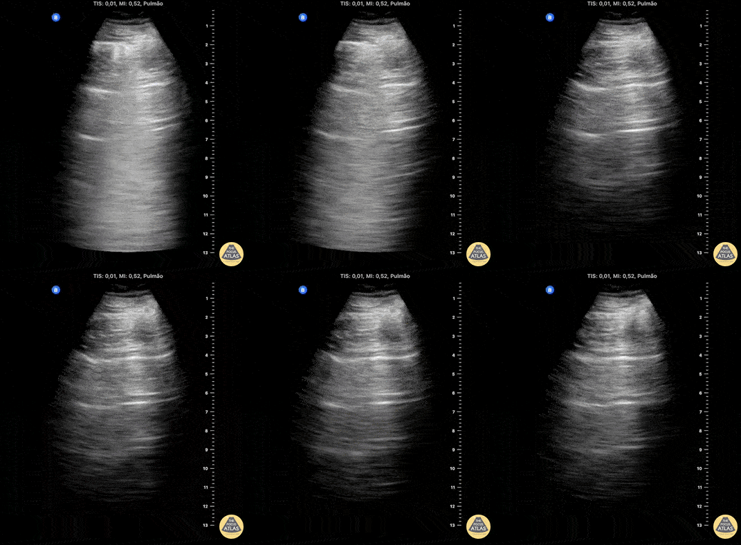
Lung ultrasound in the right apical region after puncture attempt for central venous access. The image demonstrates a LUNG POINT, a specific sign of a pneumothorax.
Contributed by: Breno Moura
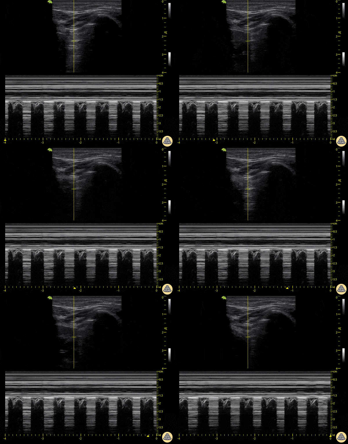
18-year-old patient who presented following a motorcycle accident in which he sustained closed chest trauma with bilateral hemopneumothoraces. In this sequence taken with a linear transducer in left pulmonary zone 1, the "pulmonary point" indicative of pneumothorax can be seen.
Libardo Valencia Chicue
Pleural Effusion
72 year old with past medical history of hiatal hernia presenting to the ED with new onset shortness of breath requiring rescue BPAP. She was diagnosed and treated with CHF exacerbation. Lung ultrasound showed a large pleural effusion with uncertain mass-like object with heterogenous fluid contents on the right (shown here), along with dense B-lines. Correlation with prior CT suggested that the structure on the right chest was a large hiatal hernia.
Contributed by: William McGill, PA-C
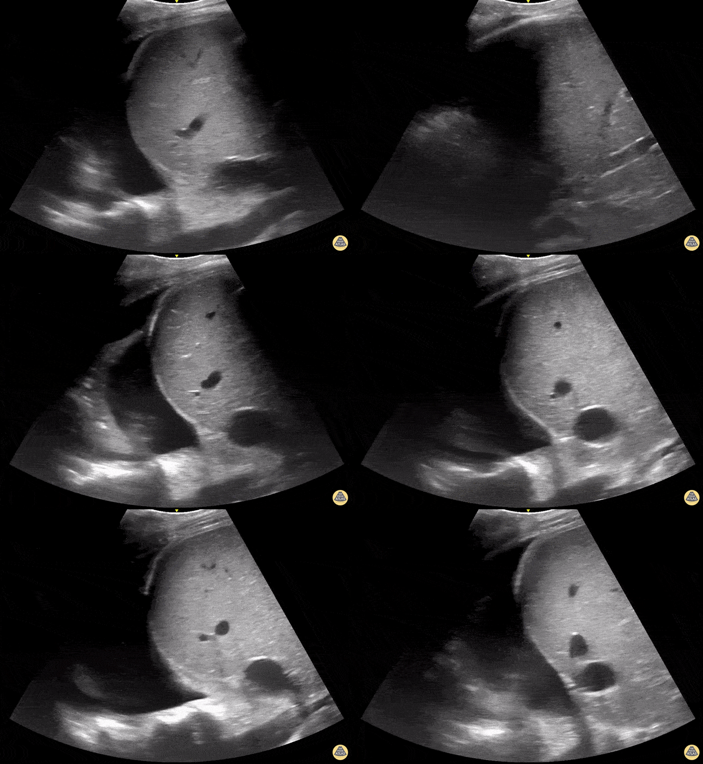
Pleural effusion with compressed lung and positive spine sign
Contributed by: Dimitri Livshits DO, Ultrasound Fellow; Jane Belyavskaya MD, Ultrasound Fellow; Chris Hanuscin MD, Ultrasound Division Director (Kings County/SUNY Downstate)
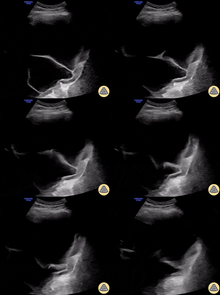
76yo male with shortness of breath on exertion, w/o fever or other signs of infection. POCUS showed a large septated unilateral pleural effusion. Note the irregular and thickened pleura adjacent to the diaphragm (right).
Thoracentesis was only partially successful with a large residual effusion after drainage of 1000ml of exudative fluid with signs of lymphocytic inflammation w/o malignant cells on cytopathologic analysis. Thoracoscopic biopsy confirmed the diagnosis of a pleural mesothelioma 60 years after exposure to asbestos.
The presence of a septated complex effusion is 94% specific for an exudative effusion and warrants further investigation (Shkolnik, 2020). Cytopathology of pleural fluid is often negative in malignant mesothelioma and pleural biopsies are needed to confirm the diagnosis (Porcel, 2014).
Victor Speidel
Langenthal Regional Hospital, Switzerland
Other Pathology
HIV+ male patient presents with cough and shortness of breath. Thoracic US demonstrated a left upper lobe cavitary lesion with calcifications.
Differentials included TB, aspergilloma, lung abscess.
Contributors: Sara Schambach, MD; Garrett Mason, MD
This is a clip of a cervical airway view during intubation attempt.
Some artifact is visible in the trachea when passing the tube demonstrating a "Snowstorm Sign" which can be used to confirm the position of an endotracheal tube. In this view it is possible to see the esophagus next to the trachea. As demonstrated there is no motion detected in esophagus during intubation therefore confirming tracheal intubation.
Contributor: Renato Tambelli (@R_Tambelli @Jedipocus)
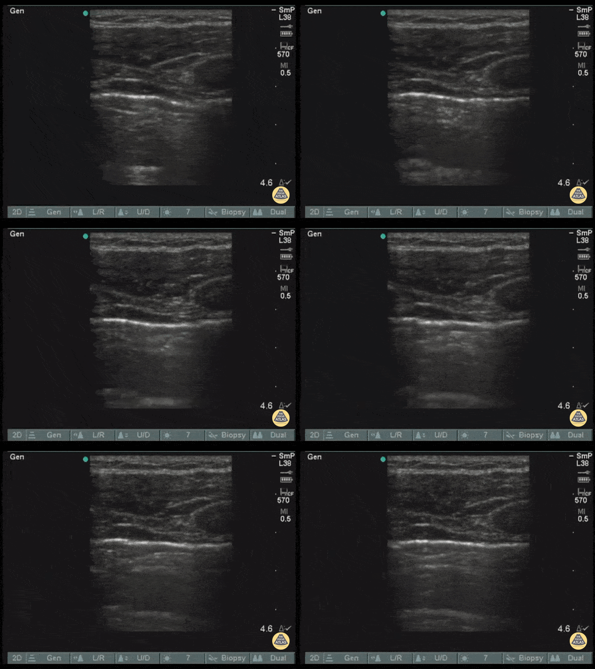
Pictured here is an amazing return of observable lung sliding as a pigtail catheter is advanced through. Reestablishment of lung sliding indicates lungs have re-expanded.
Image courtesy of Robert Jones DO, FACEP @RJonesSonoEM
Director, Emergency Ultrasound; MetroHealth Medical Center; Professor, Case Western Reserve Medical School, Cleveland, OH
View his original post here