
Aorta
Aorta
Note: These search bar results are only for the Aorta image archive. To search the entire site, click here.
Recently Added
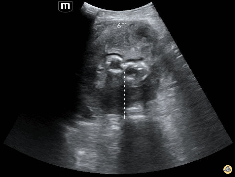
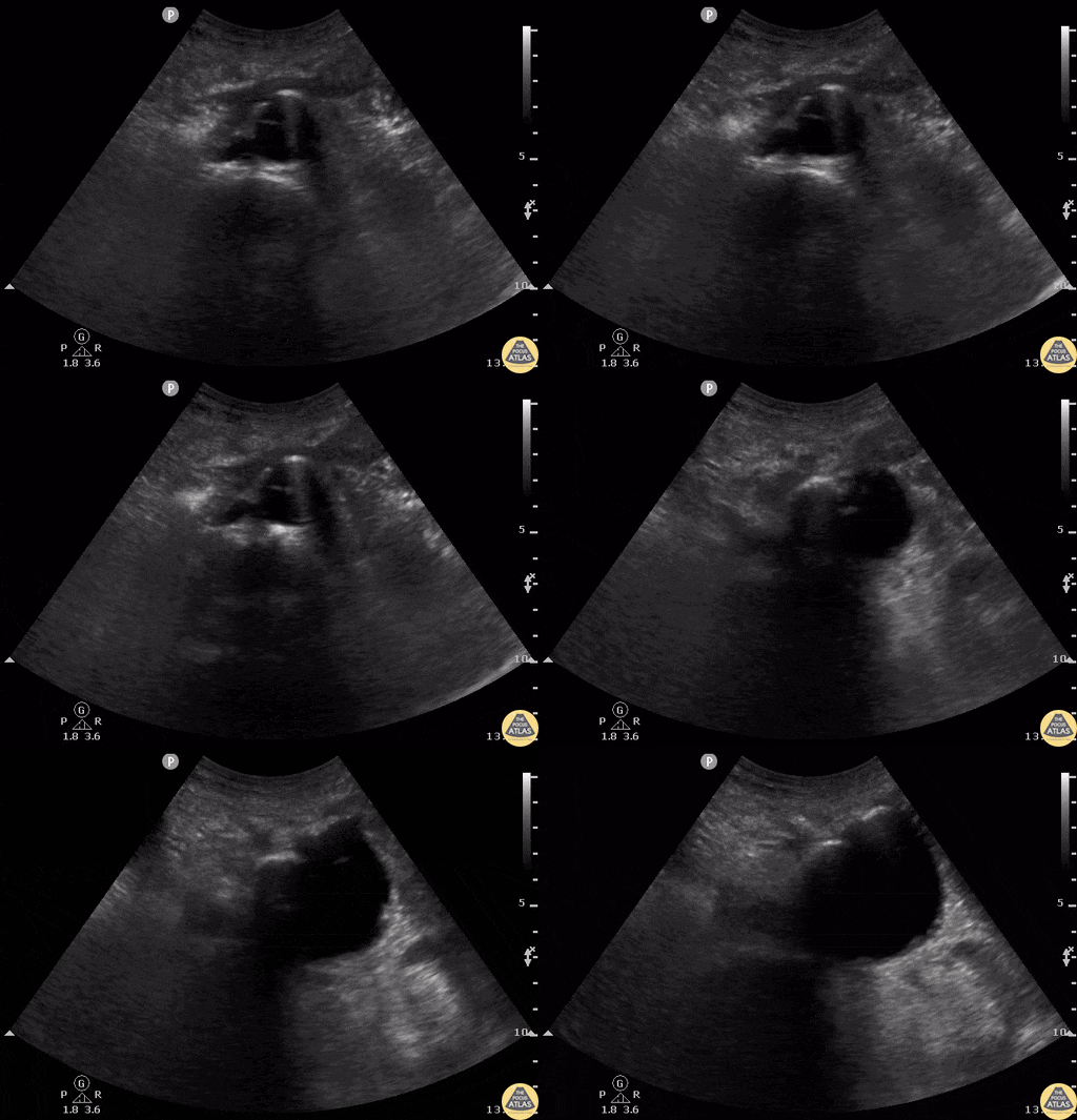
A patient in their 80s with PMH of abdominal aortic aneurysm s/p grafting with type 2 endoleak presented to the ED for dizziness. Bedside ultrasound demonstrated an 8.55 cm aortic aneurism with large thrombus. Endoleak is the hypoechoic area seen above the hyperechoic endograft.
Mehtab Galeh, MD; Bayley Espinoza, MD
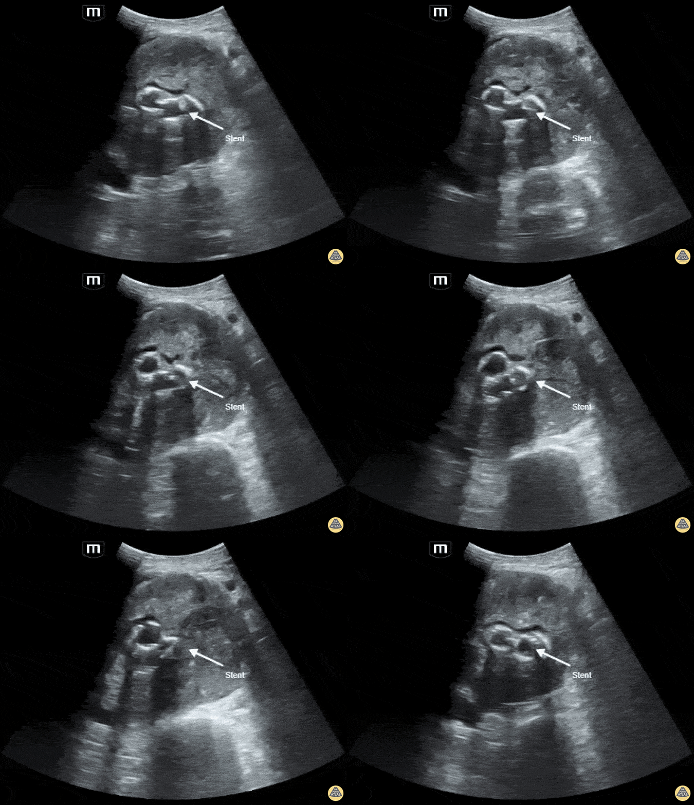
A patient in their 80s with PMH of abdominal aortic aneurysm s/p grafting with type 2 endoleak presented to the ED for dizziness. Bedside ultrasound demonstrated an 8.55 cm aortic aneurism with large thrombus. Endoleak is the hypoechoic area seen above the hyperechoic endograft.
Mehtab Galeh, MD; Bayley Espinoza, MD
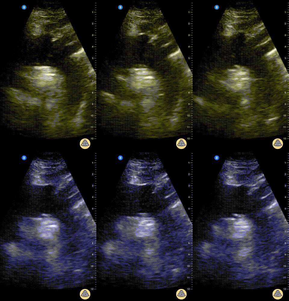
Suprasternal notch view of the aortic arch with brachiocephalic artery (BCA), left common carotid artery (LCA), and left subclavian artery (LSA) branching off. Right pulmonary artery is also visible adjacent to the ascending aorta.
Charles Jang, EM PGY-3
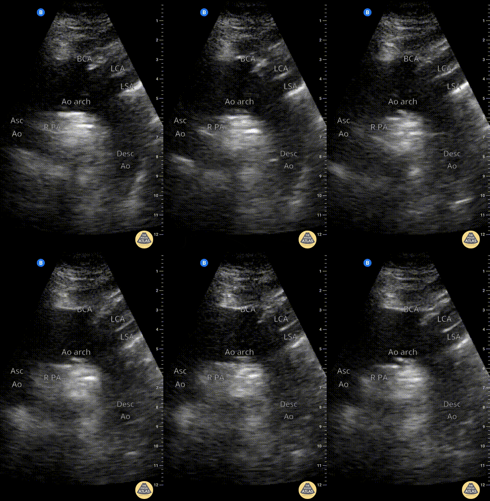
Suprasternal notch view of the aortic arch with brachiocephalic artery (BCA), left common carotid artery (LCA), and left subclavian artery (LSA) branching off. Right pulmonary artery is also visible adjacent to the ascending aorta.
Charles Jang, EM PGY-3
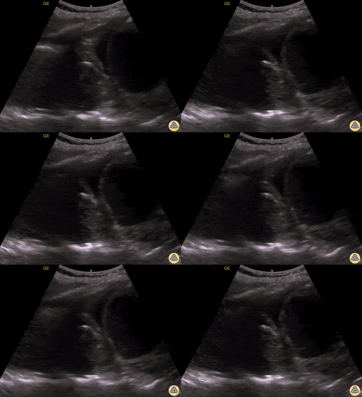
An elderly woman presented to the ED with dyspnea and left mid back pain for 1 week. Left PLAPS (Posterolateral Alveolar and/or Pleural Syndrome) view revealed a round-shaped pulsatile mass surrounded by pleural effusion & atelectatic lung. CTA later confirmed a partially ruptured, thrombosed thoracic aorta aneurysm. This case illustrates the possibility of quickly diagnosing life-threatening conditions with POCUS.
Contributed by: Caio Sangirardi, Emergency Medicine Resident Quinta D'Or Hospital, Rio de Janeiro - Brazil, @SangirardiMD
Normal Anatomy
Suprasternal notch view of the aortic arch with brachiocephalic artery (BCA), left common carotid artery (LCA), and left subclavian artery (LSA) branching off. Right pulmonary artery is also visible adjacent to the ascending aorta.
Charles Jang, EM PGY-3
Suprasternal notch view of the aortic arch with brachiocephalic artery (BCA), left common carotid artery (LCA), and left subclavian artery (LSA) branching off. Right pulmonary artery is also visible adjacent to the ascending aorta.
Charles Jang, EM PGY-3
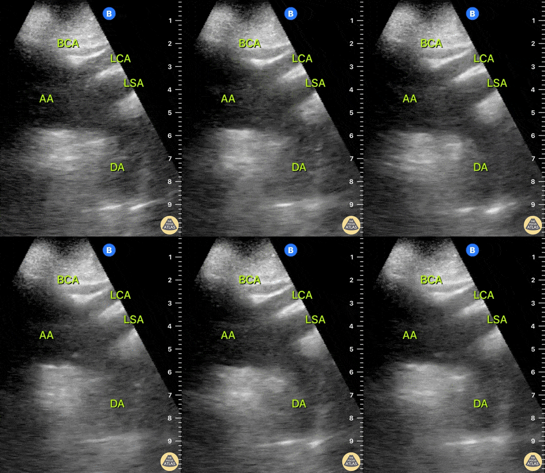
This clip represents a normal suprasternal view. The brachiocephalic artery (BCA), left carotid artery (LCA) and left subclavian artery (LSA) emerge from the aortic arch (AA). The left subclavian artery (LSA) marks the division between the proximal thoracic aorta and the distal descending aorta (DA).
Dr. Felipe Urriola P., Emergency Unit, Puerto Aysen Hospital, Chilean Patagonia.
Colorized Anatomy
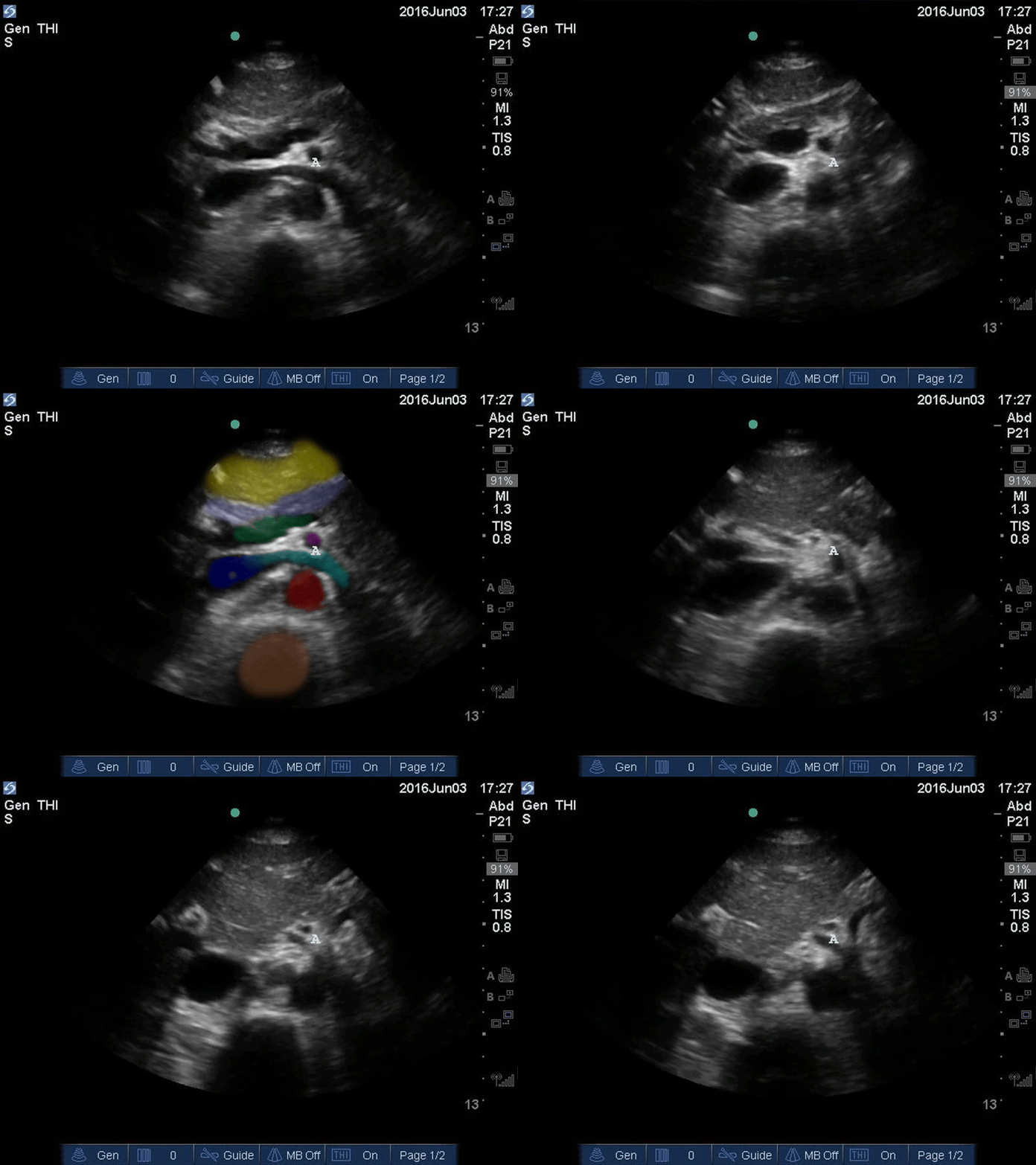
Orange: Yellow: Liver, Light blue: Pancreas, Aqua: splenic vein/portal confluence, Blue: IVC with left renal vein, Purple: SMA, Red: Aorta, Orange: Spine
Images: Dr. Lindsay Davis, Dr. Hannah Kopinski. Image Editing: Michael Amador and Dr. Matthew Riscinti
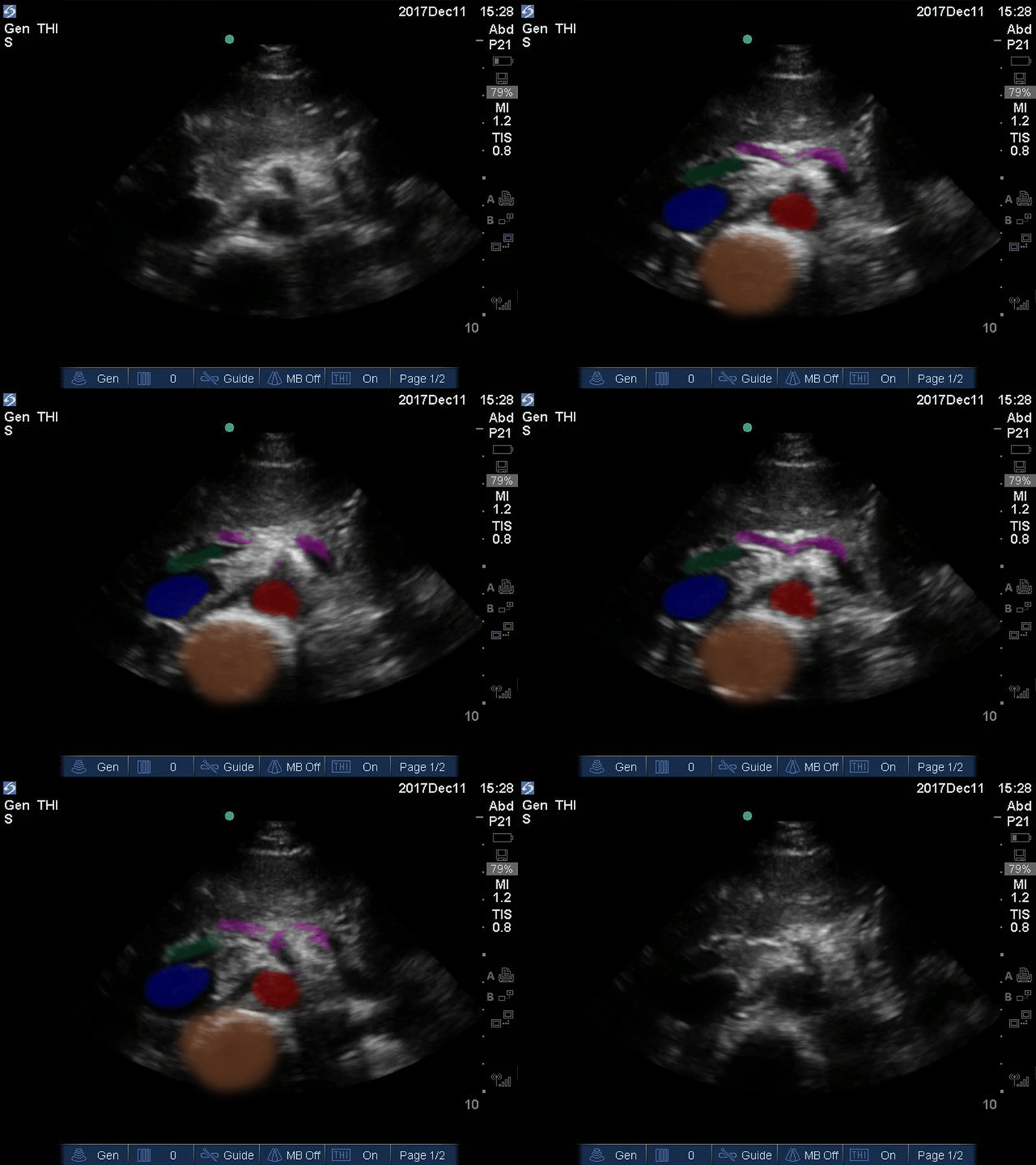
Good Seagull Sign
Orange: Spine, Red: Aorta, Blue: IVC, Green: Portal venous confluence, Pink: “Seagull sign” aka celiac trunk
Images: Dr. Lindsay Davis, Dr. Hannah Kopinski. Image Editing: Michael Amador and Dr. Matthew Riscinti
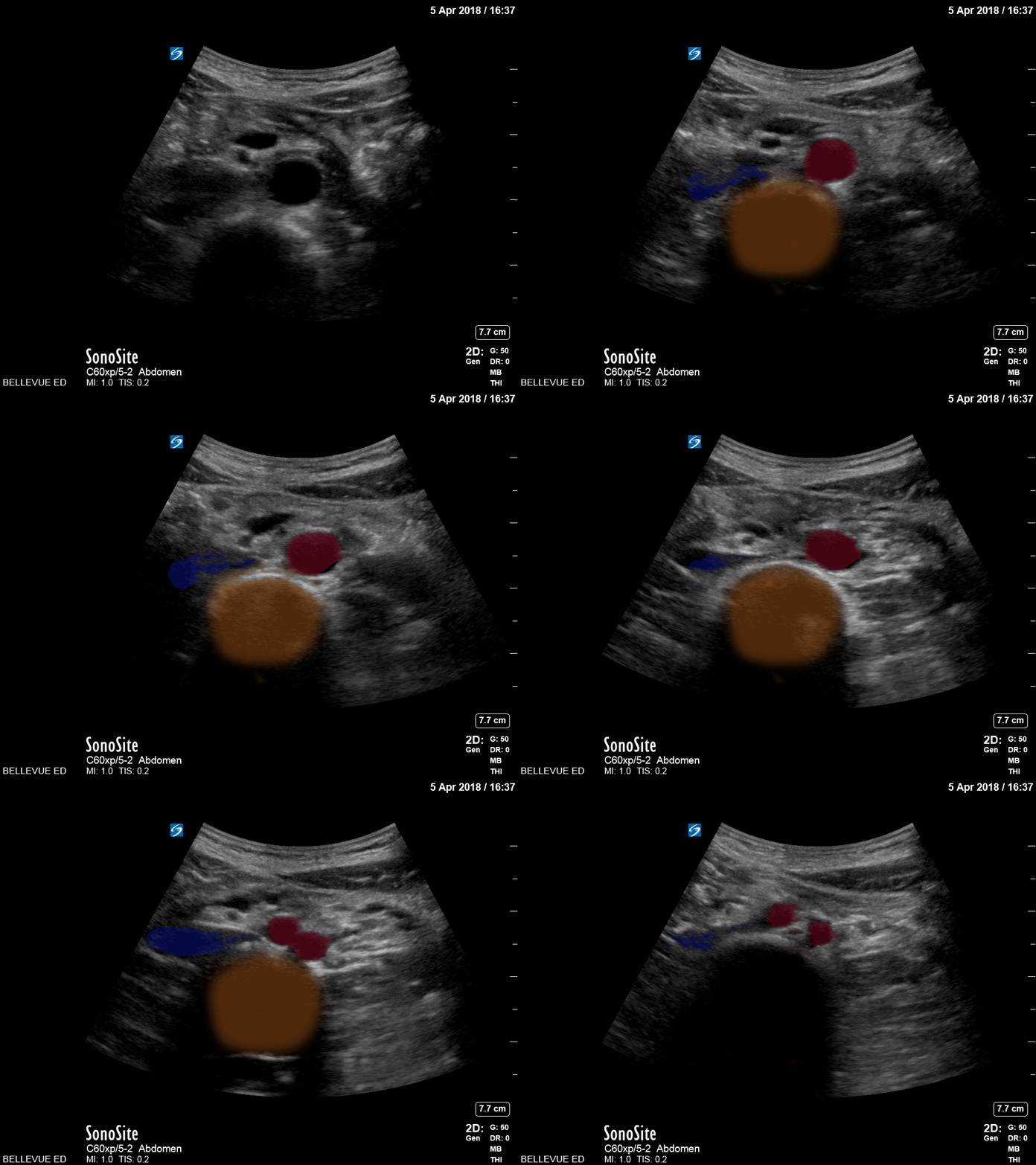
Distal Aorta
Orange: Spine, Red: Aorta and Iliacs, Blue: IVC, Green: Portal venous confluence, Pink: “Seagull sign” aka celiac trunk
Images: Dr. Lindsay Davis, Dr. Hannah Kopinski. Image Editing: Michael Amador and Dr. Matthew Riscinti
Abdominal Aortic Aneurysm
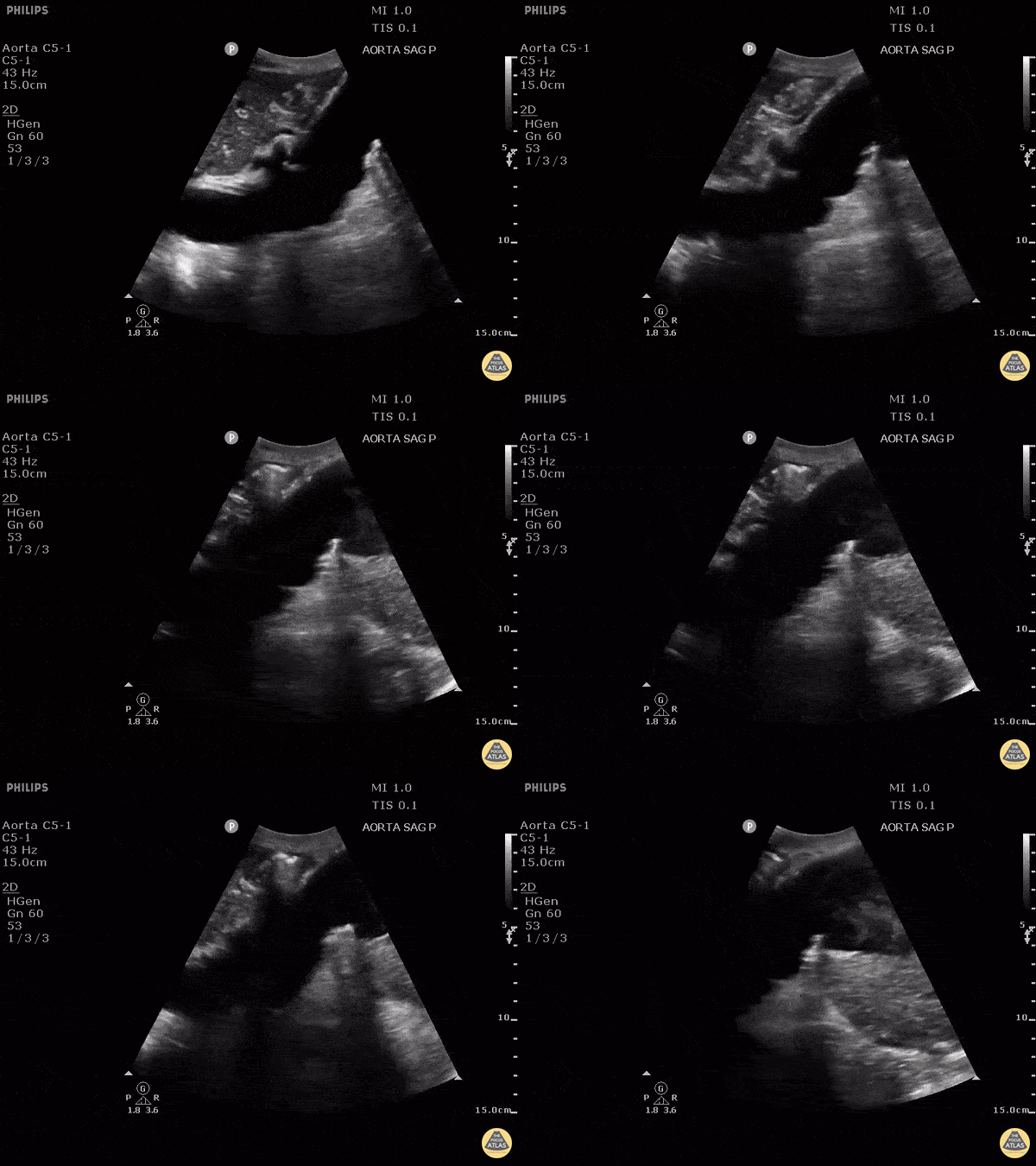
Sagittal view of large abdominal aortic aneurysm incidentally found after accidental fall.
Image courtesy of Robert Jones DO, FACEP @RJonesSonoEM
Director, Emergency Ultrasound; MetroHealth Medical Center; Professor, Case Western Reserve Medical School, Cleveland, OH
View his original post here
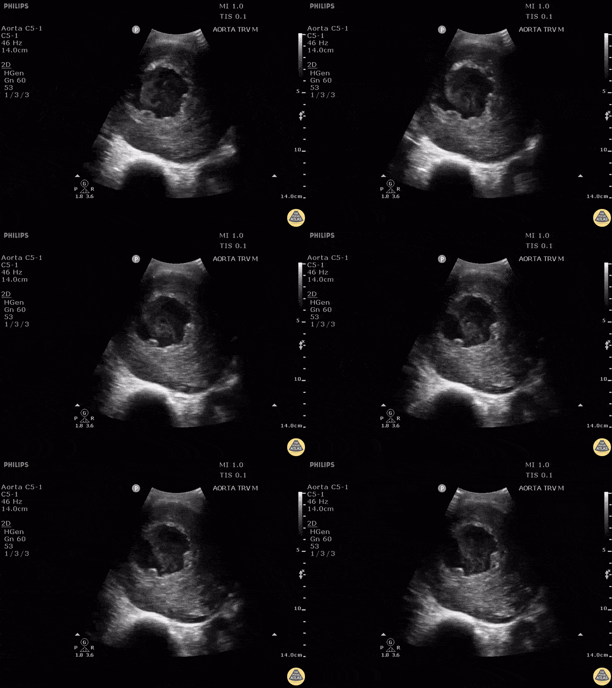
Here is another example of an abdominal aortic aneurysm that measured approximately 10.6 cm. The patient originally presented to the clinic complaining of a cough and was eventually admitted for surgical operation.
Image courtesy of Robert Jones DO, FACEP @RJonesSonoEM
Director, Emergency Ultrasound; MetroHealth Medical Center; Professor, Case Western Reserve Medical School, Cleveland, OH
View his original post here
AAA Rupture
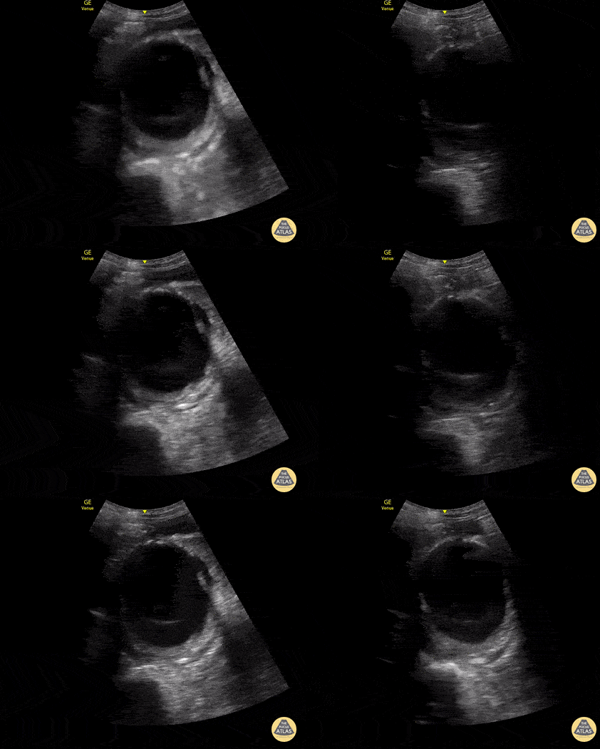
70s-year-old male brought in for altered mental status and hypotension.
As part of the initial exam, POCUS was performed. This clip, obtained using the curvilinear probe in transverse orientation over the mid-epigastric region, revealed a AAA measuring 8 cm in AP diameter at the largest point with heterogeneous echogenic material consistent with thrombus. CT abdomen/pelvis with IV contrast confirmed ruptured AAA with thrombosis and only minor extravasation.
In this case, POCUS played a critical role in the rapid diagnosis of a patient with undifferentiated shock, particularly in a patient who was unable to provide any history.
Katherine Spencer, MD
Guy Youngblood, MD, FACEP, FAAEM
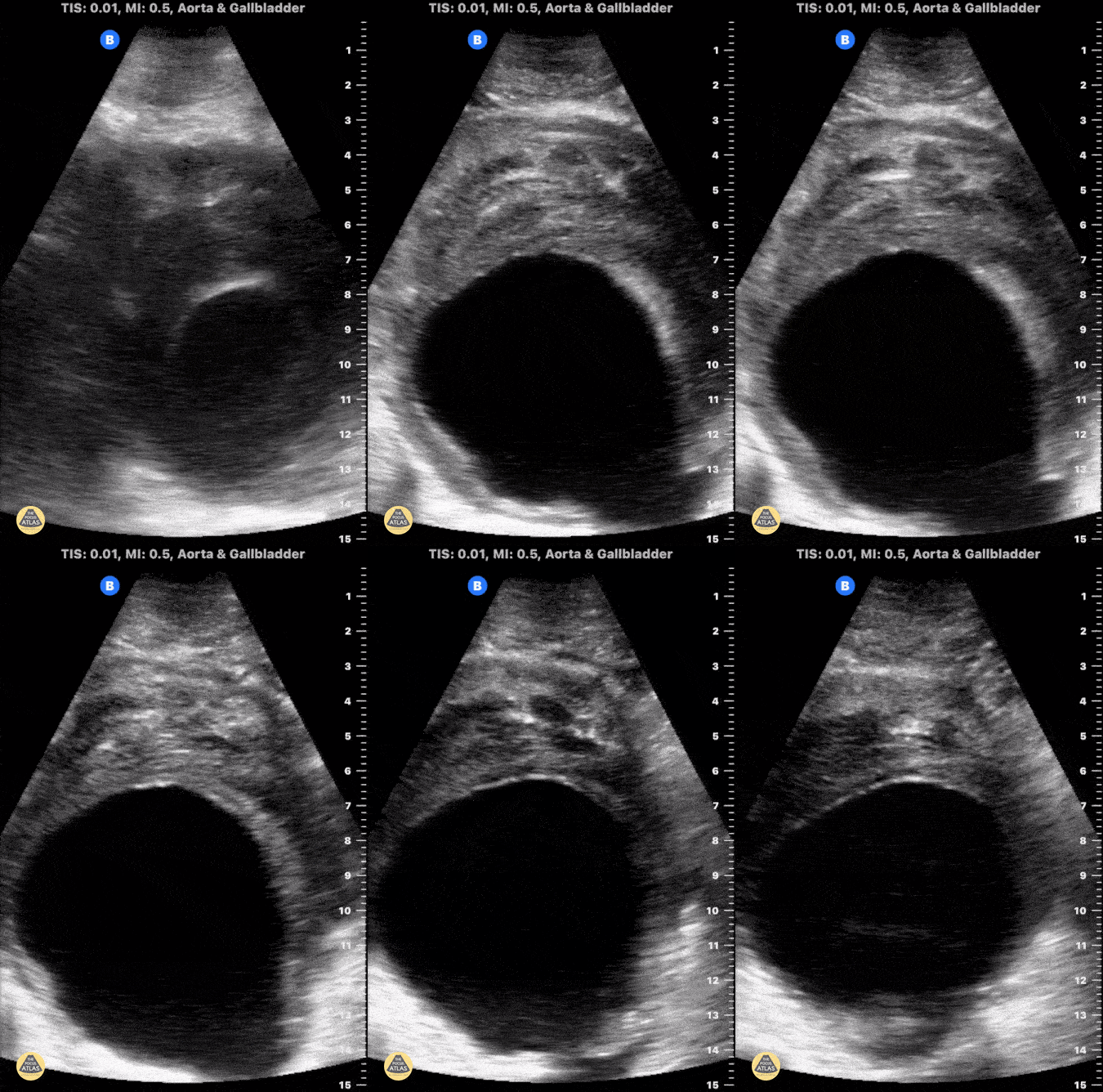
This is a clip demonstrating a large abdominal aortic aneurysm with significant intramural thrombus. Signs of rupture can be appreciated near the right lower portion of the screen as there is disruption of the aorta wall. Heterogeneity within the intramural thrombus can also be an indicator of rupture.
Michael Macias, MD
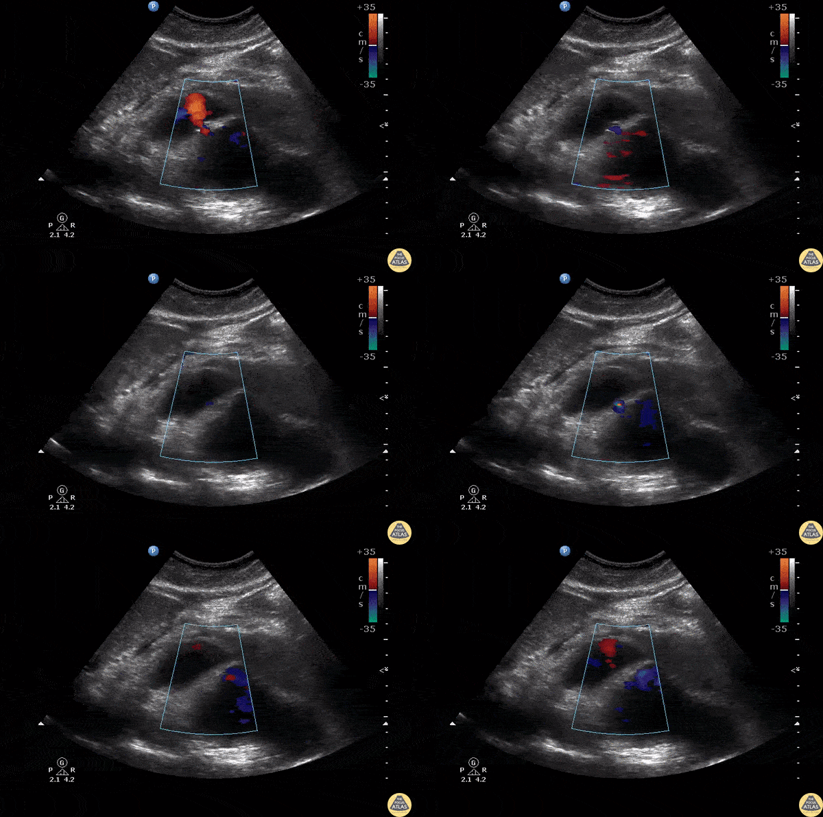
Sagittal view of an abdominal aortic aneurysm with a hemorrhage anterior to the aorta. Be sure to distinguish a AAA from a dissection by assessing the abnormal wall motion seen at the site of rupture.
Image courtesy of Robert Jones DO, FACEP @RJonesSonoEM
Director, Emergency Ultrasound; MetroHealth Medical Center; Professor, Case Western Reserve Medical School, Cleveland, OH
View his original post here
Aortic Dissection
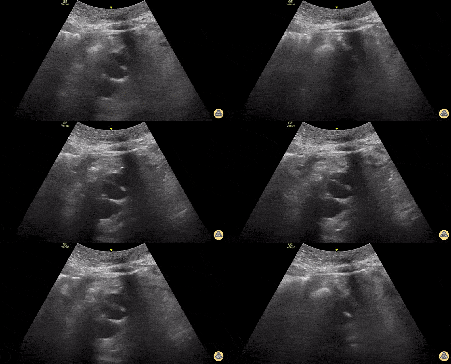
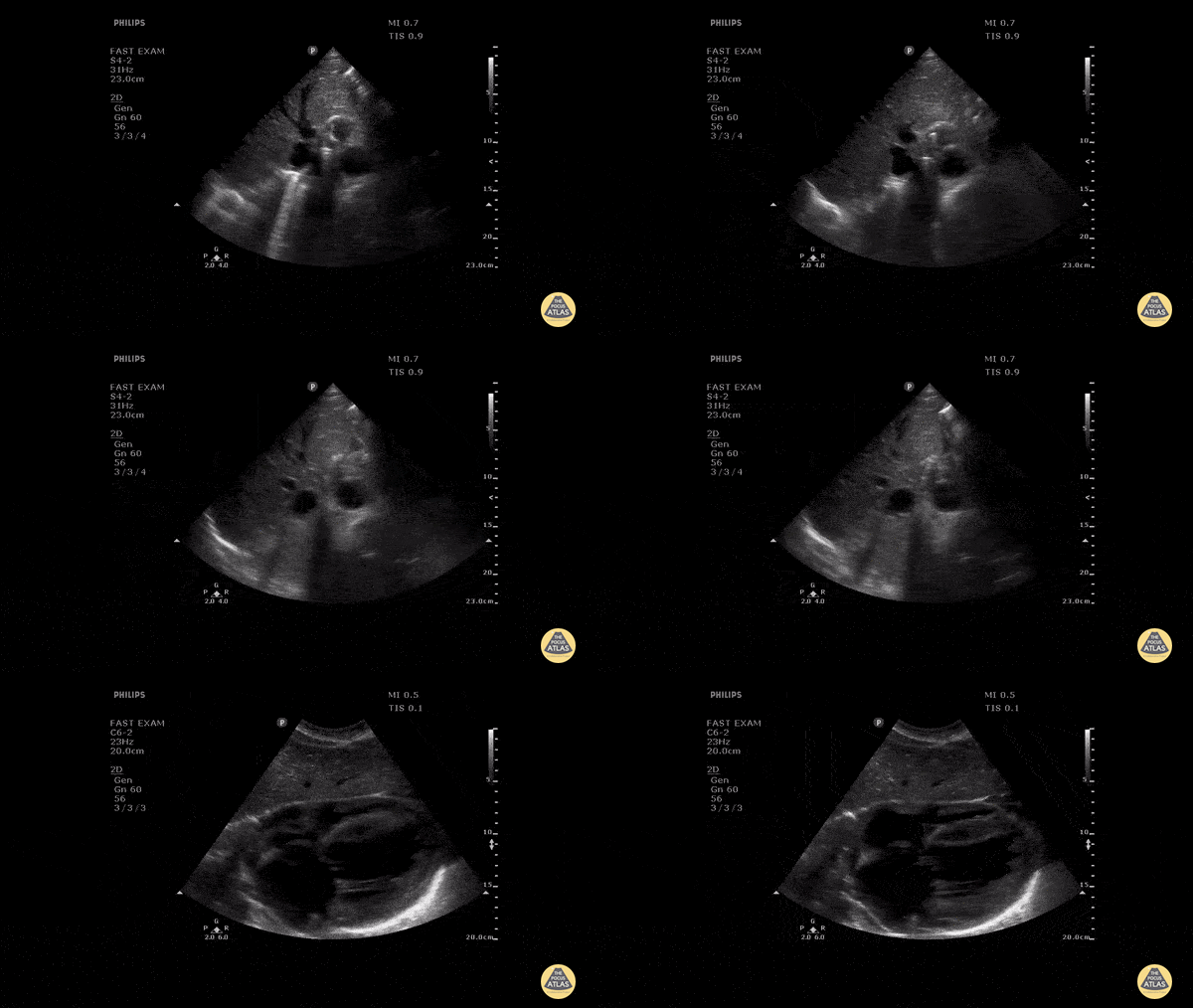
Flap seen in the descending aorta consistent with aortic dissection
Contributed by: Dimitri Livshits DO, Ultrasound Fellow; Jane Belyavskaya MD, Ultrasound Fellow; Chris Hanuscin MD, Ultrasound Division Director (Kings County/SUNY Downstate)
Patient presented with severe chest pain and concerning ECG without STEMI. A faint dissection flap can be seen in the aorta on this transverse view in the subcostal region.
Image courtesy of Robert Jones DO, FACEP @RJonesSonoEM
Director, Emergency Ultrasound; MetroHealth Medical Center; Professor, Case Western Reserve Medical School, Cleveland, OH
View his original post here
Type A Aortic Dissection
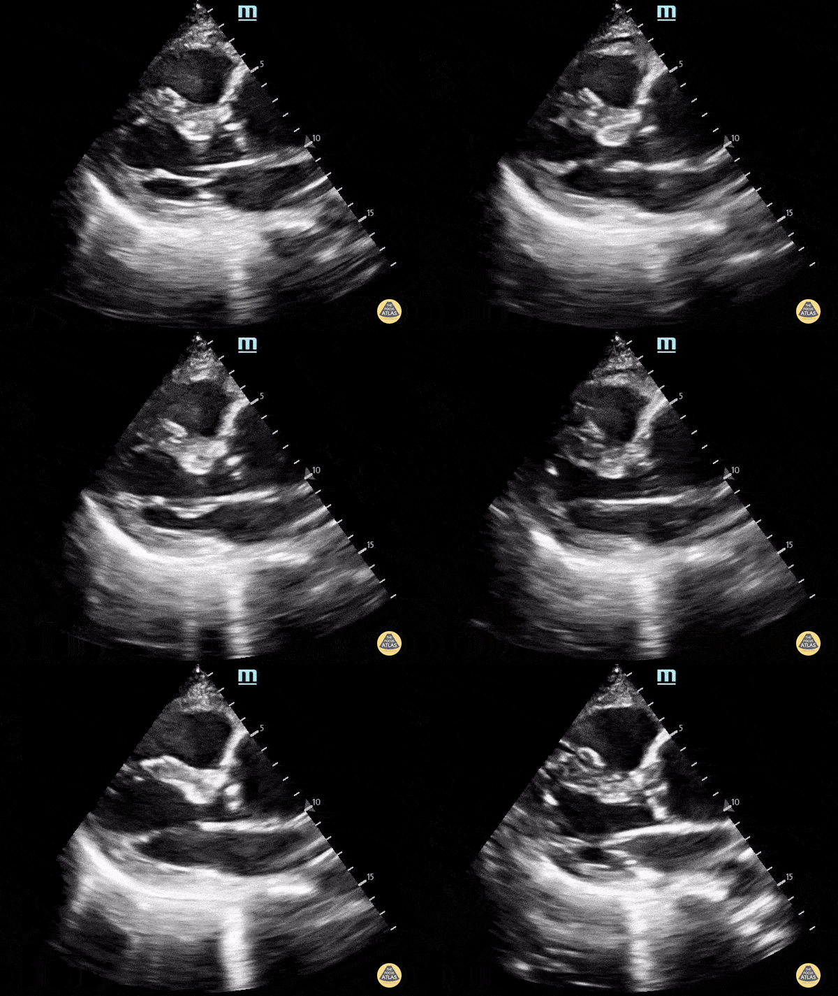
A 70 y.o. male presented with altered mental status.
POCUS echocardiogram performed in the ED revealed an ascending thoracic aortic aneurysm measuring approximately 5 cm in diameter (parasternal long-axis view shown here). A chart review revealed that the patient does indeed have a history of TAA, and a comparison of our findings to a prior CTA demonstrates no significant increase in diameter. Nevertheless, this study demonstrates the utility of POCUS in the rapid and early detection of ascending aortic abnormalities.
Alex Schlangen, D.O. PGY-1 EM Resident at Central Michigan University; Andrew Namespetra, MB BCh BAO. @AndrewNamespet1 PGY-3 EM Resident at Central Michigan University
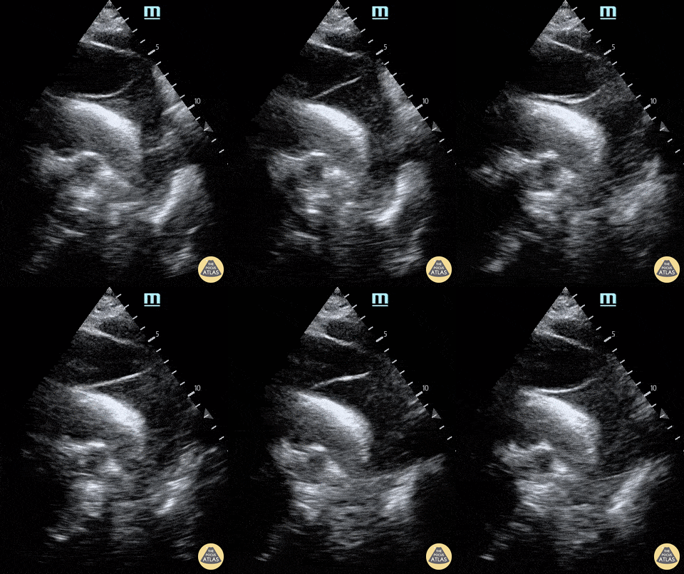
A 65-year-old male presents with shortness of breath (no chest pain) and was found to have a dilated aortic root on CT pulmonary angiogram. POCUS (supra sternal view) showed a dissection flap in the arch of aorta; a finding subsequently confirmed on CT aortagram. Patient was sent for emergency surgical intervention.
Dr.Rajasutharsan Kathirgamanathan, Emergency Physician
The Northern Hospital, Melbourne, Australia
@raj_kathir007
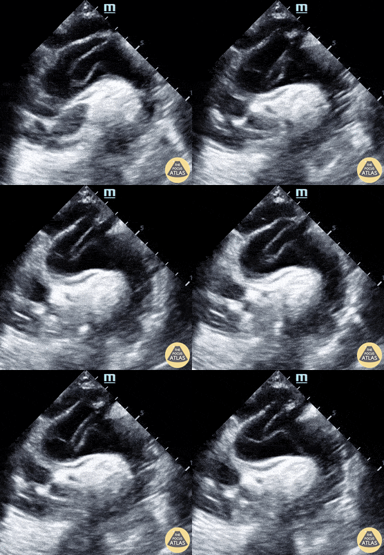
Suprasternal notch view shows a mobile intimal dissection flap in the aortic arch.
Michael Cover, MD
@michaelc0ver
Other Pathology
A patient in their 80s with PMH of abdominal aortic aneurysm s/p grafting with type 2 endoleak presented to the ED for dizziness. Bedside ultrasound demonstrated an 8.55 cm aortic aneurism with large thrombus. Endoleak is the hypoechoic area seen above the hyperechoic endograft.
Mehtab Galeh, MD; Bayley Espinoza, MD
A patient in their 80s with PMH of abdominal aortic aneurysm s/p grafting with type 2 endoleak presented to the ED for dizziness. Bedside ultrasound demonstrated an 8.55 cm aortic aneurism with large thrombus. Endoleak is the hypoechoic area seen above the hyperechoic endograft.
Mehtab Galeh, MD; Bayley Espinoza, MD
An elderly woman presented to the ED with dyspnea and left mid back pain for 1 week. Left PLAPS (Posterolateral Alveolar and/or Pleural Syndrome) view revealed a round-shaped pulsatile mass surrounded by pleural effusion & atelectatic lung. CTA later confirmed a partially ruptured, thrombosed thoracic aorta aneurysm. This case illustrates the possibility of quickly diagnosing life-threatening conditions with POCUS.
Contributed by: Caio Sangirardi, Emergency Medicine Resident Quinta D'Or Hospital, Rio de Janeiro - Brazil, @SangirardiMD
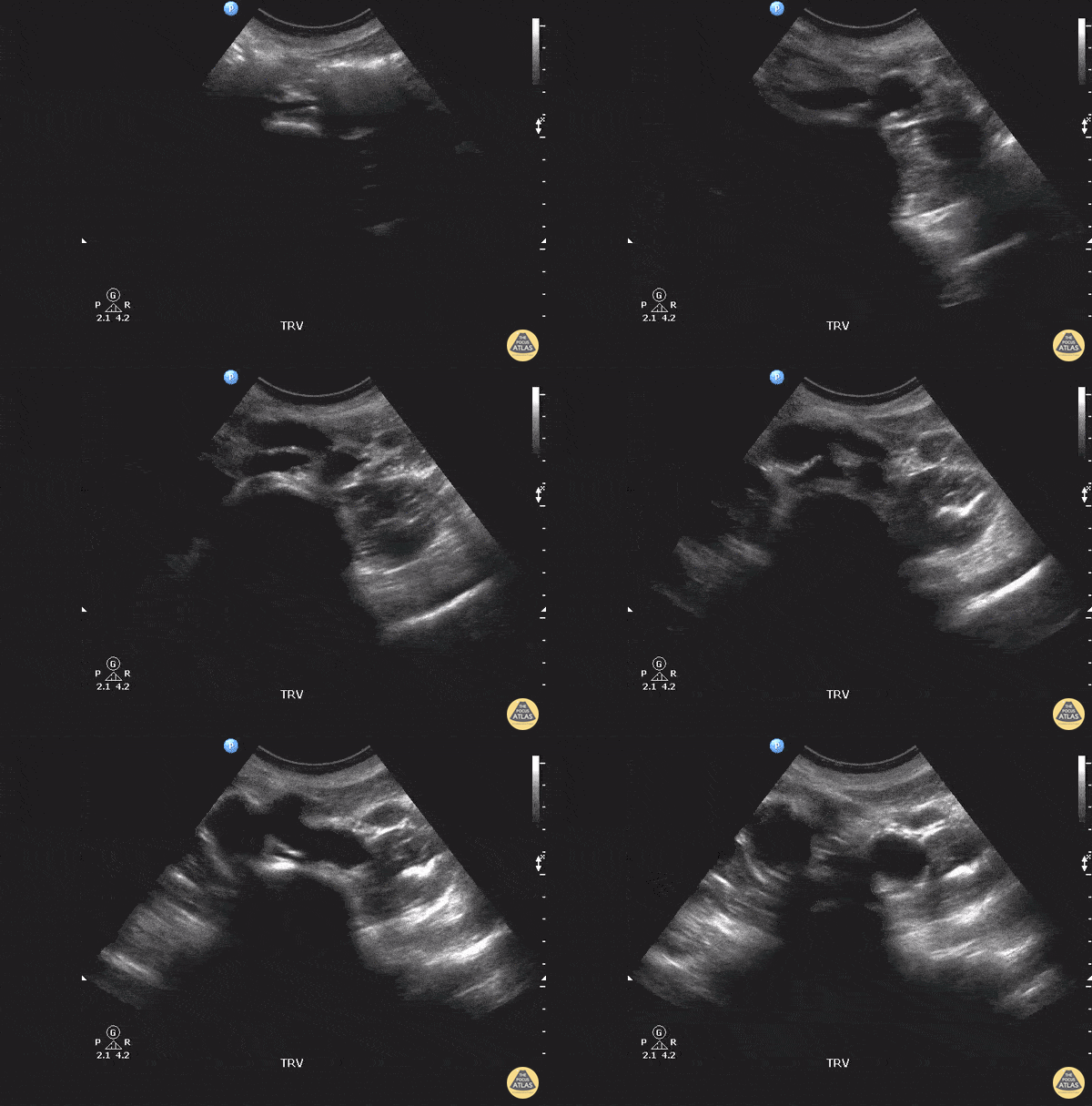
This patient originally complained of right back pain as well as numbness along the thigh and knee. Starting from the abdominal artery, gliding inferiorly will eventually reveal the bifurcation of the abdominal aorta into the left and right common iliac arteries. In this scan, you can see both iliac arteries have enlarged diameters, indicating aneurysms in both.
Image courtesy of Robert Jones DO, FACEP @RJonesSonoEM
Director, Emergency Ultrasound; MetroHealth Medical Center; Professor, Case Western Reserve Medical School, Cleveland, OH
View his original post here
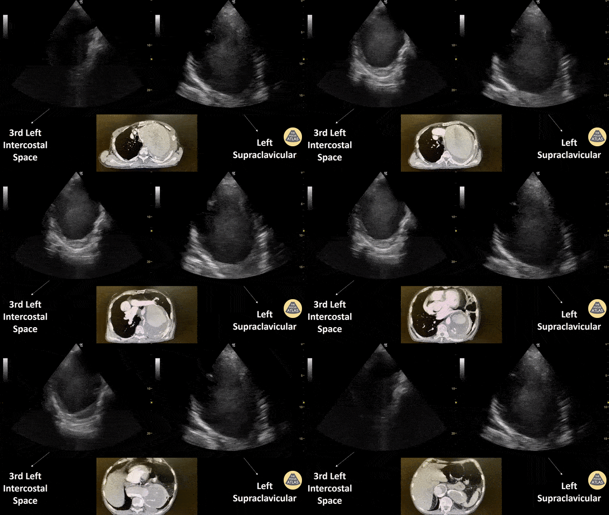
Patient with a subacute history of chest trauma (occurred approximately 30 days prior) presented reporting intermittent chest pain. He was noted in the ED to have persistent pain and to be sweating. Bedside evaluation included lung ultrasound that identified the cystic structure seen here with a hypoechoic interior with visible swirling. Chest CT shown confirmed this to be sequelae of aortic trauma.
Renato Melo, Emergency Physician Brazil. PocusJedi affiliated.
@Renato_Melo_
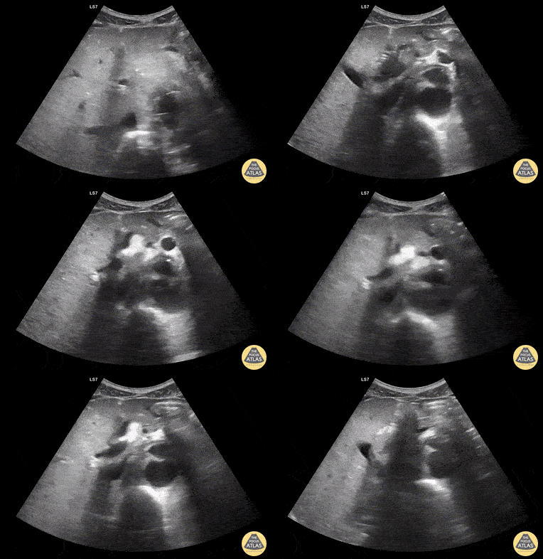
Aortic Endograft Leak - Dr. Lindsay Howe Dr. Tim Scheel Dr. Paul Pelletier - Denver Health