Gastrointestinal
Gastrointestinal
Note: These search bar results are only for the Gastrointestinal image archive. To search the entire site, click here.
Recently Added
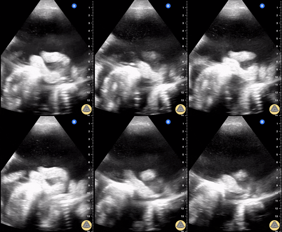
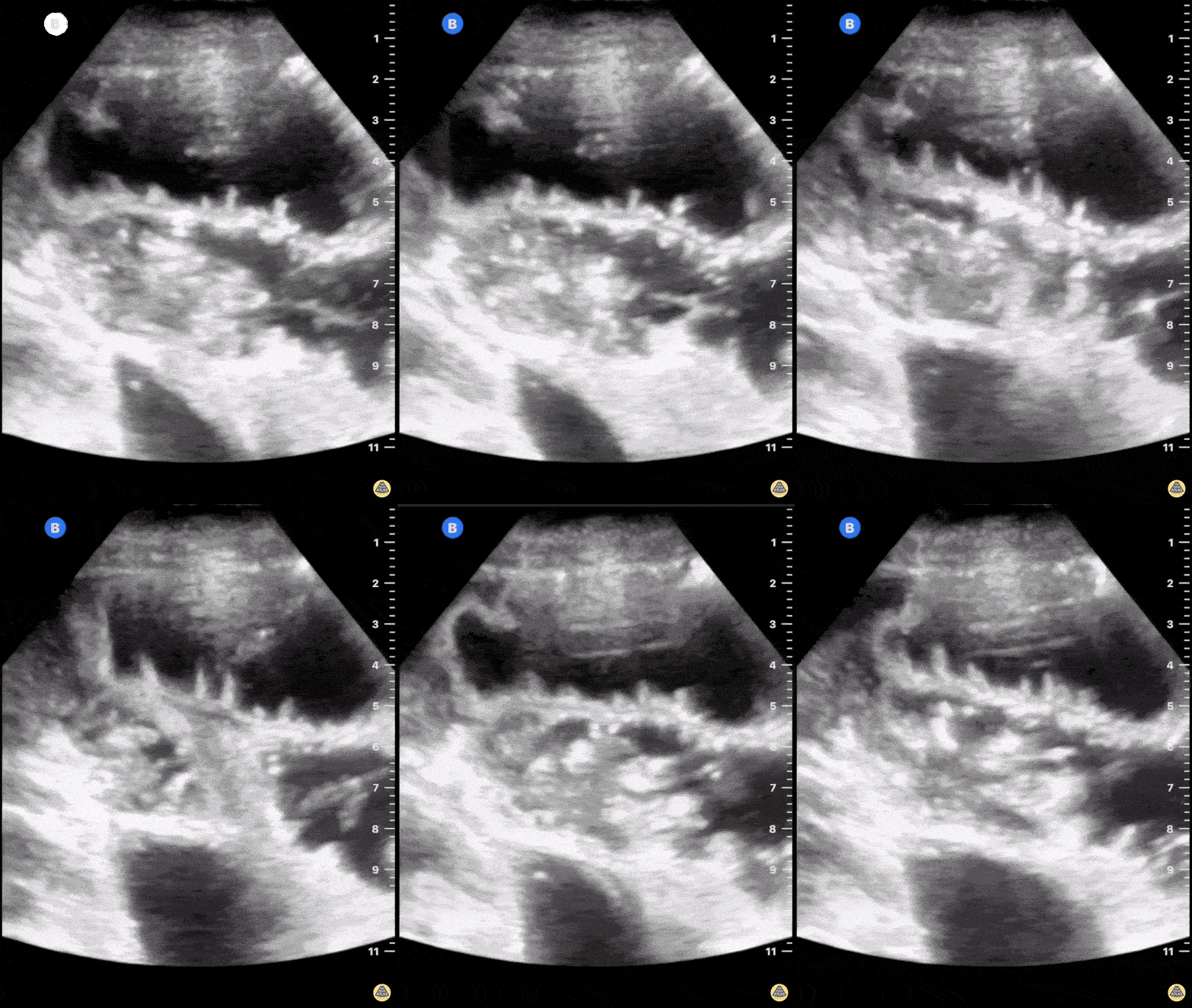
Malignant ascites with plankton sign in woman with metastatic ovarian cancer.
Samuel Eglin, MD
A 24 year old male comes to ER with abdominal pain, vomiting, and fever.
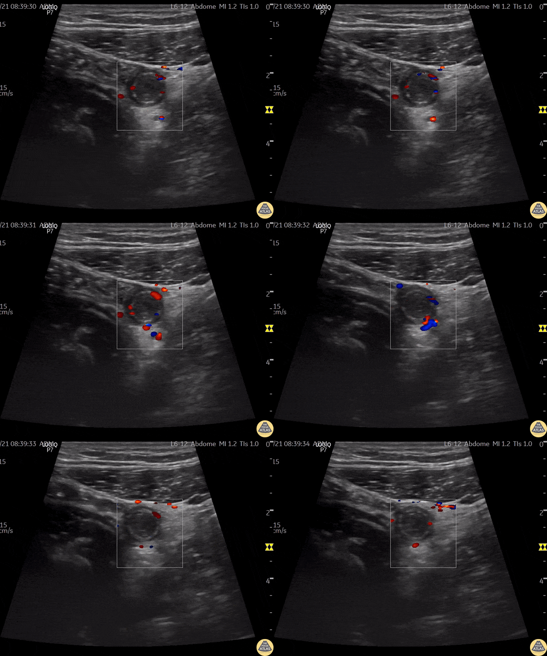
A directed scan with a high-frequency linear transducer in the right iliac fossa shows a tubular, non-compressible structure, a "target like" image, with increased echogenicity of the fat around it. When applied Color Doppler - we can observe increased vascular flow around the appendix - a sign known as "The Ring of Fire". This finding suggests the diagnosis of acute appendicitis.
Contributor: Renato Tambelli (@R_Tambelli @Jedipocus)
Fatigue for the past 3–4 months with mild disturbance of bowel habits. The patient reported visualizing proglottids in the stool.
POCUS findings: Ultrasound performed in the left lower quadrant revealed a structure consistent with parasitic segments.
Diagnosis: Laboratory confirmation a few days later identified Taenia saginata.
Dr. Guillaume Schramme
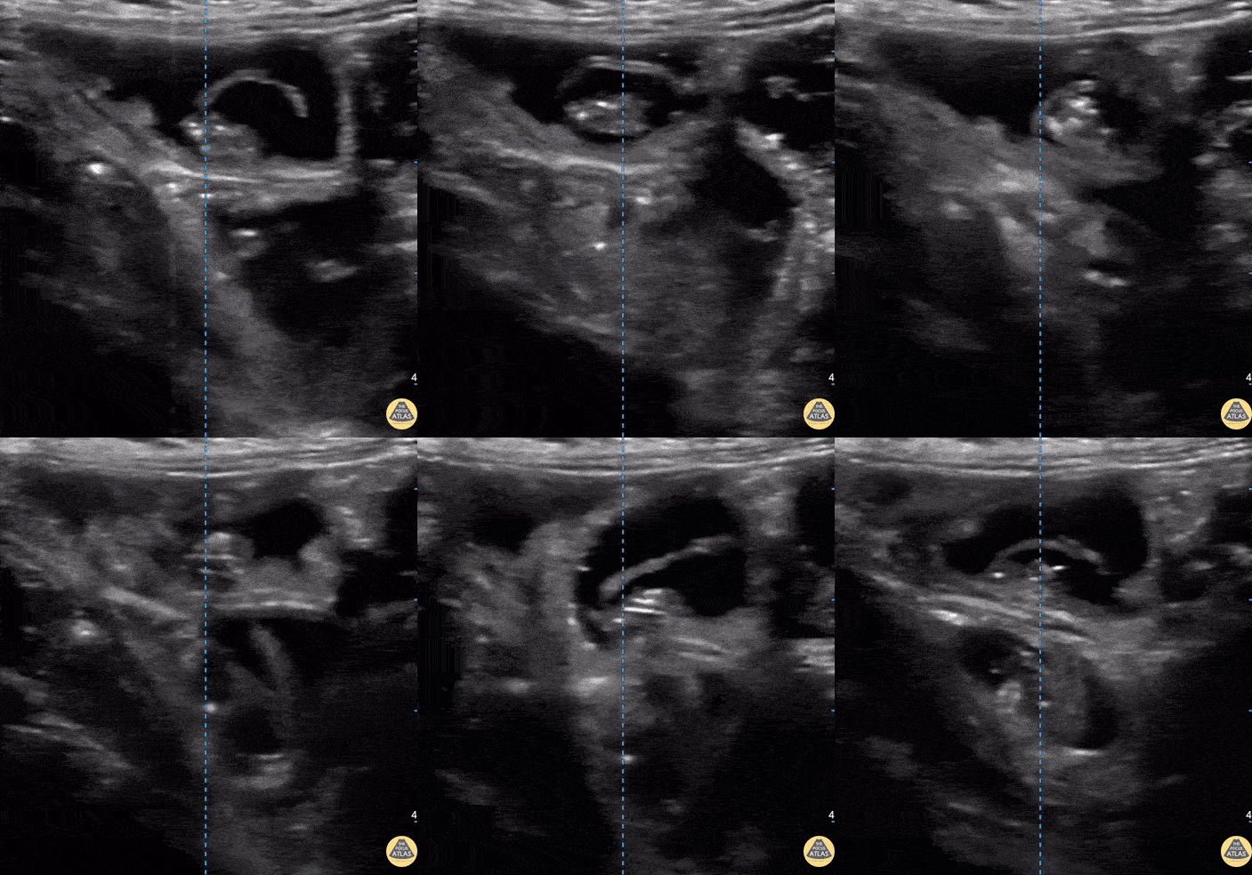
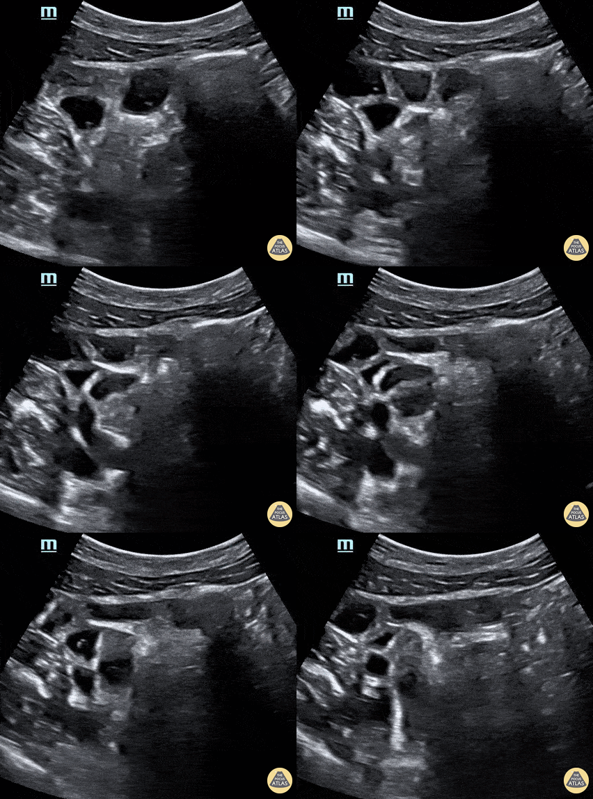
Fecal material can be seen moving forward and backwards through dilated bowels in the patient with a bowel obstruction.
Contributed by: Brittany Garza, DO and Saleem Nasseh, MD and Sadie Ellenson, MS4
Free fluid demonstrated at the liver tip in a patient with ascites.
The liver tip is the most sensitive part of the RUQ for free fluid. In the context of blunt trauma this would be concerning for bleeding.
Contributors: Dimitri Livshits, DO; Jane Belyavskaya, MD; Chris Hanuscin, MD
Kings County/SUNY Downstate
Sign of small bowel obstruction. We see heterogeneous material inside the loop of bowel with a back and forth movement, characterizing the obstruction. The patient in question had a paralytic ileus due to chronic antipsychotic use.
Felipe Teles; Internal Medicine at Fortaleza General Hospital
@drfelipemoraisteles
Normal Anatomy
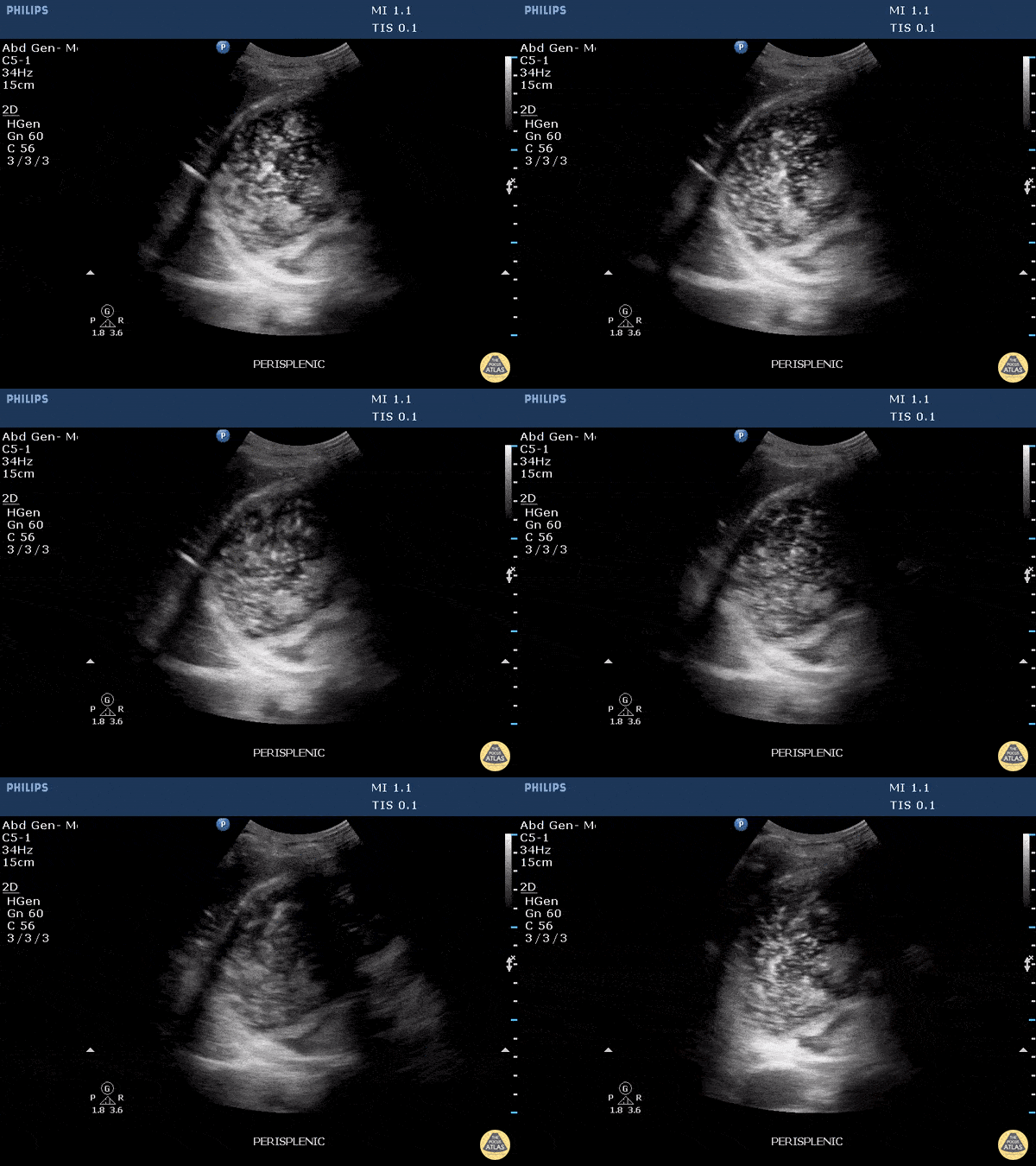
This clip demonstrates a full stomach. Gastric contents with internal air signatures can be seen swirling around within the stomach.
Image courtesy of Robert Jones DO, FACEP @RJonesSonoEM
Director, Emergency Ultrasound; MetroHealth Medical Center; Professor, Case Western Reserve Medical School, Cleveland, OH
View his original post here
51 year old male presented with a chief complaint of abdominal pain for 3 days with nausea and vomiting.
The curvilinear probe was used to evaluate the aorta with an incidental finding (shown) of clear peristalsis of the bowel contents with hypoechoic and anechoic contents. In this segment it is clear there is no obstruction.
Lindsay Davis, DO, MPH, @Lindsadavis18
Lydia Mansour, DO
Emily Nagourney, MS4
Central Michigan University
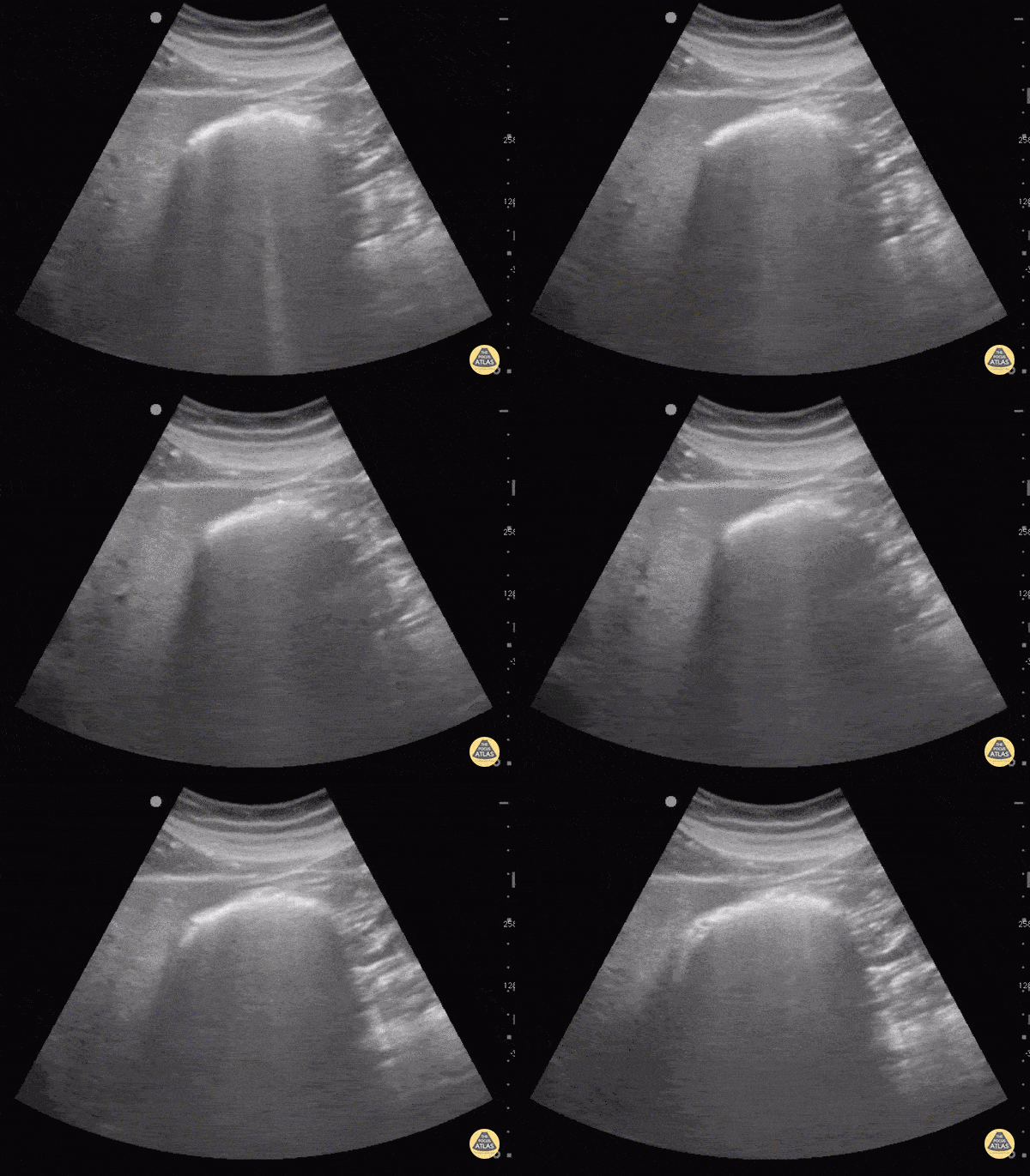
Bowel gas scatters ultrasound waves making the structure containing the gas as well as posterior structures difficult to visualize. Applying more gentle graded pressure to the probe may displace the gas and improve visualization.
Sukh Singh, MD
Colorized Anatomy
Appendix
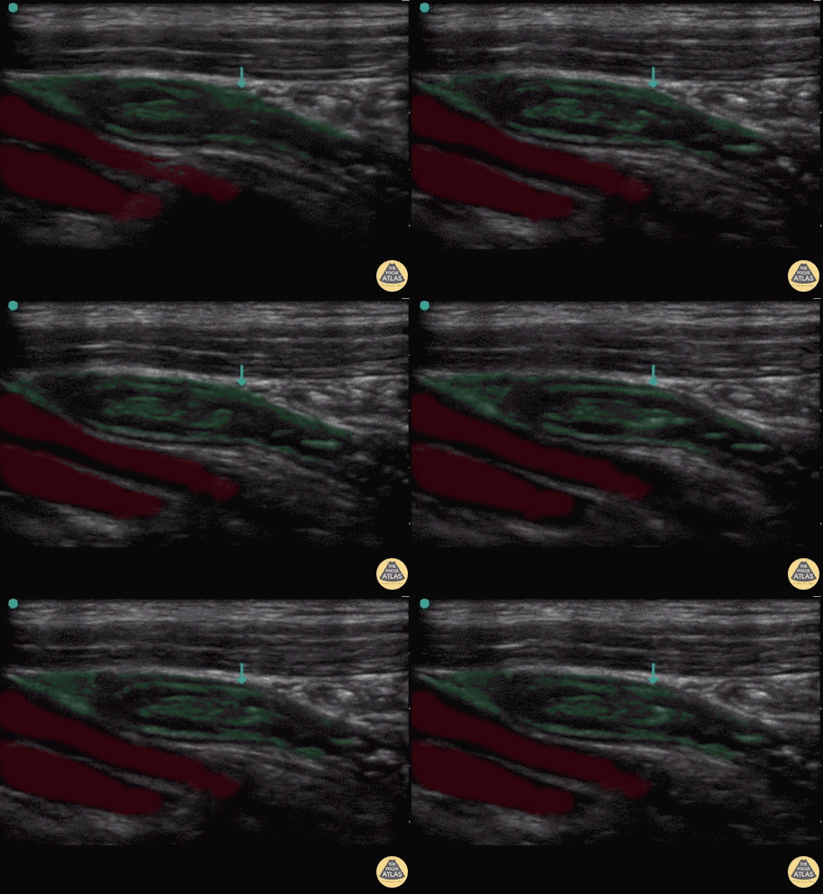
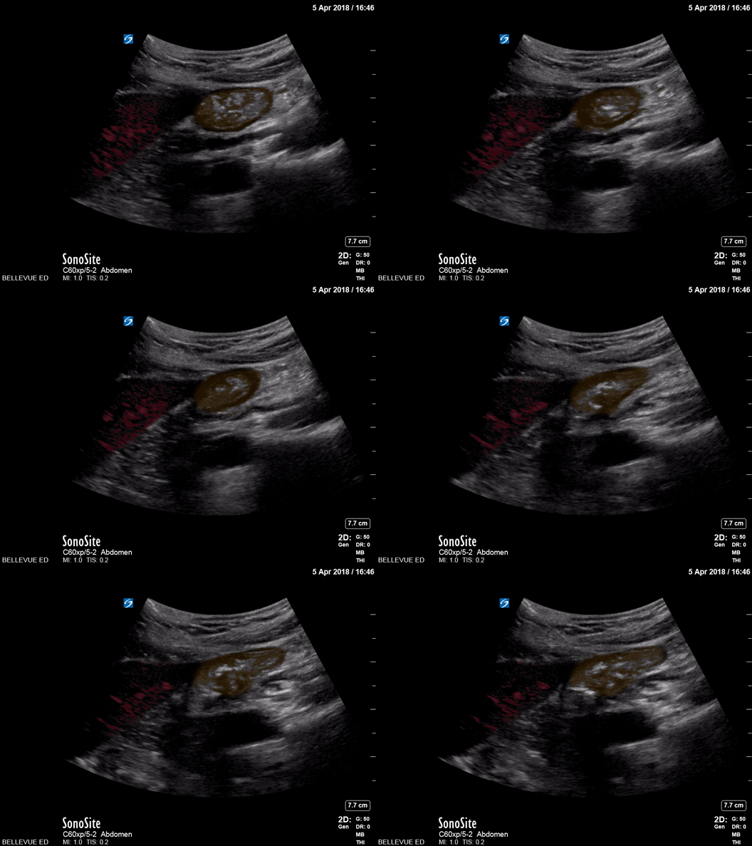
Red: Iliac vessels, Green: Appendix
Images: Dr. Lindsay Davis, Dr. Hannah Kopinski. Image Editing: Michael Amador and Dr. Matthew Riscinti
Large Bowel
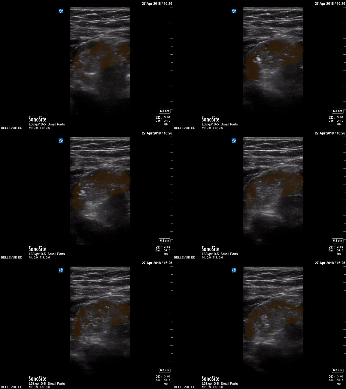
Orange - Large bowel lumen
Images: Dr. Lindsay Davis, Dr. Hannah Kopinski. Image Editing: Michael Amador and Dr. Matthew Riscinti
Duodenum and Liver
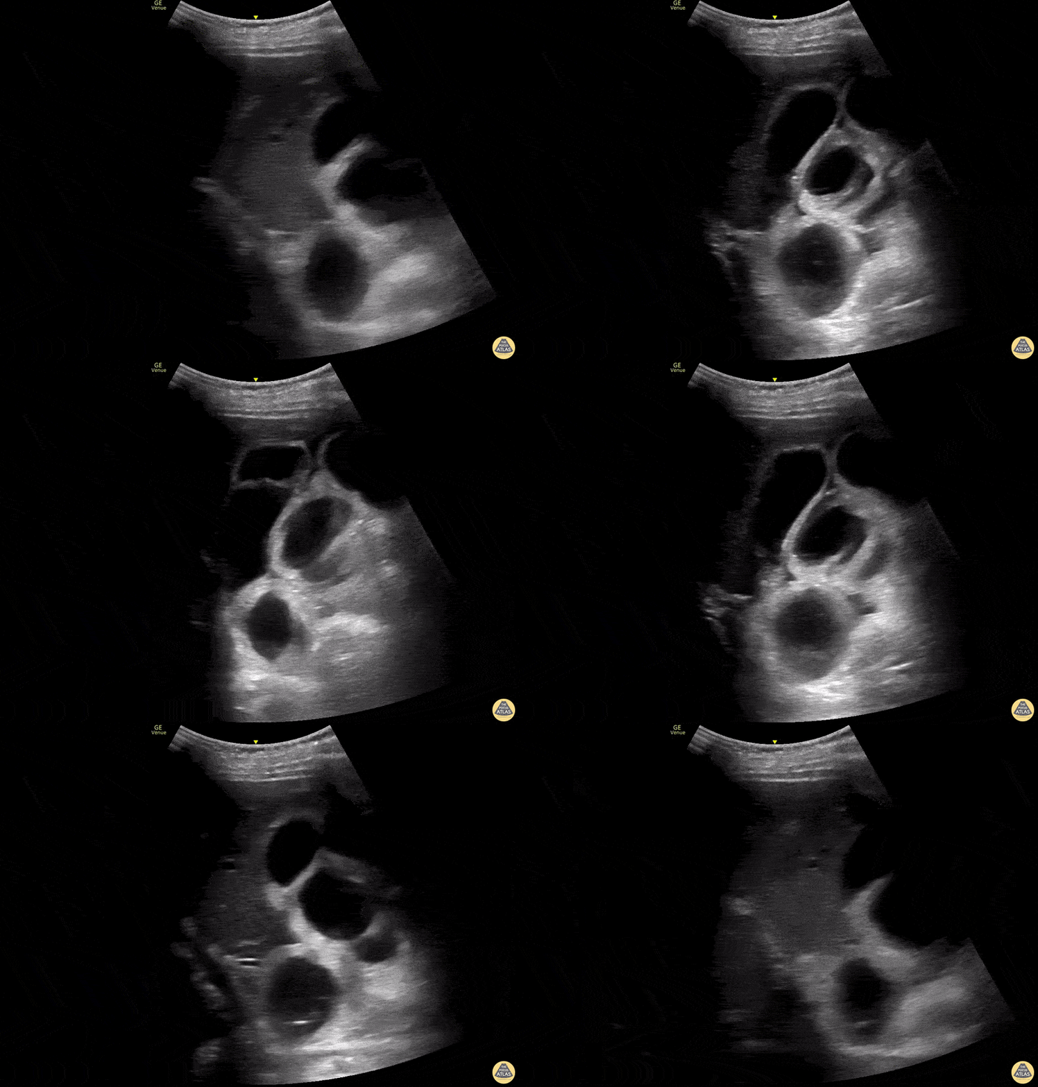
Orange: Duodenum, Red: Liver
Images: Dr. Lindsay Davis, Dr. Hannah Kopinski. Image Editing: Michael Amador and Dr. Matthew Riscinti
Small Bowel Obstruction
Fecal material can be seen moving forward and backwards through dilated bowels in the patient with a bowel obstruction.
Contributed by: Brittany Garza, DO and Saleem Nasseh, MD and Sadie Ellenson, MS4
Sign of small bowel obstruction. We see heterogeneous material inside the loop of bowel with a back and forth movement, characterizing the obstruction. The patient in question had a paralytic ileus due to chronic antipsychotic use.
Felipe Teles; Internal Medicine at Fortaleza General Hospital
@drfelipemoraisteles
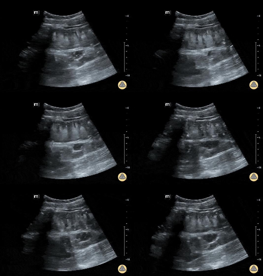
Dilated loops of small bowel with free fluid adjacent to them (Tanga sign), CT confirmed SBO.
Dimitri Livshits DO, Ultrasound Fellow; Jane Belyavskaya MD, Ultrasound Fellow; Chris Hanuscin MD, Ultrasound Division Director (Kings County/SUNY Downstate)
Ascites
Malignant ascites with plankton sign in woman with metastatic ovarian cancer.
Samuel Eglin, MD
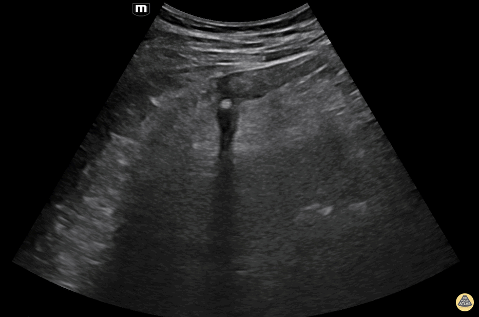
Free fluid demonstrated at the liver tip in a patient with ascites.
The liver tip is the most sensitive part of the RUQ for free fluid. In the context of blunt trauma this would be concerning for bleeding.
Contributors: Dimitri Livshits, DO; Jane Belyavskaya, MD; Chris Hanuscin, MD
Kings County/SUNY Downstate
22 year old G3P2 female presented to the ER, 18 weeks pregnant, with central abdominal pain with radiation to her back. She was found to have large volume ascites of unknown etiology on US. She was admitted to the hospital and underwent paracentesis. A reminder to keep a broad differential for abdominal pain, even if the patient is pregnant.
Michael Cannova, DO; Mara McMurray, DO
Appendicitis
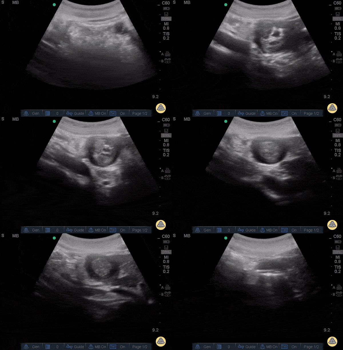
A 24 year old male comes to ER with abdominal pain, vomiting, and fever.
A directed scan with a high-frequency linear transducer in the right iliac fossa shows a tubular, non-compressible structure, a "target like" image, with increased echogenicity of the fat around it. When applied Color Doppler - we can observe increased vascular flow around the appendix - a sign known as "The Ring of Fire". This finding suggests the diagnosis of acute appendicitis.
Contributor: Renato Tambelli (@R_Tambelli @Jedipocus)
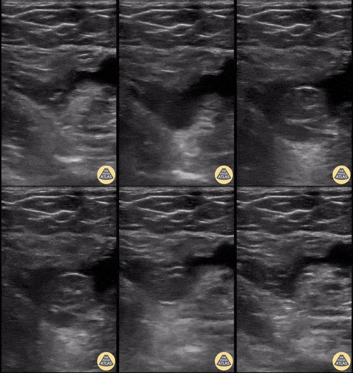
A 3 year old M presented with 3 days of fever, vomiting and abdominal pain. He was lethargic on arrival and found to be septic. POCUS shows ruptured appendicitis with a significant amount of hypoechoic fluid in the abdomen.
Paul Khalil, MD. Assistant PEM POCUS director at University of Louisville/Norton Children’s
@khalil3paul
A 2 year old F with 1 week of fever, vomiting, diarrhea and abdominal pain presented to the emergency department. A work up was initiated including POCUS of the right lower quadrant which revealed a ruptured appendix with abscess formation.
Paul Khalil, MD. Assistant PEM POCUS director at University of Louisville/Norton Children’s
Hernia
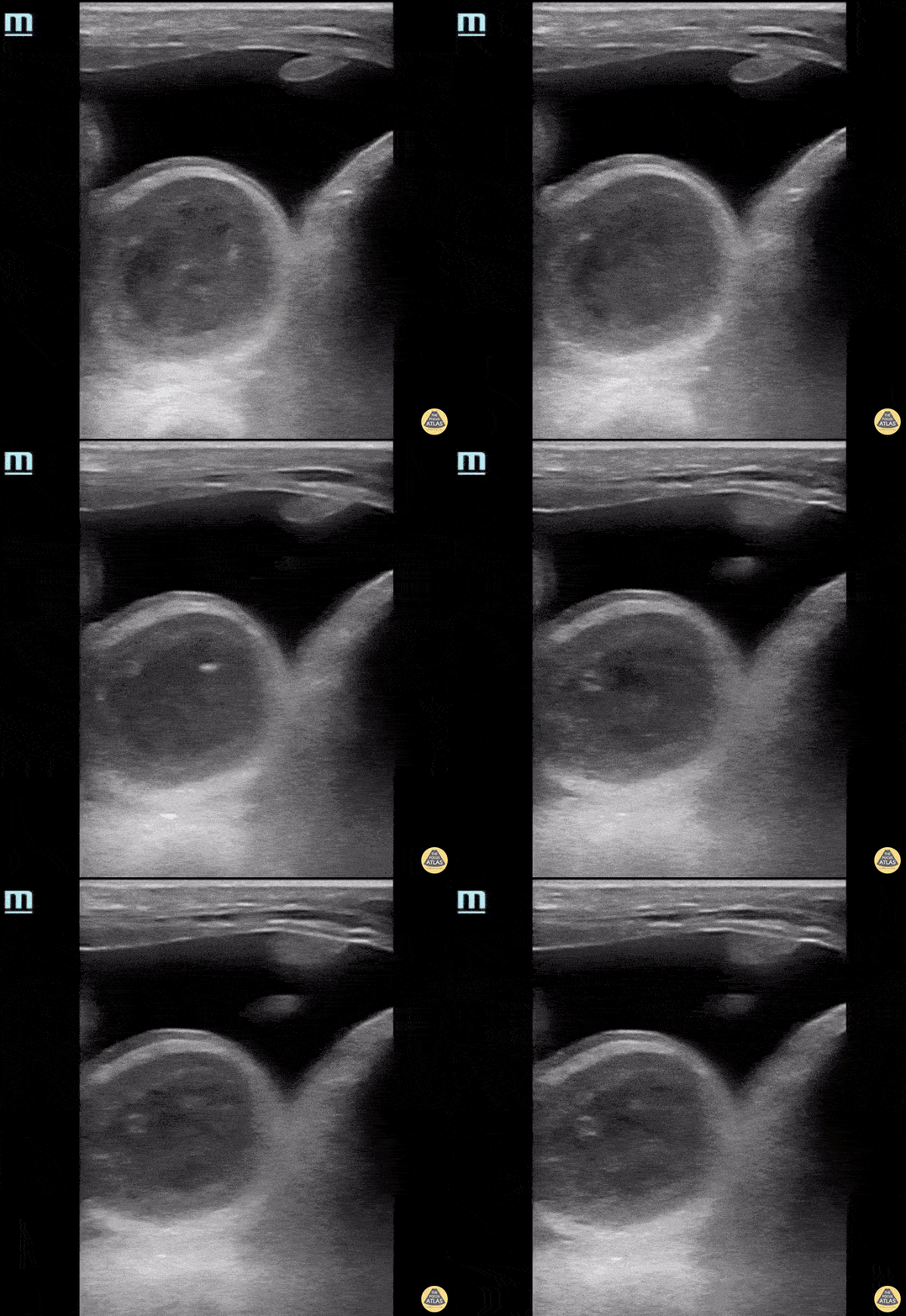
This patient presented with acute onset, severe groin pain. On physical exam, a large inguinal hernia was appreciated, and the scrotum was enlarged, erythematous, and exquisitely tender to palpation. POCUS exam was performed and there were multiple bowel loops with active peristalsis and a large anechoic fluid collection. CT imaging confirmed this fluid to be a herniated portion of the bladder, which likely provided a good acoustic window for clear visualisation of the bowel loops.
Catherine Barrington MD, MSc, PGY1 Emergency Medicine, CMU; Andrew Namespetra MB BCh BAO, MSc, PGY3 Emergency Medicine, CMU
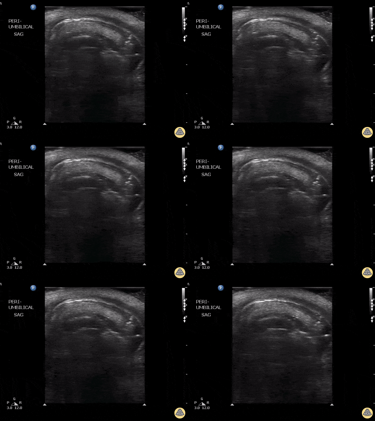
This patient presented with a known paraumbilical hernia however what should be noted here are is the lack of peristalsis as well as the presence of air in the within the wall of the bowel. These findings suggest that necrosis has resulted from strangulation of a hernia. Other findings with this diagnosis that could also be found include hyperechoic fat, fluid within the sac, thickening of the hernial sac, dilated bowel and edema within the walls.
Image courtesy of Robert Jones DO, FACEP @RJonesSonoEM
Director, Emergency Ultrasound; MetroHealth Medical Center; Professor, Case Western Reserve Medical School, Cleveland, OH
View his original post here
An elderly female presented to the emergency department with left groin pain that had been intermittent for 6 months. At time of initial evaluation, she noted a “bump” in the left groin. On examination with POCUS, the inguinal canal was evaluated and revealed a direct left inguinal hernia. As the patient performs a Valsalva maneuver, bowel is seen moving into the canal. Upon release of Valsalva, the bowel reduces spontaneously.
Daniel Coffey, Emergency Medicine PGY-2; Jocelyn Garcia MS-3
Central Michigan University College of Medicine
Intussusception
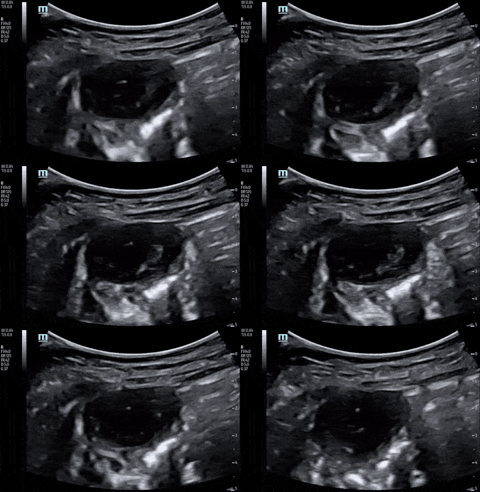
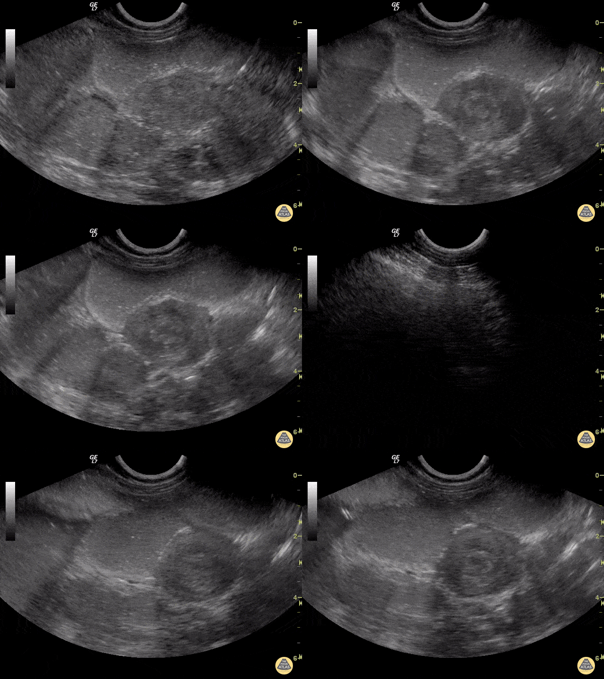
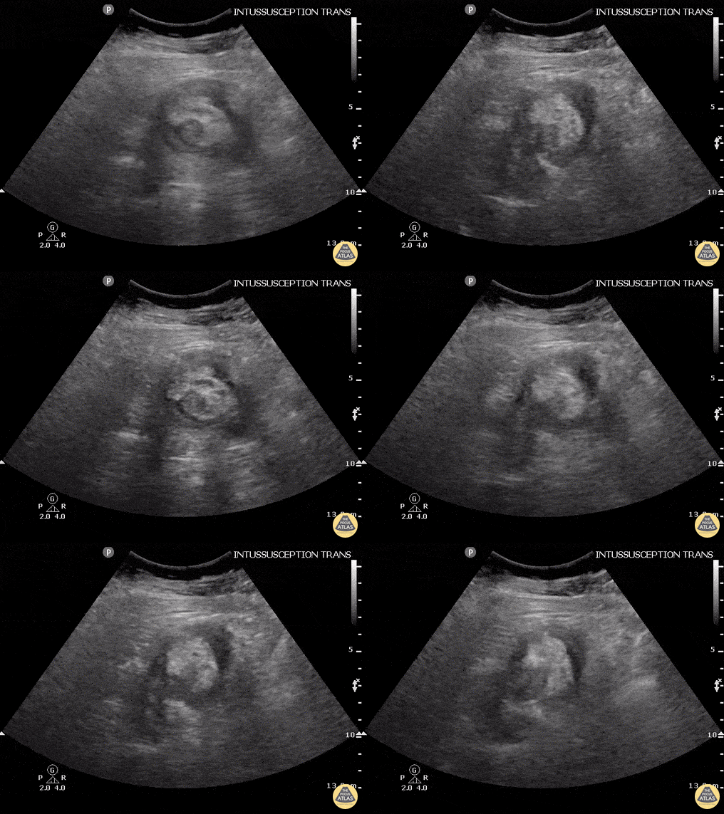
Here we can see an intussusception. Multiple layers and concentric rings, target configuration on cross-sectional imaging.
Evgeny Domanin
Intussusception is typically a disease of the young (6 months to 6 years) but it can occur in adults. Adults will often present similar to bowel obstruction, with vomiting, constipation, and rectal bleeding. Ultrasound can still be used to work them up although most of the time CT will be used first.
In kids, the sensitivity and specificity approach 100% but can be operator dependent. It is usually performed with a linear probe in children but in this image, a curvilinear probe was used.
Justin Bowra MBBS, FACEM, CCPU Emergency Physician, RNSH et al.
Pyloric Stenosis
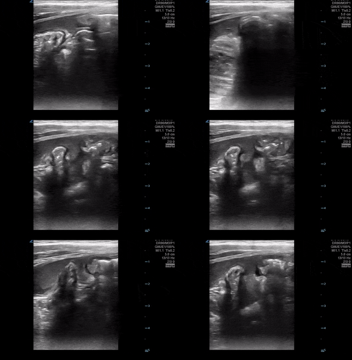
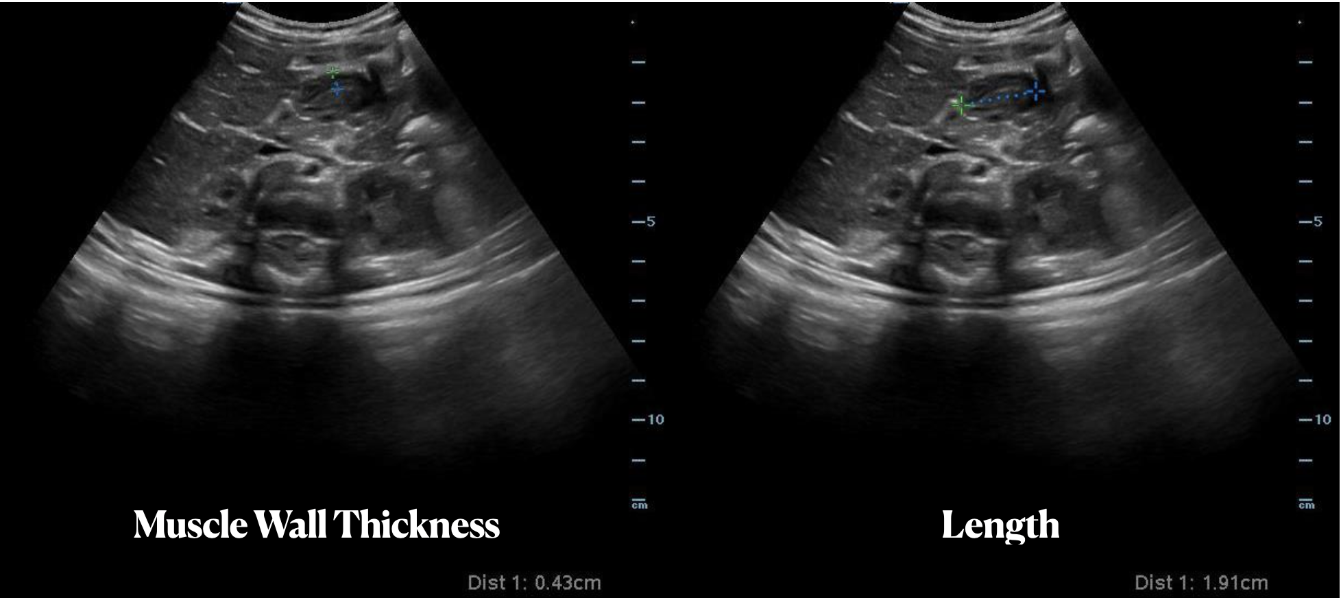
A 5 week old female previously healthy presents with 1 day of projectile vomiting. A bedside ultrasound was performed demonstrating muscle wall thickness of 4.3 mm (> 3 mm is abnormal) and a length of length of 19 mm (abnormal is >15 mm) consistent with a diagnosis of hypertrophic pyloric stenosis.
Paul Khalil, MD @Khalil3Paul
Assistant PEM POCUS Director at University of Louisville/Norton Children’s
28 y/o F no significant PMH with 1 month epigastric abdominal pain associated with daily emesis in the mornings and postprandial discomfort.. Patient notes she feels a firmness in her epigastrium. POCUS with an enlarged pylorus with gastric outlet obstruction, confirmed with formal sono. Admitted and evaluated with EGD by GI, found to have peptic ulcer in the antrum with edema and degree of gastric outlet obstruction.
Dr. Adrian Aurrecoechea, Dr. Andrew Aherne - Kings County Hospital
Other Pathology
Fatigue for the past 3–4 months with mild disturbance of bowel habits. The patient reported visualizing proglottids in the stool.
POCUS findings: Ultrasound performed in the left lower quadrant revealed a structure consistent with parasitic segments.
Diagnosis: Laboratory confirmation a few days later identified Taenia saginata.
Dr. Guillaume Schramme
60 year old man presenting with left lower quadrant pain and fever.
POCUS was performed and revealed a thick-walled colon and associated diverticula with surrounding hyperechoic fat stranding. Inside the pouch there's an echogenic structure casting deep shadowing consistent with a fecalith.
Alessandro Lena
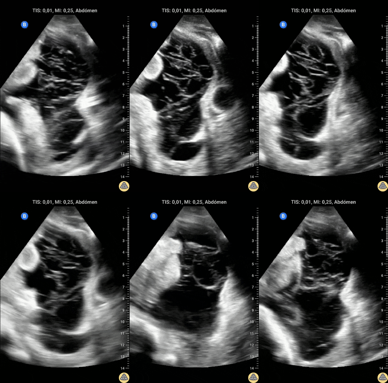
A 19 year old male, with hx of cocaine abuse presented to the ICU for respiratory insufficiency due to cardiogenic pulmonary edema. POCUS evaluation of the abdomen demonstrated ascites with Spider Web Sign, which is not expected in cardiogenic ascites. Because of this, peritoneal tuberculosis was suspected. It was later discovered that the patient had close contact with a person who was being treated for a confirmed tuberculosis diagnosis.
Contributor: Dr. Alessandro Ferreira
Hospital Metropolitano de Alagoas
In this case an elderly patient presented with severe back pain and an ultrasound exam was performed to evaluate the aorta. Instead in this transverse view, the pancreas was identified which appears enlarged with surrounding fluid. Besides appearing enlarged, an important finding in this scan is the hypoechoic regions which are indicative of necrosis.
Image courtesy of Robert Jones DO, FACEP @RJonesSonoEM
Director, Emergency Ultrasound; MetroHealth Medical Center; Professor, Case Western Reserve Medical School, Cleveland, OH
View his original post here
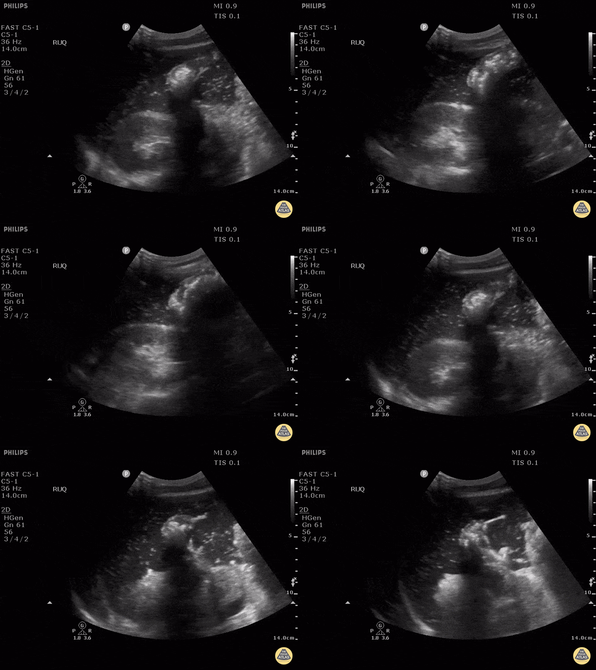
Patient presented with altered mental status, hypotension and a rigid abdomen. It was unclear at the time if this patient’s presentation was due to trauma. Right-upper quadrant ultrasound used to view for potential cause using the perihepatic window. Here we can see air within fluid and a hyperechoic peritoneal stripe, indicating a perforated viscus. In this case, due to a duodenal ulcer.
Image courtesy of Robert Jones DO, FACEP @RJonesSonoEM
Director, Emergency Ultrasound; MetroHealth Medical Center; Professor, Case Western Reserve Medical School, Cleveland, OH
View his original post here
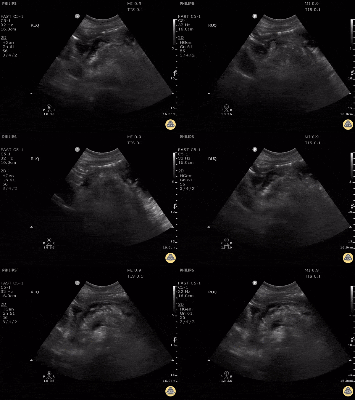
RUQ scan revealed an isoechoic region adjacent to the hepatic flexure indicative of an abscess formation secondary to a colonic perforation.
Image courtesy of Robert Jones DO, FACEP @RJonesSonoEM
Director, Emergency Ultrasound; MetroHealth Medical Center; Professor, Case Western Reserve Medical School, Cleveland, OH
View his original post here