
Ocular
Orbit
Note: These search bar results are only for the Orbit image archive. To search the entire site, click here.
Recently Added
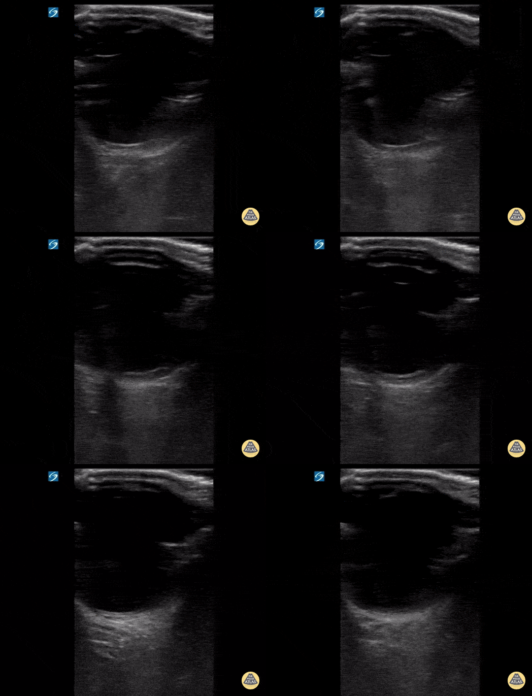
17 y/o female with sudden onset bilateral blurry vision after prolonged indoor cat exposure. Fundoscopy showed retinal pigment epithelium loss consistent with bilateral neuroretinitis from cat scratch disease (Bartonella henselae).
The hyperechoic retina, normally flush with the posterior aspect of the globe, is seen separated by a hypoechoic band of edema.
Retinal edema from cat scratch disease is rare (Gen Opth 19th ed. 2017:378-422), but can be diagnosed by POCUS.
Contributed by: John Hipskind, MD, Ultrasound Director, Kaweah Health EM Residency
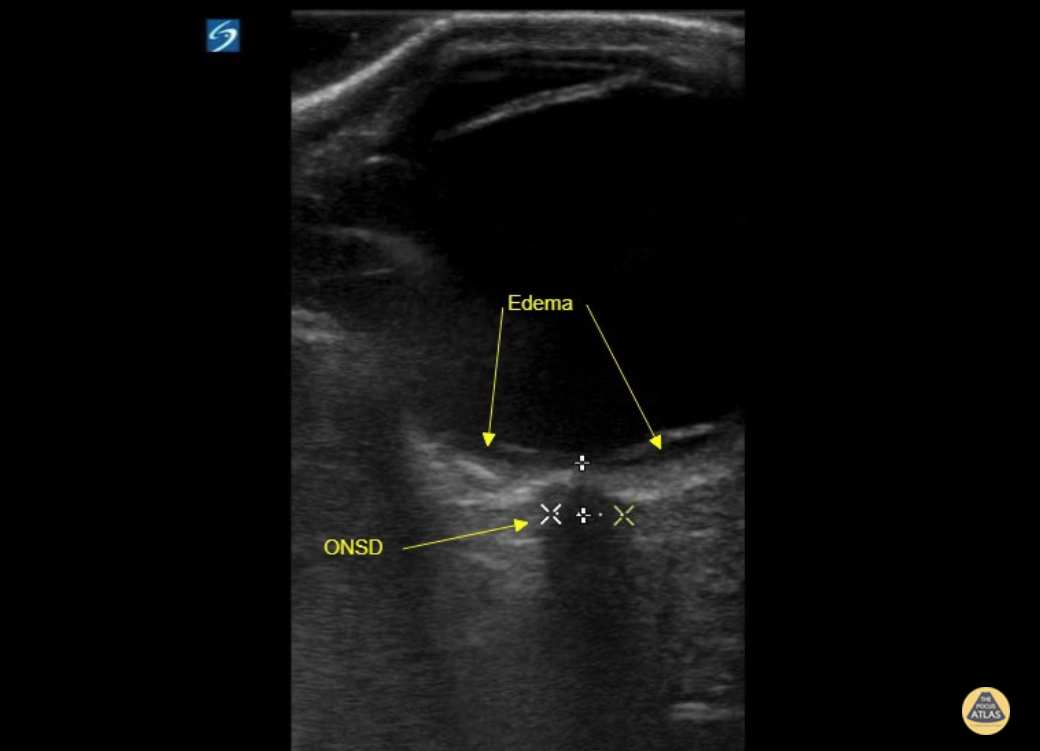
17 y/o female with sudden onset bilateral blurry vision after prolonged indoor cat exposure. Fundoscopy showed retinal pigment epithelium loss consistent with bilateral neuroretinitis from cat scratch disease (Bartonella henselae).
The hyperechoic retina, normally flush with the posterior aspect of the globe, is seen separated by a hypoechoic band of edema.
Retinal edema from cat scratch disease is rare (Gen Opth 19th ed. 2017:378-422), but can be diagnosed by POCUS.
Contributed by: John Hipskind, MD, Ultrasound Director, Kaweah Health EM Residency
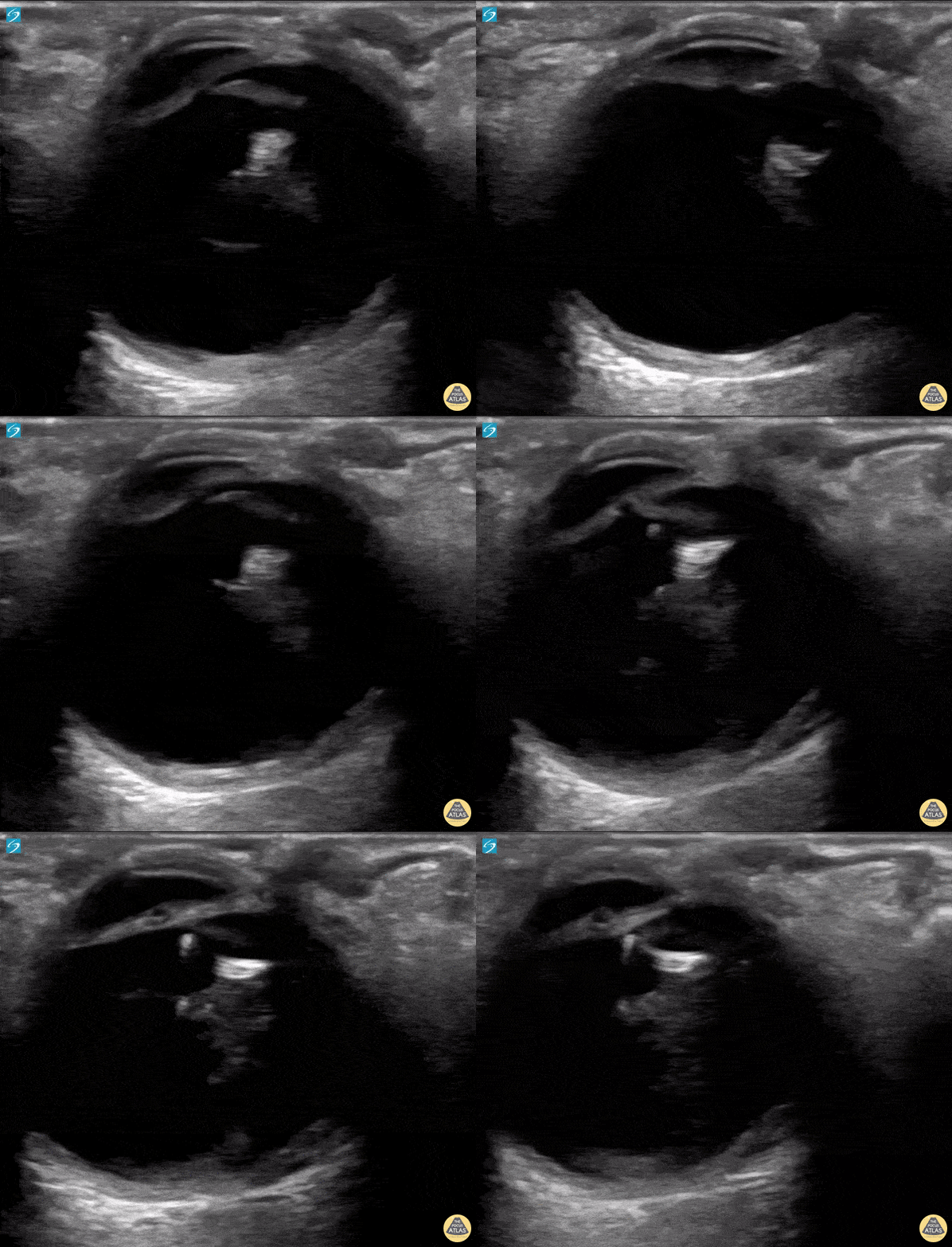
A 50 year old male with a prior history of bilateral intraoccular lens (IOL) transplants presented to our ED with sudden onset foggy vision in his right eye while getting out of the shower. He was unable to participate in visual acuity due to the extent of his blurred. POCUS demonstrated “iridodonesis” and a provisional diagnosis of IOL subluxation/dislocation was made. Ophthalmology was consulted and confirmed the diagnosis.
Dr. Piaseczny, PGY4 Emergency Medicine, Queen's University, Kingston, Ontario, Canada
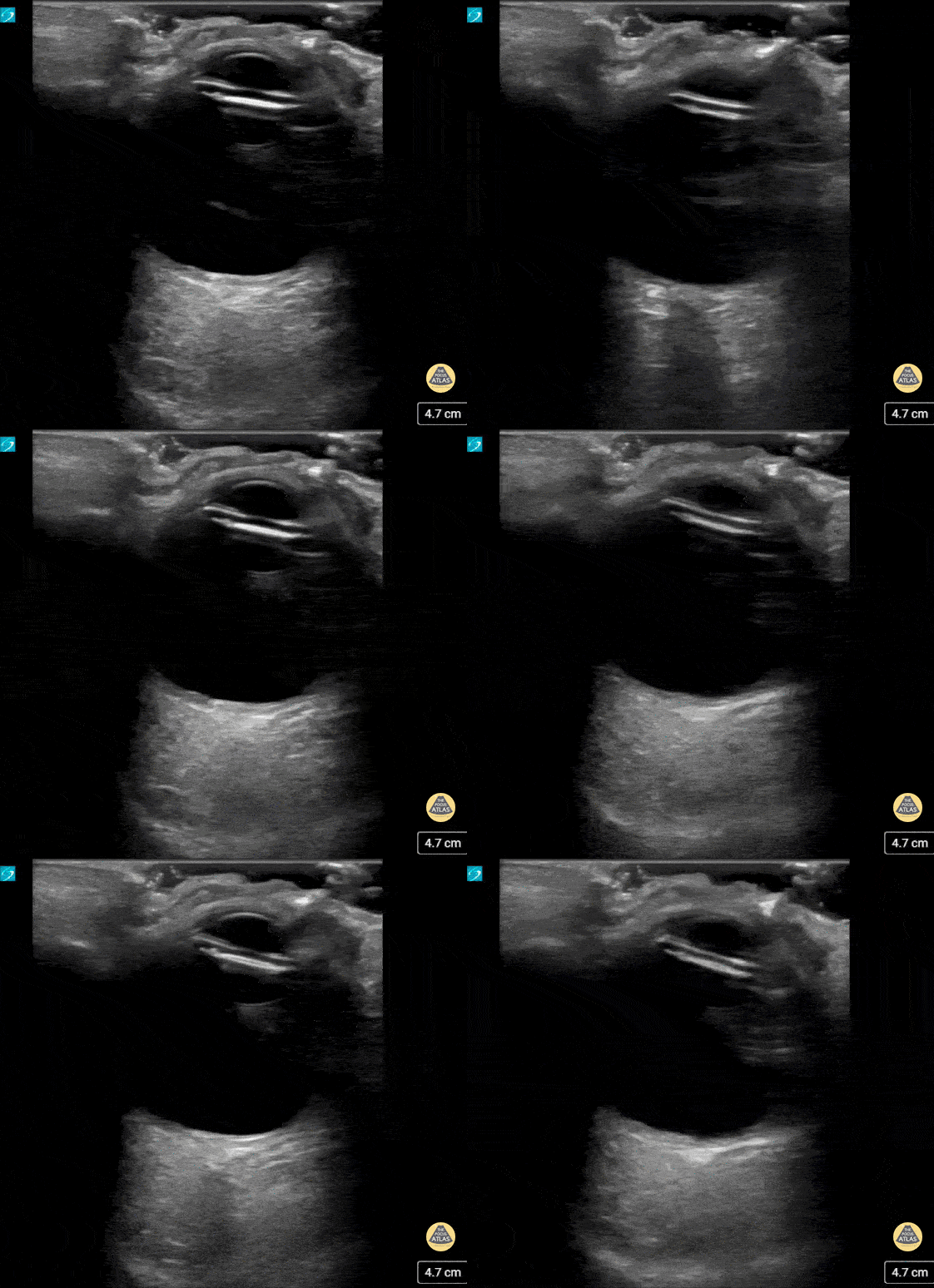
This patient presented with decreased vision on a background of advanced macular degeneration. VA in the eye had decreased from 20/150 to 20/400 on presentation.
Interestingly the patient stated "I think my ocular lens has displaced."
Contributed by: Colin Bell, FRCPC, DPSPC
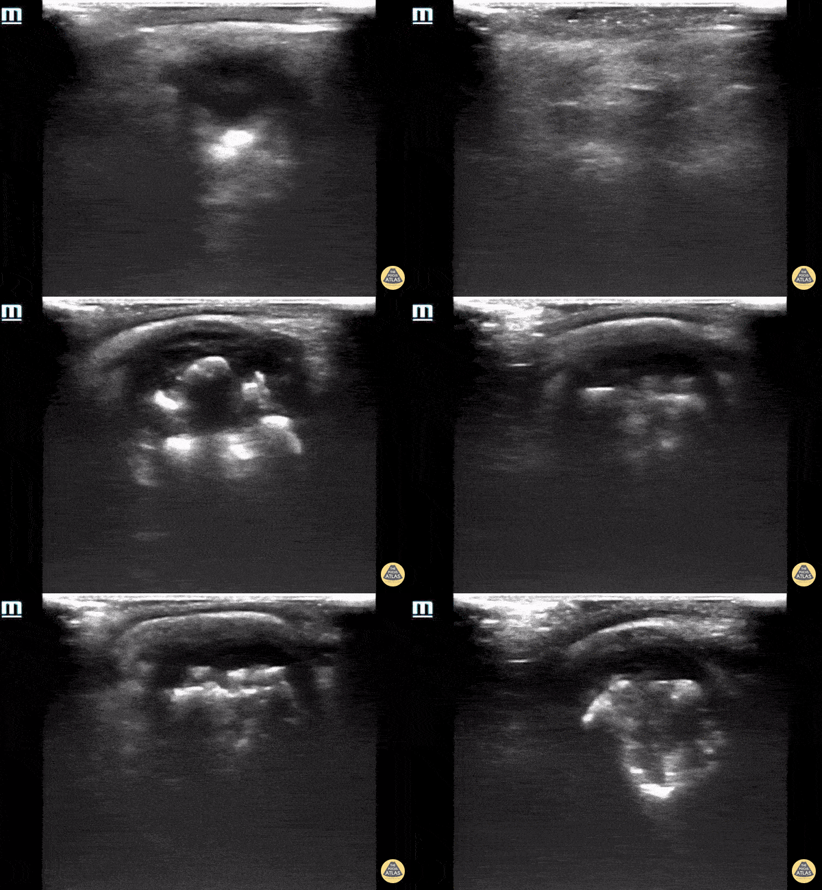
93 year old male with established end stage eye disease found incidentally to have prominent Phthisis Bulbi made evident by a heterogeneous hyper-echoic structures (calcifications) within the posterior chamber of the left eye.
Contributed by: Davis, Lindsay; Schlangen, Alex; Welch, Matthew
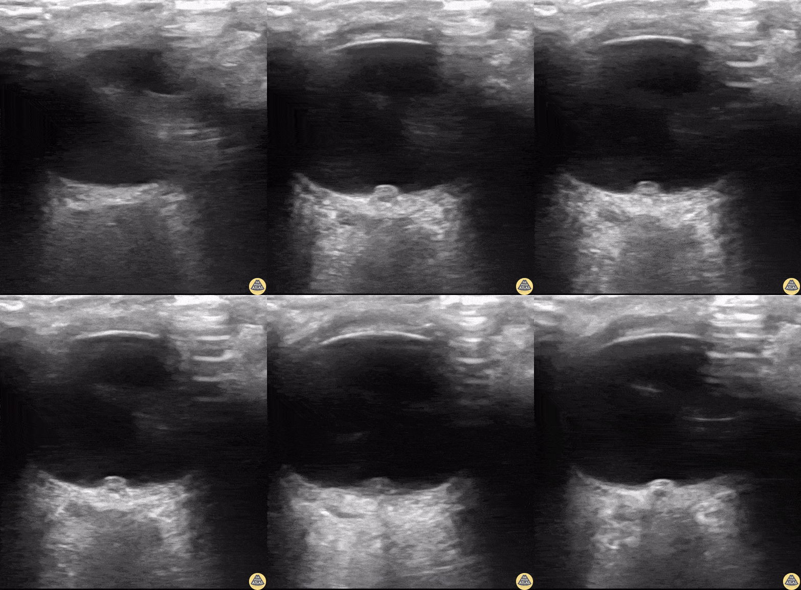
A 24-year old male presented to the ED with a six-month history of progressive total right vision loss. A bedside ocular ultrasound examination was performed that revealed an elevated optic disc with enlarged ONSD measuring 9.7mm, consistent with papilledema. An MRI of the head confirmed an enlarged intraconal portion of the right optic nerve, consistent with glioma.
Marko Lubardic, MS4; Tom Taugher, DO, PGY3; Michael Bernard, DO, PGY1; Central Michigan University Emergency Medicine Residency
Normal Anatomy
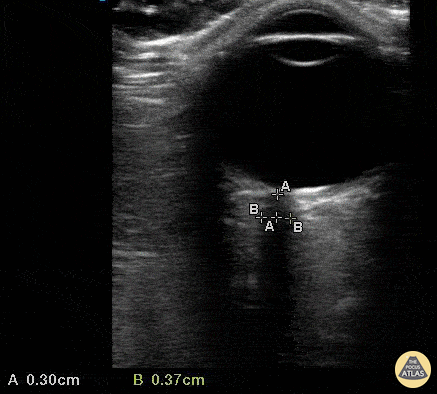
A 35 yo female presented to a rural ED with worsening headache accompanied by nausea/vomiting after failed treatment for sinusitis. The department did not have immediate access to CT scanning, therefore Optic Nerve Sheath Diameter (ONSD) was used to assess for associated increased intracranial pressure. The optic nerve sheath is contiguous with the subarachnoid space, making ONSD an indirect measure of intracranial pressure. Visualize the hypoechoic optic nerve posterior to the globe and measure 3mm back from the globe (calipers A) to determine site of measurement . Then measure to the edges of the sheath (calipers B) horizontally. Value >5mm is concerning for increased ICP. This ONSD is normal.
Mandy Peach, MD @mandy_peach
Saint John Regional Hospital. NB, Canada
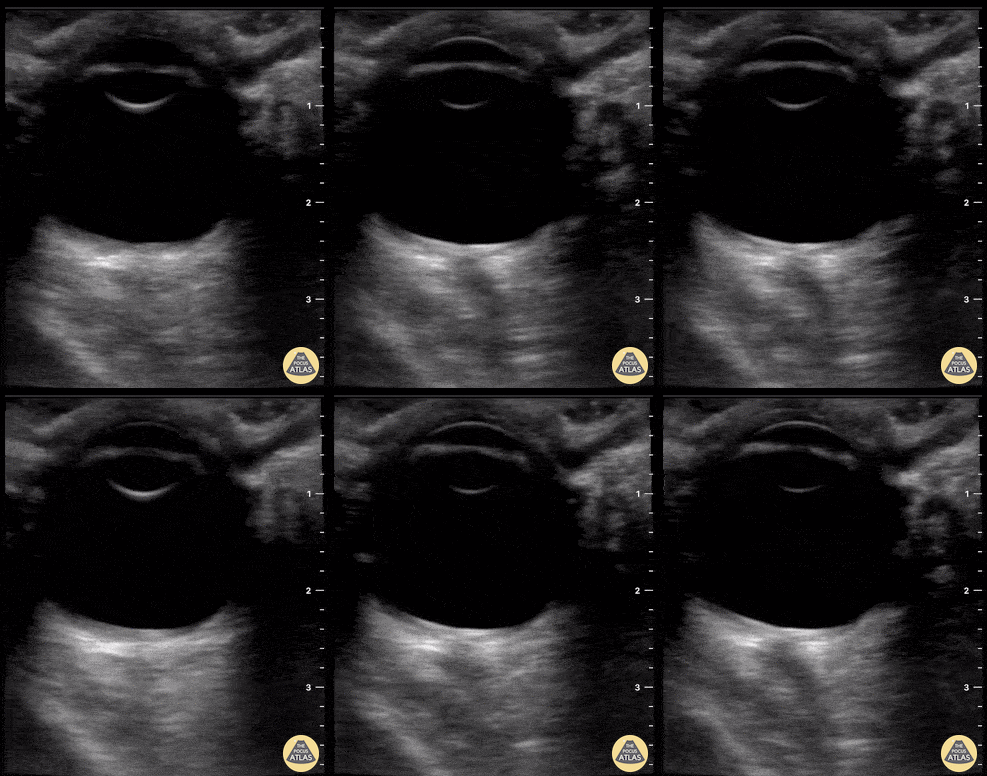
Patient presented with fall from standing height with resultant traumatic subarachnoid hemorrhage (SAH). POCUS of ONSD is reassuring in that ONSD is <5 mm, suggestive of no associated elevated intracranial pressure.
Tessa W. Damm, DO. Intensivist. Milwaukee, WI
@DrDamm
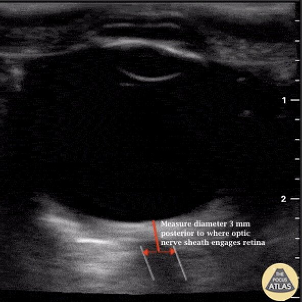
When evaluating for elevated intracranial pressure using optic nerve sheath diameter, (ONSD), proper measurement should be obtained 3 mm posterior to where the optic nerve engages the retina.
Image courtesy of Dr. Tessa Damm (@DrDamm)
Colorized Normal Anatomy
Coming Soon…
Retinal Detachment
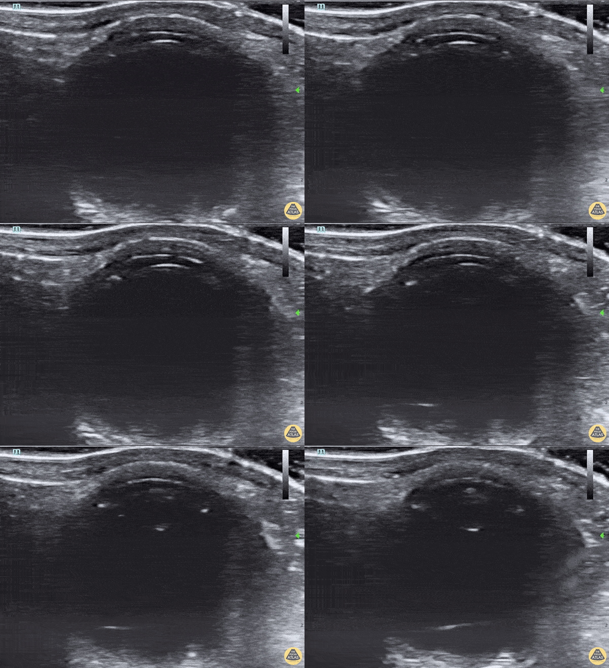
46 yo woman pmh multiple right eye surgeries presenting with atraumatic left eye pain, decreased vision, and mydriasis, which was initially concerning for acute angle-closure glaucoma. POCUS revealed anterior lens subluxation given the very shallow anterior chamber. Around 5% of acute angle-closure glaucoma is induced by lens subluxation. Retinal detachment, the hyperechoic line in the far field, can also be seen on POCUS. Ophthalmology successfully treated her with lensectomy and secondary intraocular lens implantation.
Robert Adrian
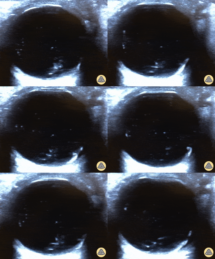
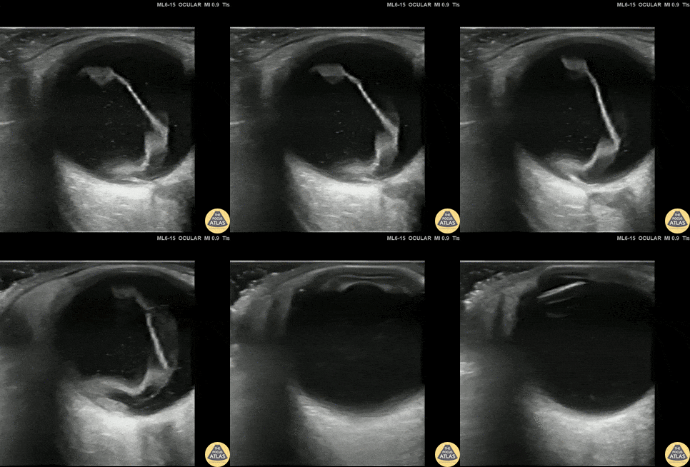
30s M with no known medical history or ocular history presented with 2 days of progressive monocular visual field deficits and floaters without antecedent trauma or other injury. Visual acuity was limited to finger counting at 3 feet in the affected eye. POCUS of the affected eye is shown here, demonstrating a macula-off retinal detachment. The patient was seen by ophthalmology and had urgent surgery the next morning.
Dr. Amy Allen, PGY1
Denver Health Residency in Emergency Medicine
Vitreous Detachment
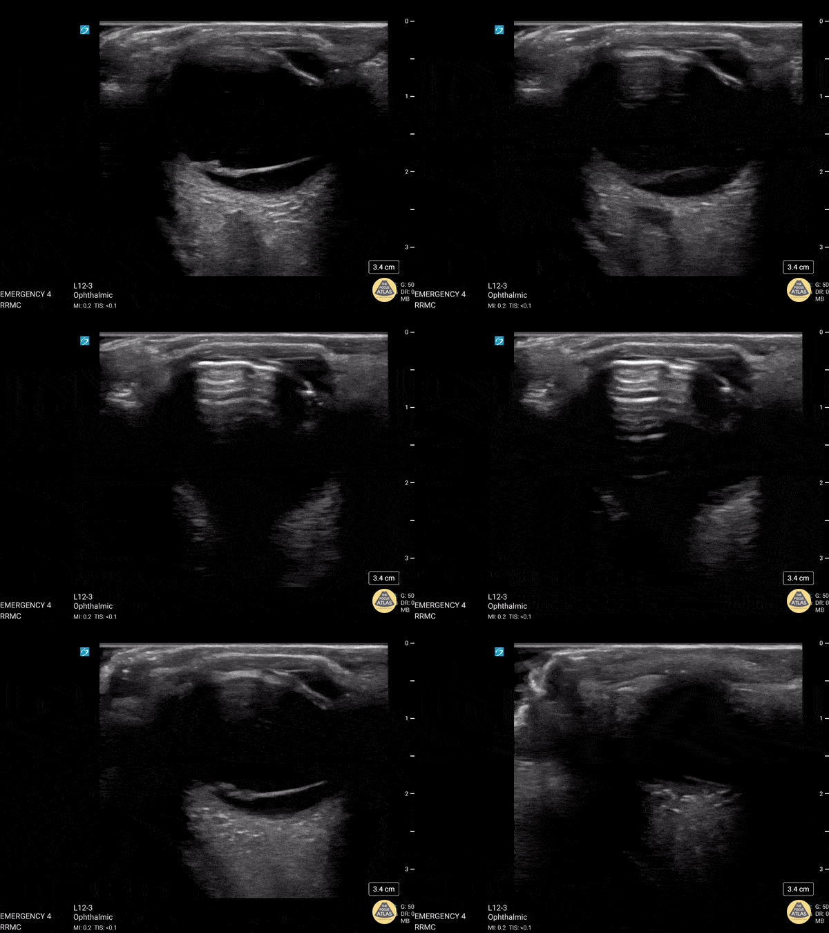
This was from a 52 year old male with a recent surgical history of vitreous detachment that became a retinal detachment. He presented to the emergency room for vision changes in same eye after an object fell and hit his head. Ocular ultrasound was performed at the bedside which revealed a new vitreous detachment. Important to note here that it is crucial to fan/tilt through the entire eye, making sure to visualize the optic nerve in order to differentiate between a vitreous and retinal detachment. In this case, notice how the lesion spotted in the chamber does not connect to the optic nerve, which is consistent with a vitreous detachment.
Dr. Christopher Paulo, DO, PGY-1
Riverside Regional Medical Center Emergency Medicine Program (Newport News, VA)
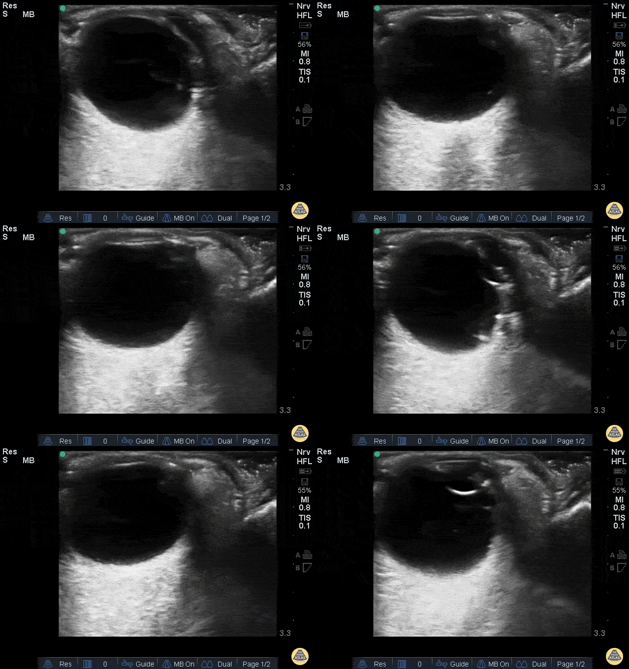
A 44 year old man came to ED with a shimmering effect in his left eye and unilateral temporary painless visual disturbance. He had previously been treated for a retinal tear.
Ocular PoCUS shows a frond-like linear structure lifting away from the posterior surface of the globe.
Posterior vitreous detachment (PVD) was suspected while retinal detachment (RD) was also considered. PVD was confirmed by the specialist team and the patient was treated conservatively.
Dr. Cian McDermott, Emergency Physician - University Hospital Geelong, Australia
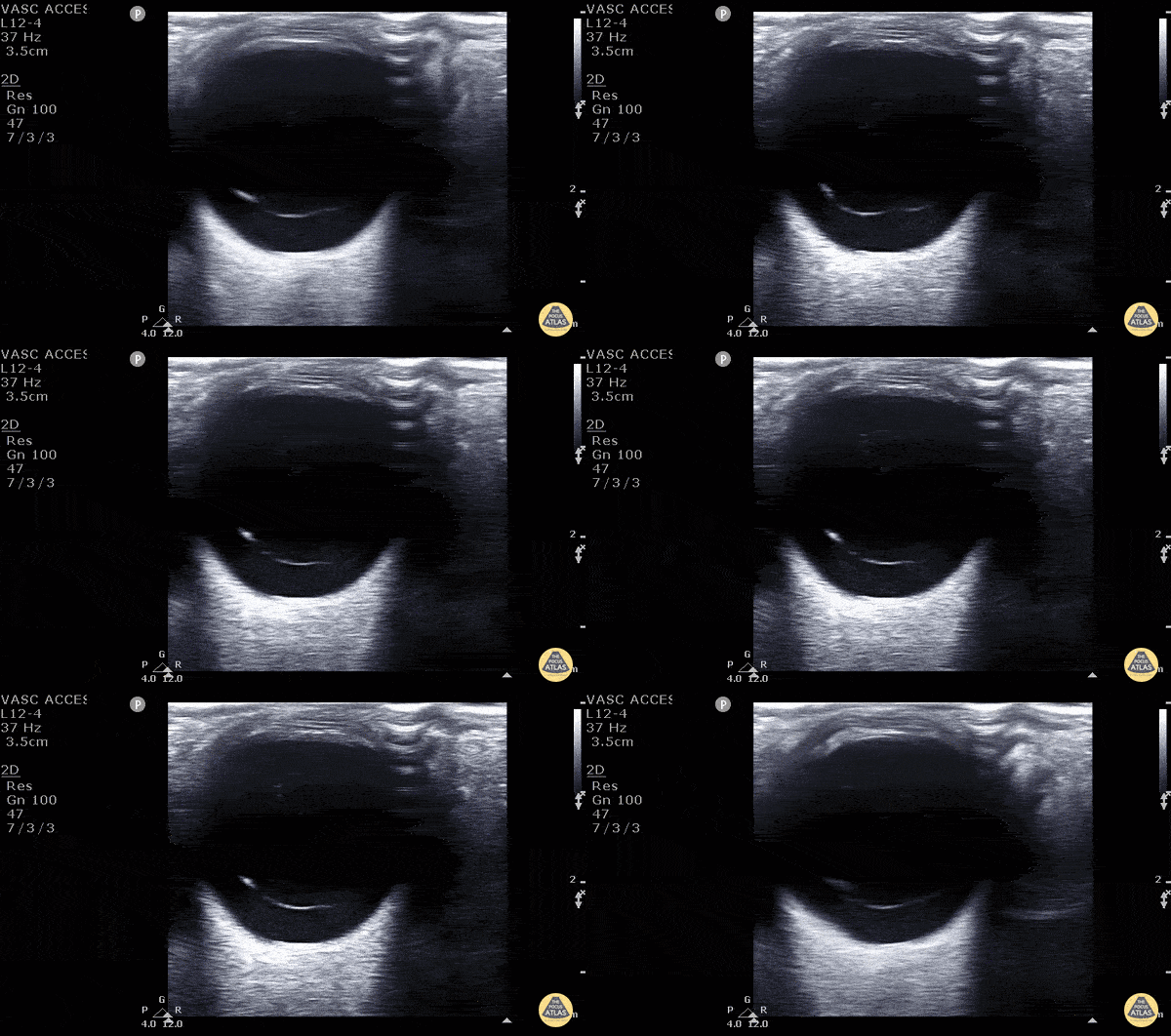
40 year old male with 6 months of decreased vision from his left eye. No pmh or history of trauma. VA was obviously decreased relative to the contralateral eye. POCUS rapidly revealed a freely floating hyperechoic membrane consistent with vitreal detachment. To differentiate between a vitreous and retinal detachment you must look to see if the membrane is attached to the hypoechoic optic nerve. The retinal is continuous with the optic nerve therefore stays attached to the optic nerve in a retinal detachment. A vitreous detachment would be completely untethered as seen in this image. You can also see the “washing machine sign” better pictured in other images in this gallery.
Matt Riscinti, MD Bryan Flores, MD - Kings County Emergency Medicine
Vitreous Hemorrhage
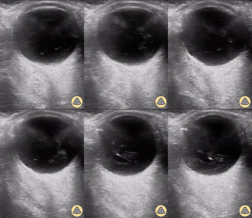
An 81 yo female on ASA presented with 6 hour hx of blurred vision in her right eye. Ocular POCUS demonstrated a hyperechoic clot, seen here as 'swirling' movement, with debris settling near the bottom of the globe when eye is not in motion (end of clip). In early presentation of vitreous hemorrhage when blood is still liquified, increasing the ultrasound gain can greatly assist with identification of pathology.
Mandy Peach, MD @mandy_peach
Saint John Regional Hospital. NB, Canada
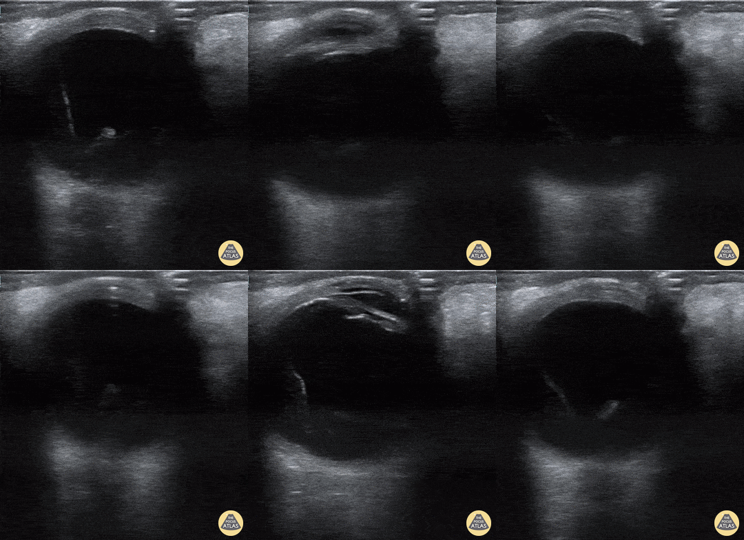
A 70-year-old man presented with loss of vision to his right eye (hand motion and light perception). He takes warfarin and has diabetes. Monocular vision loss triggered a POCUS that revealed a hazy echogenic substance (consistent with blood) seen best when patient activates extra-ocular muscle movements. Also note the associated bright linear structure within one portion of the bleed, the hyaloid membrane. This patient was subsequently assessed by the Eye Hospital and diagnosed with vitreous hemorrhage due to proliferative diabetic retinopathy.
Peter Cheng
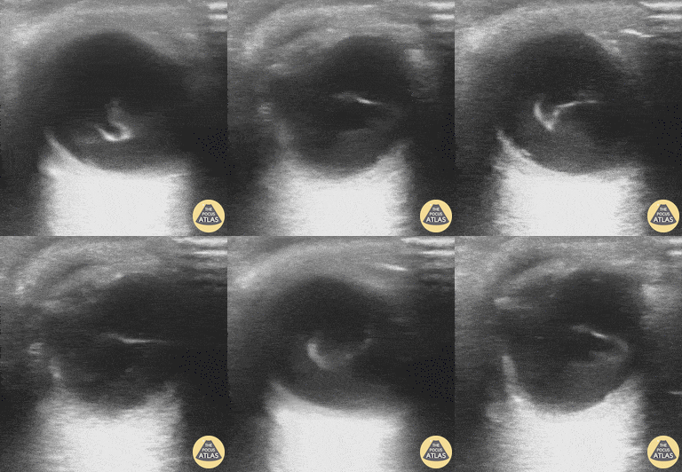
A 71-year-old male with a history of hypertension and diabetes presented to the ED reporting gradual vision loss in his right eye over a 3-day period. Physical exam was notable for a 3 mm and nonreactive pupil with ipsilateral visual acuity reduced to patient only being able to detect movement. POCUS revealed swirling echodensity within the right eye, most appreciable with extraocular muscle movements. Ophthalmology subsequently confirmed our suspected diagnosis of vitreous hemorrhage.
Richard Cunningham, MD @HappyDays_EM
Maricopa Medical Center
Dilated Optic Nerve
A 24-year old male presented to the ED with a six-month history of progressive total right vision loss. A bedside ocular ultrasound examination was performed that revealed an elevated optic disc with enlarged ONSD measuring 9.7mm, consistent with papilledema. An MRI of the head confirmed an enlarged intraconal portion of the right optic nerve, consistent with glioma.
Marko Lubardic, MS4; Tom Taugher, DO, PGY3; Michael Bernard, DO, PGY1; Central Michigan University Emergency Medicine Residency
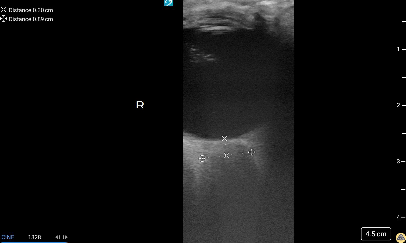
A 75 year old male with history of ocular melanoma and blindness of the right eye presented for right sided headache and nausea for the past 2 days.
Ocular US was performed demonstrating significant enlargement of the optic nerve sheath.
Contributors: Michael Bernard, DO; Thomas Taugher, DO; Marko Lubardic
Central Michigan University Residency of Emergency Medicine
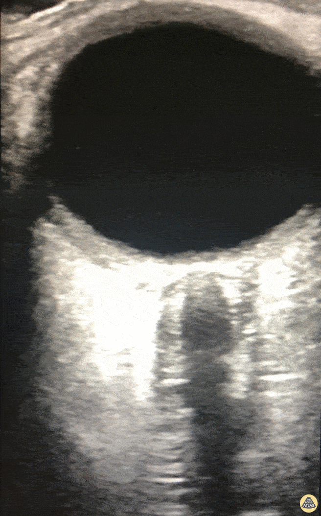
We used OSND to evaluate a patient with COVID-19 encephalitis and found evidence of increased intracranial pressure (ICP). Note, due to the high frequency probe used, we were able to appreciate fine detail including two hypoechoic boarders at each lateral edge of the optic nerve. It is the most lateral outer edge/ hypoechoic structure that represents the optic nerve sheath.
Eric M. Siegal, MD, SFHM, FCCM
Aurora Critical Care Service; Milwaukee, WI
Adjunct Clinical Professor of Medicine University of Wisconsin School of Medicine and Public Health
Other Pathology
17 y/o female with sudden onset bilateral blurry vision after prolonged indoor cat exposure. Fundoscopy showed retinal pigment epithelium loss consistent with bilateral neuroretinitis from cat scratch disease (Bartonella henselae).
The hyperechoic retina, normally flush with the posterior aspect of the globe, is seen separated by a hypoechoic band of edema.
Retinal edema from cat scratch disease is rare (Gen Opth 19th ed. 2017:378-422), but can be diagnosed by POCUS.
Contributed by: John Hipskind, MD, Ultrasound Director, Kaweah Health EM Residency
17 y/o female with sudden onset bilateral blurry vision after prolonged indoor cat exposure. Fundoscopy showed retinal pigment epithelium loss consistent with bilateral neuroretinitis from cat scratch disease (Bartonella henselae).
The hyperechoic retina, normally flush with the posterior aspect of the globe, is seen separated by a hypoechoic band of edema.
Retinal edema from cat scratch disease is rare (Gen Opth 19th ed. 2017:378-422), but can be diagnosed by POCUS.
Contributed by: John Hipskind, MD, Ultrasound Director, Kaweah Health EM Residency
A 50 year old male with a prior history of bilateral intraoccular lens (IOL) transplants presented to our ED with sudden onset foggy vision in his right eye while getting out of the shower. He was unable to participate in visual acuity due to the extent of his blurred. POCUS demonstrated “iridodonesis” and a provisional diagnosis of IOL subluxation/dislocation was made. Ophthalmology was consulted and confirmed the diagnosis.
Dr. Piaseczny, PGY4 Emergency Medicine, Queen's University, Kingston, Ontario, Canada
This patient presented with decreased vision on a background of advanced macular degeneration. VA in the eye had decreased from 20/150 to 20/400 on presentation.
Interestingly the patient stated "I think my ocular lens has displaced."
Contributed by: Colin Bell, FRCPC, DPSPC
93 year old male with established end stage eye disease found incidentally to have prominent Phthisis Bulbi made evident by a heterogeneous hyper-echoic structures (calcifications) within the posterior chamber of the left eye.
Contributed by: Davis, Lindsay; Schlangen, Alex; Welch, Matthew
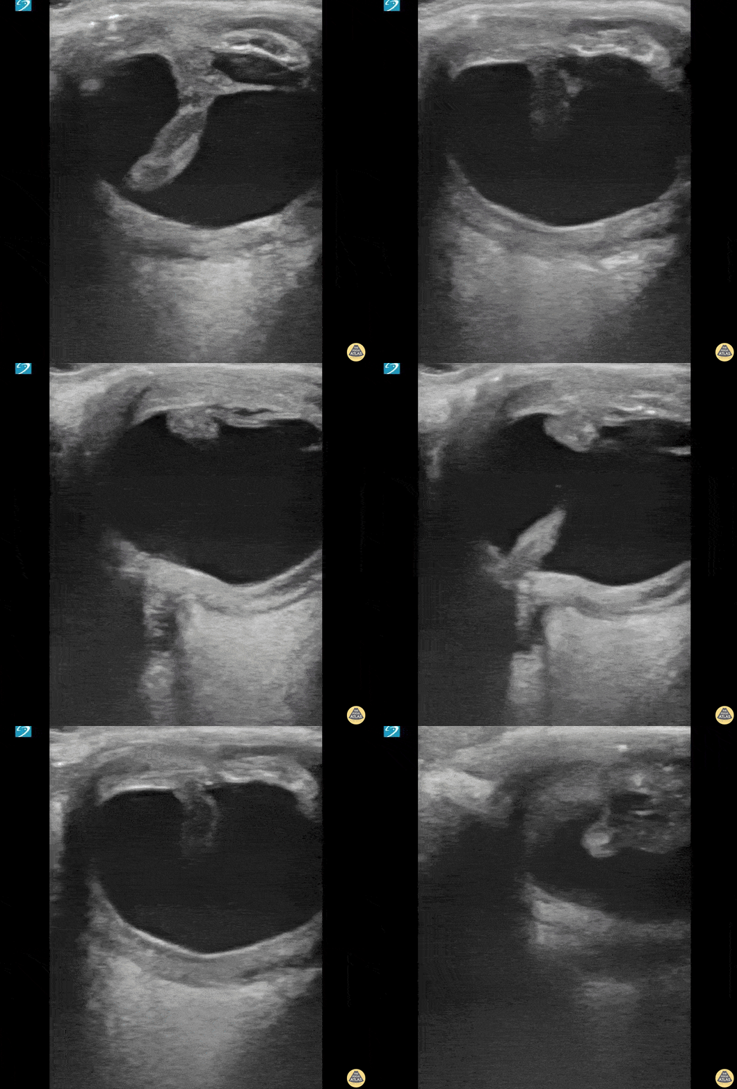
Irregularly shaped eye with internal dense structure concerning for intraocular hematoma. This patient had a history of previous rupture of the globe.
A CT Head without IV contrast described the left eye as follows: Small left globe with high density concerning for intraocular hemorrhage. Direct examination is recommended.
Halimah Hamidu-Egiebor Central Michigan University College of Medicine MD-MBA '24, Arthur Sieron Central Michigan University Emergency Medicine Resident, Eric 'Dax' Spencer Central Michigan University Emergency Medicine Resident