
Trauma
Trauma
Note: These search bar results are only for the Trauma image archive. To search the entire site, click here.
Recently Added
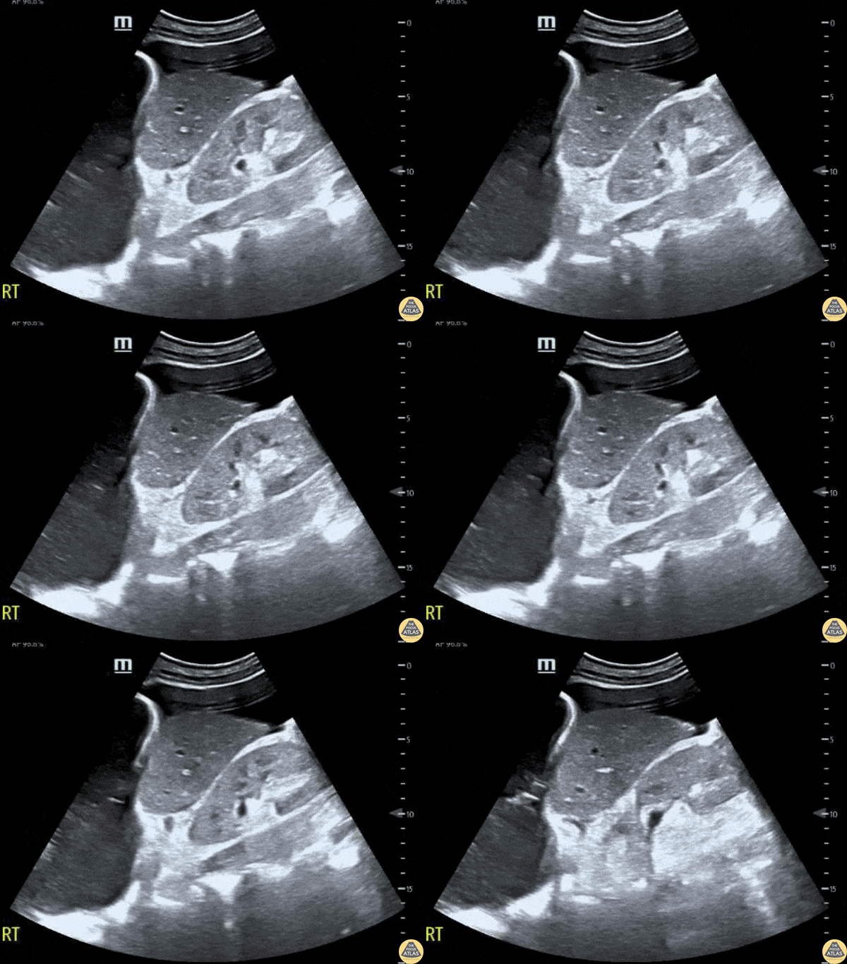
Patient presented to the emergency department with atraumatic acute onset of shortness of breath. An EFAST was performed to evaluate for pneumothorax and assess for free fluid in the abdomen, thorax, and pericardium.
A significant right pleural effusion was present. Additionally, US of RUQ demonstrated a liver which appeared small in size with prominent ascites. This demonstrates the utility of POCUS to identify free fluid in a quick and efficient manner as compared to other common studies such as a CT scan, even in non-traumatic patients.
Contributors: Lauren Lowes, DO; Krishna Patel, DO; Julia Tu, MS-4
Central Michigan University Residency of Emergency Medicine
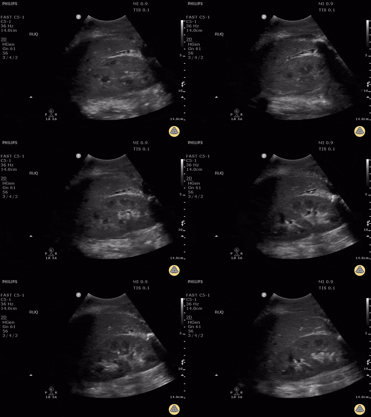
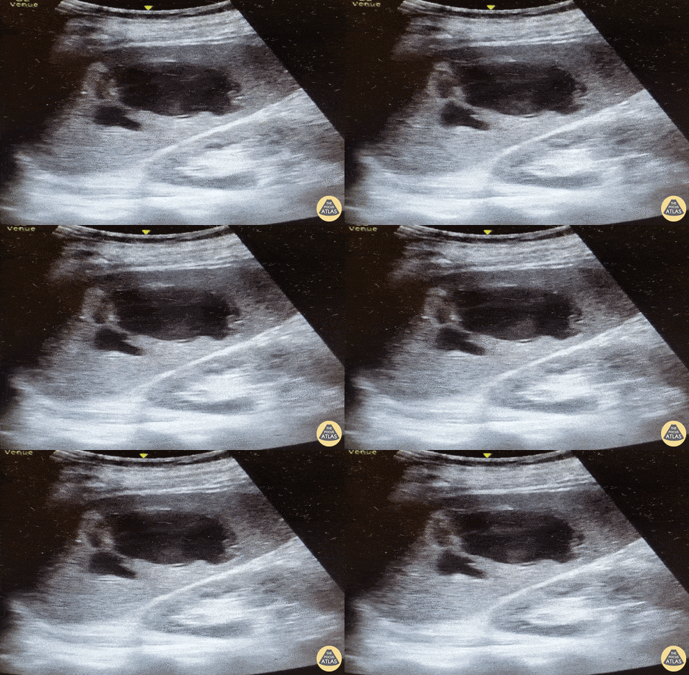
This is RUQ view from a patient who presented to the emergency department after falling from a roof and with low blood pressures. Although there appears to be perihepatic fluid, an interesting learning point form this case is whether or not we can discern if fluid observed is truly in the retroperotineal space or in the peritoneal cavity. Because the fluid is in direct contact with perinephric fat, this is actually in the retroperitoneal space and not in the peritoneal cavity. This may be difficult to distinguish in patients with very little perinephric fat.
Image courtesy of Robert Jones DO, FACEP @RJonesSonoEM
Director, Emergency Ultrasound; MetroHealth Medical Center; Professor, Case Western Reserve Medical School, Cleveland, OH
View his original post here
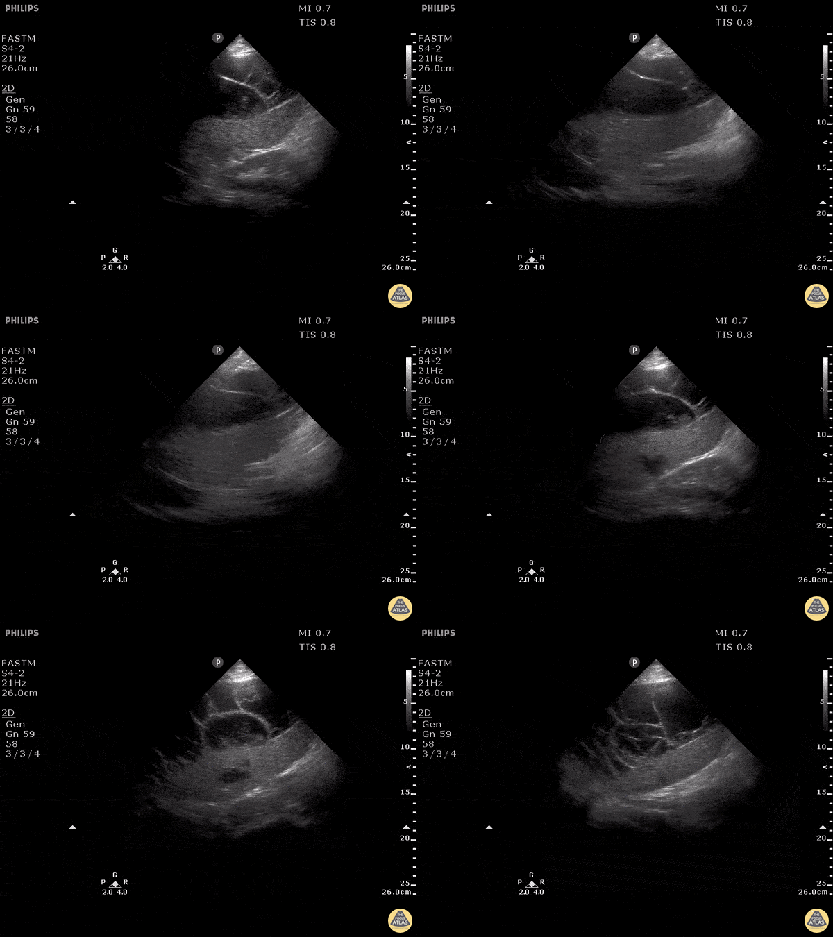
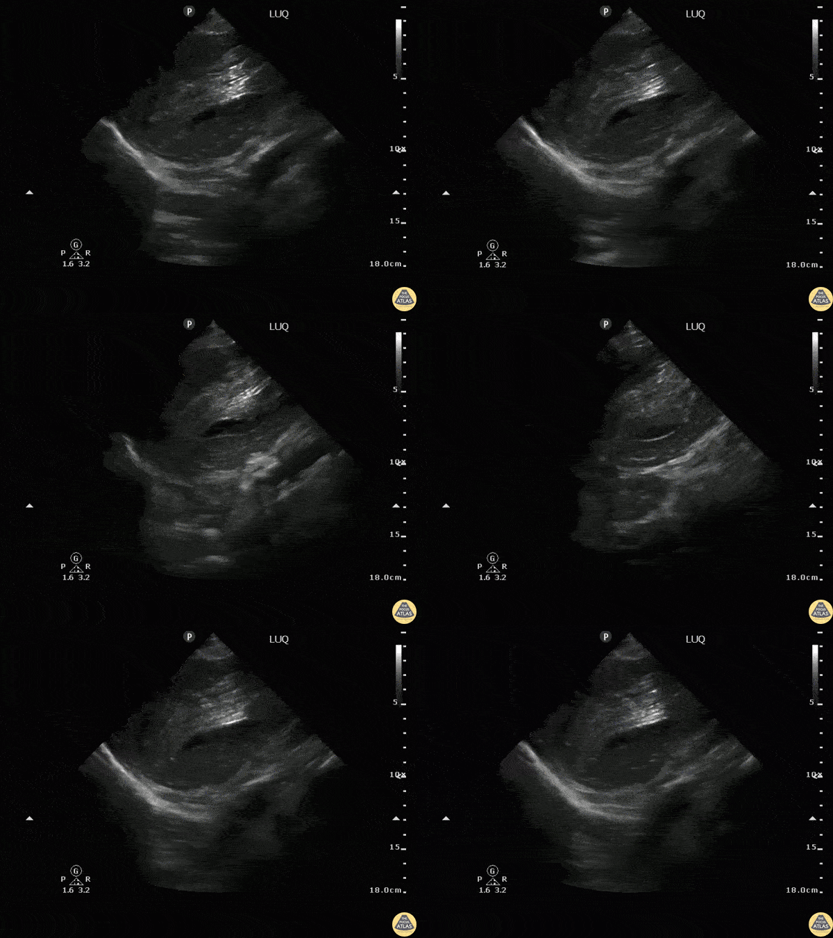
This is a clip of the LUQ performed during FAST exam following blunt trauma. It demonstrates loculated fluid around the spleen concerning for blood. CT imaging followed demonstrating a large subcapsular splenic hematoma.
Image courtesy of Robert Jones DO, FACEP @RJonesSonoEM
Director, Emergency Ultrasound; MetroHealth Medical Center; Professor, Case Western Reserve Medical School, Cleveland, OH
View his original post here
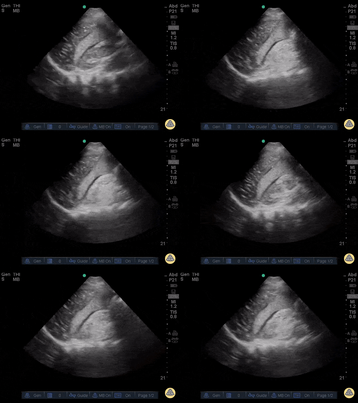
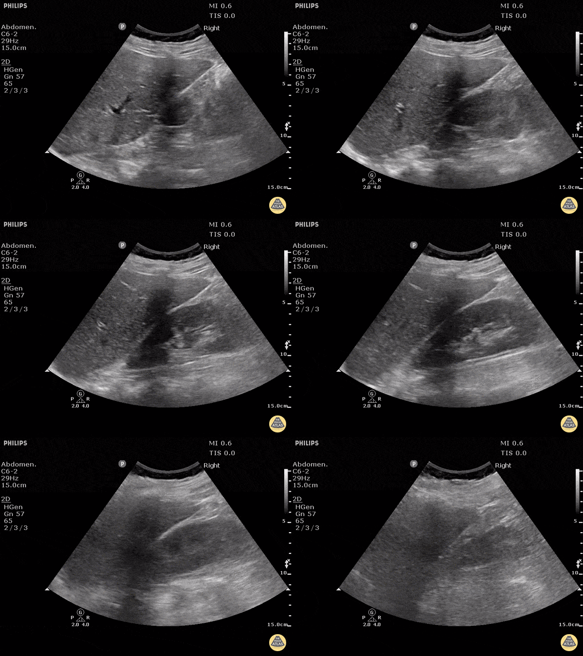
This clip was obtained in a patient presenting after a fall from roof. This hepatic window demonstrates an echogenic clot surrounded by a thin sliver of anechoic free fluid. This patient had massive acute hemoperitoneum. Remember that not all acute hemorrhage is anechoic!
Image courtesy of Robert Jones DO, FACEP @RJonesSonoEM
Director, Emergency Ultrasound; MetroHealth Medical Center; Professor, Case Western Reserve Medical School, Cleveland, OH
View his original post here
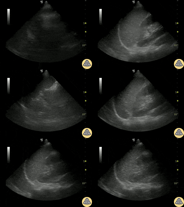
Positive eFAST exam in a blunt trauma patient reveals abnormal splenic architecture and perisplenic fluid.
Image courtesy of Robert Jones DO, FACEP @RJonesSonoEM
Director, Emergency Ultrasound; MetroHealth Medical Center; Professor, Case Western Reserve Medical School, Cleveland, OH
View his original post here
72 year-old male on Eliquis presented to the ED hypotensive after a fall. He was found to have left-sided rib fractures and ecchymosis. FAST was negative for intra-abdominal free fluid, pulmonary effusion, and pneumothorax but did show a splenic hematoma with a fluid jet and swirl (seen here). IR read the CTA as no active extravasation. After describing the ultrasound images the pt was taken to IR and had splenic artery angiography which confirmed extravasation, and was subsequently embolized with resultant hemodynamic stabilization.
Dr Alec Glucksman PGY-III @alecglucksman, Dr Anna Van Tuyl, Dr Norman Ng, Dr Joshua Greenstein
Northwell Health - Staten Island University Hospital
Normal E-FAST Views
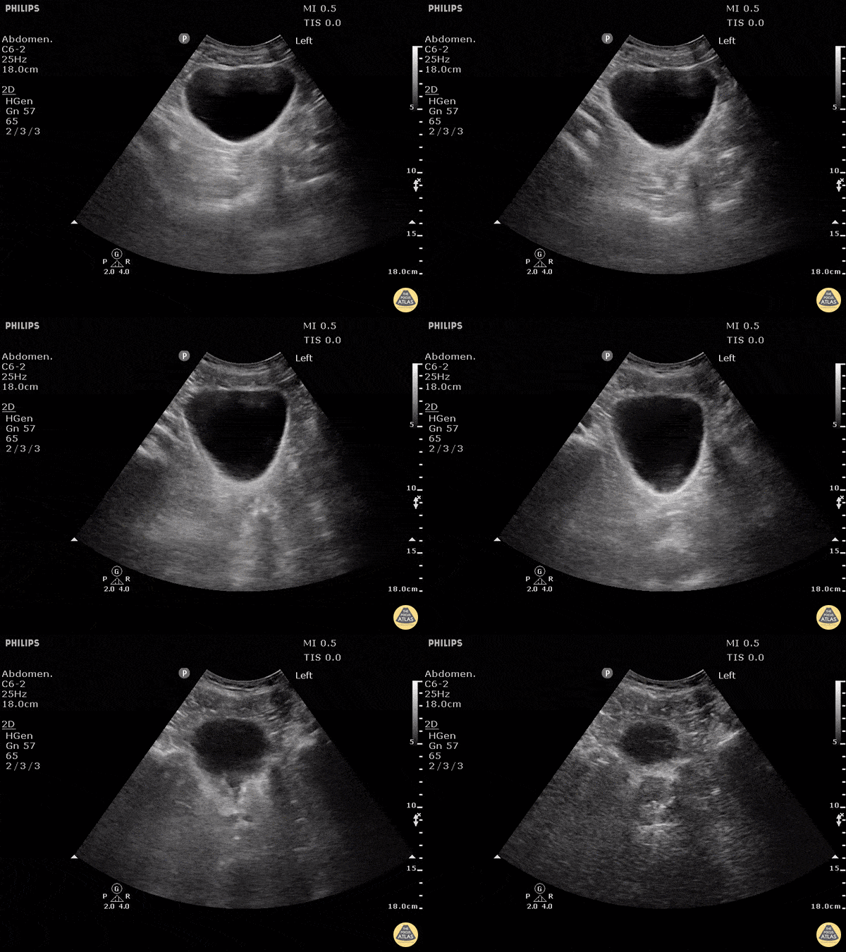
No free fluid is seen around or behind the bladder in this negative FAST.
Dr. Justin Bowra
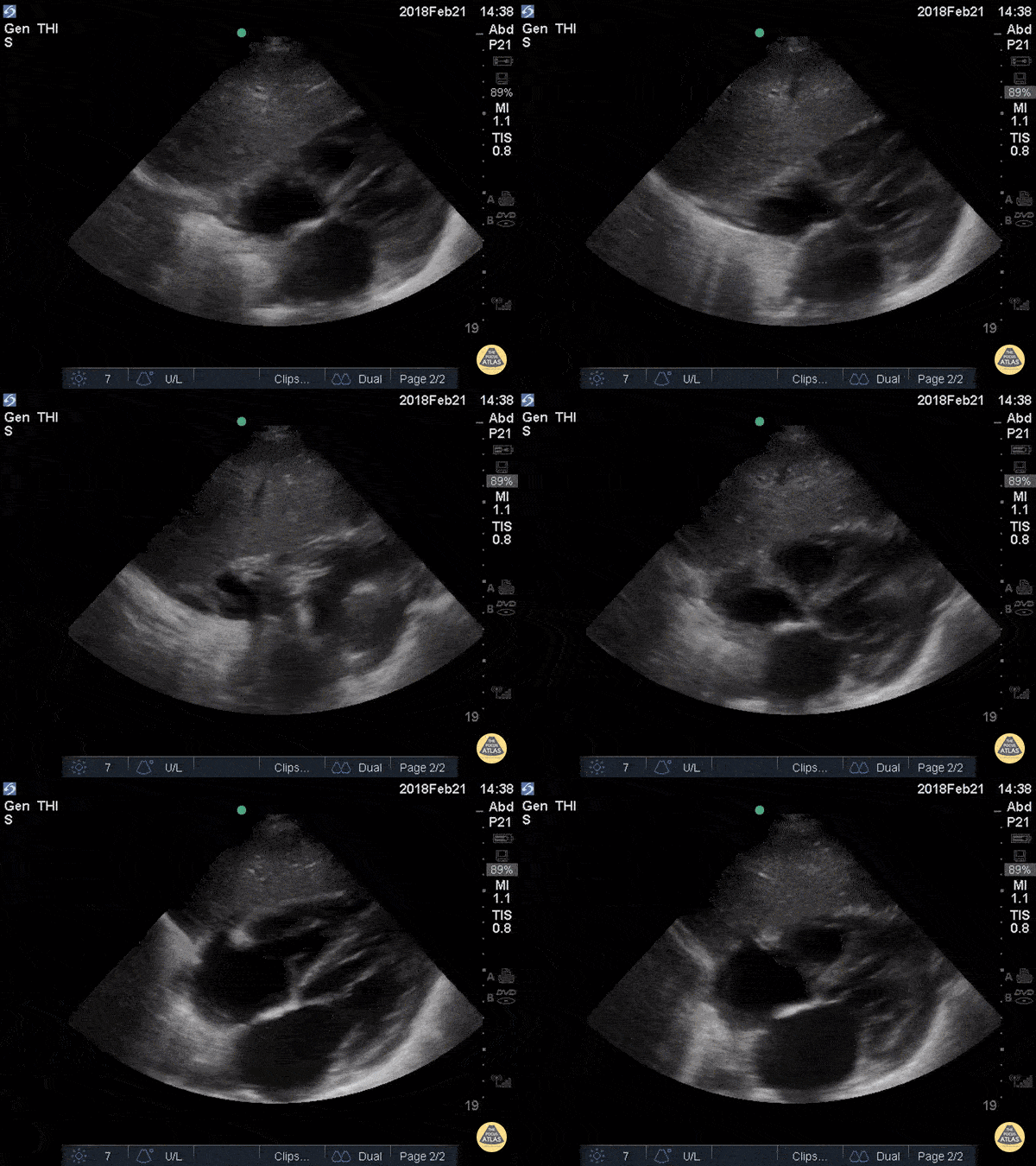
The most superficial structure we see is the liver. Immediately deep to that we see the heart separated from the liver by the diaphragm. Closest to the liver is the right atrium, tricuspid valve, and right ventricle. Deeper to that, we see the left atrium, mitral valve, and left ventricle. There is no anechoic fluid between the bright hyperechoic pericardium and the myocardium, indicating absence of pericardial effusion.
Hannah Kopinksi and Dr. Lindsay Davis - NYU Emergency Medicine
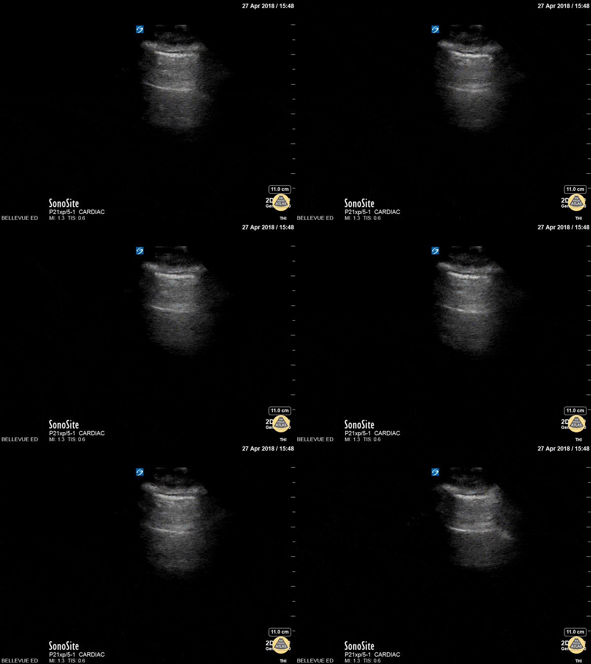
This is a clip demonstrating lung sliding. The most superficial hyperechoic layers are the soft tissue and muscular layers of the chest wall. Immediately deep to that is a bright, thin hyperechoic line which appears to be in motion - this is the pleural line. The parietal pleura rubbing against the visceral pleura as the patient breathes creates this shimmery appearance of lung sliding, also often described as “ants marching”. Lung sliding indicates that there is no pneumothorax.
Hannah Kopinksi and Dr. Lindsay Davis - NYU Emergency Medicine
Positive RUQ View
Patient presented to the emergency department with atraumatic acute onset of shortness of breath. An EFAST was performed to evaluate for pneumothorax and assess for free fluid in the abdomen, thorax, and pericardium.
A significant right pleural effusion was present. Additionally, US of RUQ demonstrated a liver which appeared small in size with prominent ascites. This demonstrates the utility of POCUS to identify free fluid in a quick and efficient manner as compared to other common studies such as a CT scan, even in non-traumatic patients.
Contributors: Lauren Lowes, DO; Krishna Patel, DO; Julia Tu, MS-4
Central Michigan University Residency of Emergency Medicine
This is RUQ view from a patient who presented to the emergency department after falling from a roof and with low blood pressures. Although there appears to be perihepatic fluid, an interesting learning point form this case is whether or not we can discern if fluid observed is truly in the retroperotineal space or in the peritoneal cavity. Because the fluid is in direct contact with perinephric fat, this is actually in the retroperitoneal space and not in the peritoneal cavity. This may be difficult to distinguish in patients with very little perinephric fat.
Image courtesy of Robert Jones DO, FACEP @RJonesSonoEM
Director, Emergency Ultrasound; MetroHealth Medical Center; Professor, Case Western Reserve Medical School, Cleveland, OH
View his original post here
This clip was obtained in a patient presenting after a fall from roof. This hepatic window demonstrates an echogenic clot surrounded by a thin sliver of anechoic free fluid. This patient had massive acute hemoperitoneum. Remember that not all acute hemorrhage is anechoic!
Image courtesy of Robert Jones DO, FACEP @RJonesSonoEM
Director, Emergency Ultrasound; MetroHealth Medical Center; Professor, Case Western Reserve Medical School, Cleveland, OH
View his original post here
Positive LUQ View
This is a clip of the LUQ performed during FAST exam following blunt trauma. It demonstrates loculated fluid around the spleen concerning for blood. CT imaging followed demonstrating a large subcapsular splenic hematoma.
Image courtesy of Robert Jones DO, FACEP @RJonesSonoEM
Director, Emergency Ultrasound; MetroHealth Medical Center; Professor, Case Western Reserve Medical School, Cleveland, OH
View his original post here
Positive eFAST exam in a blunt trauma patient reveals abnormal splenic architecture and perisplenic fluid.
Image courtesy of Robert Jones DO, FACEP @RJonesSonoEM
Director, Emergency Ultrasound; MetroHealth Medical Center; Professor, Case Western Reserve Medical School, Cleveland, OH
View his original post here
72 year-old male on Eliquis presented to the ED hypotensive after a fall. He was found to have left-sided rib fractures and ecchymosis. FAST was negative for intra-abdominal free fluid, pulmonary effusion, and pneumothorax but did show a splenic hematoma with a fluid jet and swirl (seen here). IR read the CTA as no active extravasation. After describing the ultrasound images the pt was taken to IR and had splenic artery angiography which confirmed extravasation, and was subsequently embolized with resultant hemodynamic stabilization.
Dr Alec Glucksman PGY-III @alecglucksman, Dr Anna Van Tuyl, Dr Norman Ng, Dr Joshua Greenstein
Northwell Health - Staten Island University Hospital
Positive Pelvic View
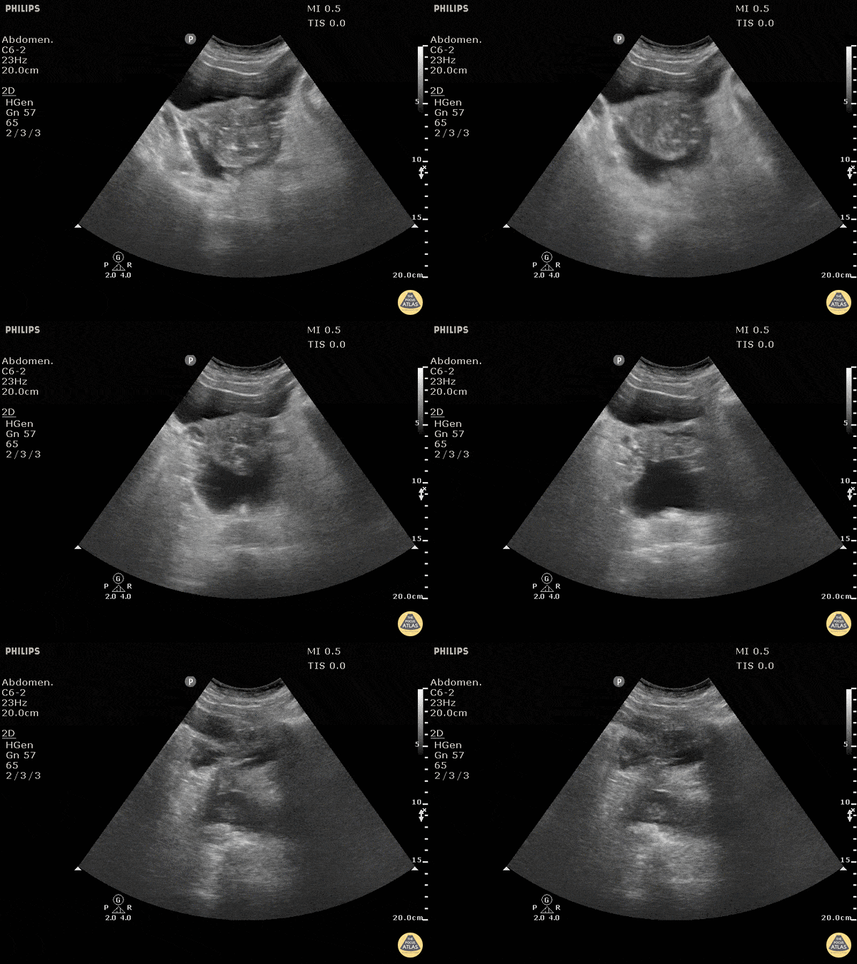
Blunt trauma patient with POSITIVE FAST scan. The uterus can been seen floating in free fluid.
Dr. Justin Bowra
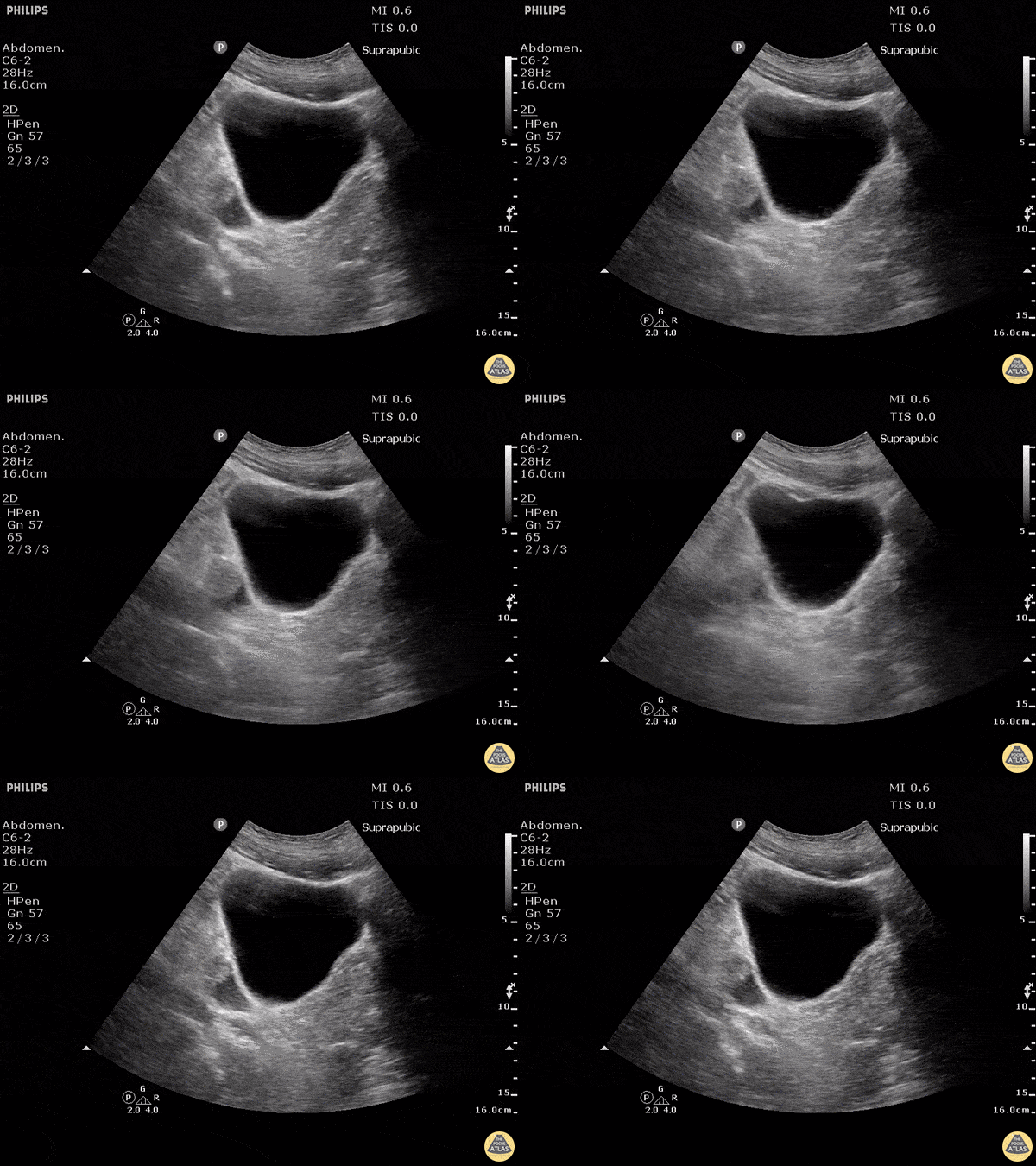
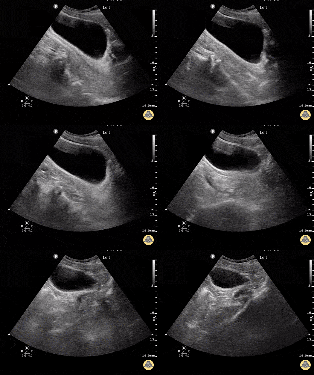
Blunt trauma patient with POSITIVE FAST scan. Free fluid can be seen posterior and lateral to the bladder in this sagittal view.
Dr. Justin Bowra
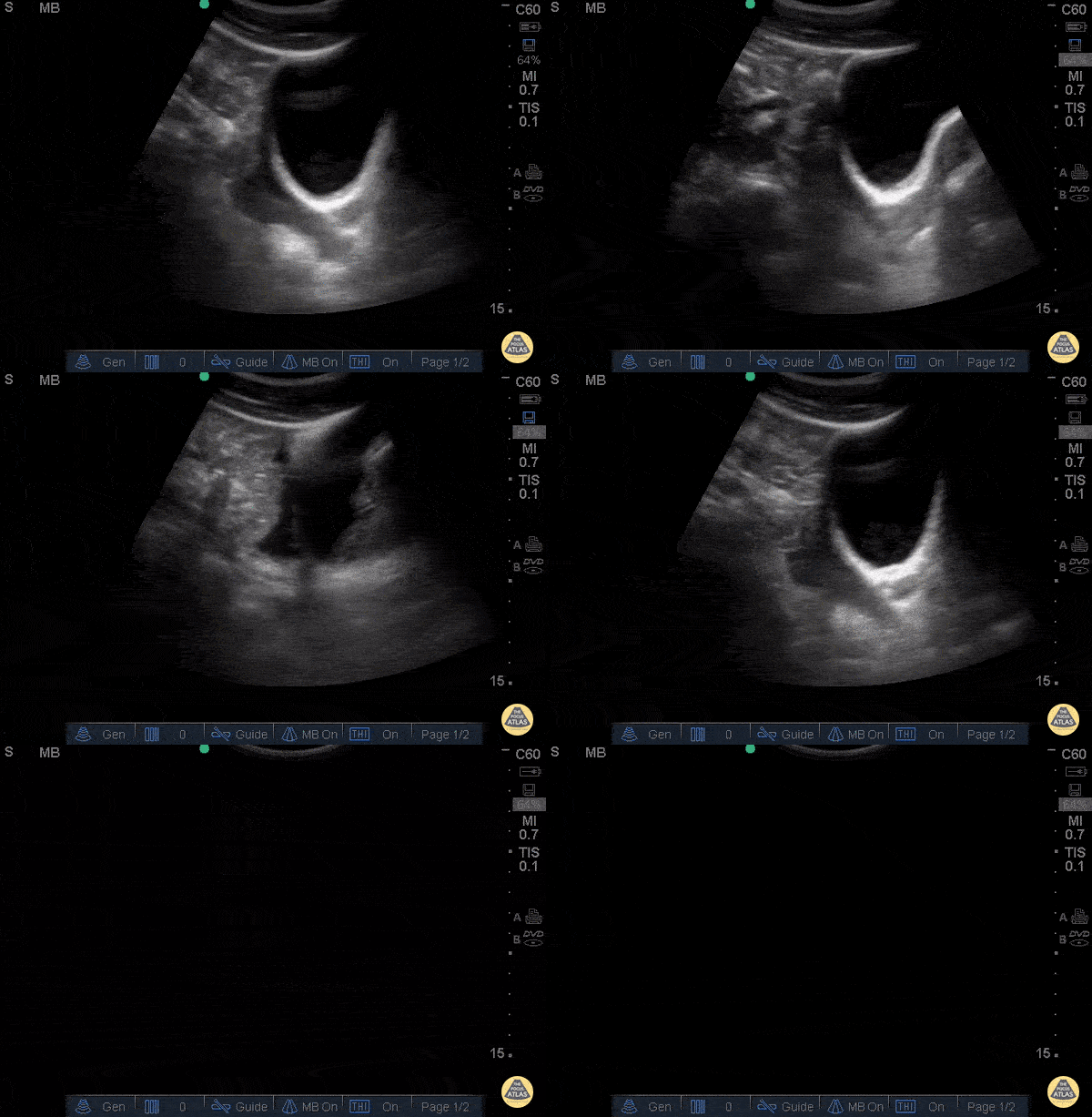
30 y/o pedestrian struck by car, hemodynamically unstable, tachycardic. FAST performed after primary survey revealed free fluid in all four abdominal views of the FAST exam.
Free fluid seen superior and posterior to the bladder in this sagittal view.
Dr. Catharine Bon - Kings County Emergency Medicine
Cardiac Trauma
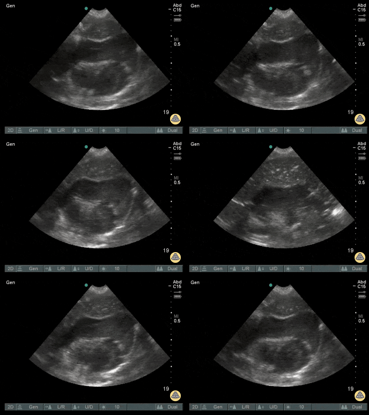
This subxiphoid view of the heart was performed on a patient after a singular stab wound to the chest. From here, we can observe a large collection of blood within the pericardial sac (hemopericardium) with visible cardiac activity still occurring.
Image courtesy of Robert Jones DO, FACEP @RJonesSonoEM
Director, Emergency Ultrasound; MetroHealth Medical Center; Professor, Case Western Reserve Medical School, Cleveland, OH
View his original post here
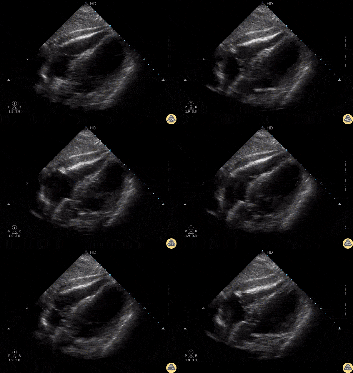
27-year-old woman presents with gunshot wound to right chest. Patient phonating on arrival but tachycardic in 150s with BP 60/palp. EFAST revealed pericardial effusion with hematoma and near complete RV diastolic collapse, consistent with cardiac tamponade. Decision was made to take patient directly for OR thoracotomy based on EFAST findings. Patient lost pulses for 10 sec intra-operatively prior to receiving pericardial window. ROSC was achieved with removal of pericardial hematoma. LV laceration was identified and repaired. LAD laceration identified for which patient received emergent CABG by CT surgery. Patient was successfully extubated the next day, discharged one week later.
EFAST aids in rapid diagnosis and expedites care in hemodynamically unstable trauma patients. POCUS is a useful tool for determining etiology of undifferentiated shock.
Quinn Fujii DO, William Hamrick DO, Ulysses Garcia DO, Robert Rigby DO
Desert Regional Medical Center, Emergency Medicine
POCUS revealed isoechoic fluid within the pericardium indicative of a hemopericardium in a patient suffering from a gunshot wound.
Image courtesy of Robert Jones DO, FACEP @RJonesSonoEM
Director, Emergency Ultrasound; MetroHealth Medical Center; Professor, Case Western Reserve Medical School, Cleveland, OH
View his original post here
Pulmonary Trauma
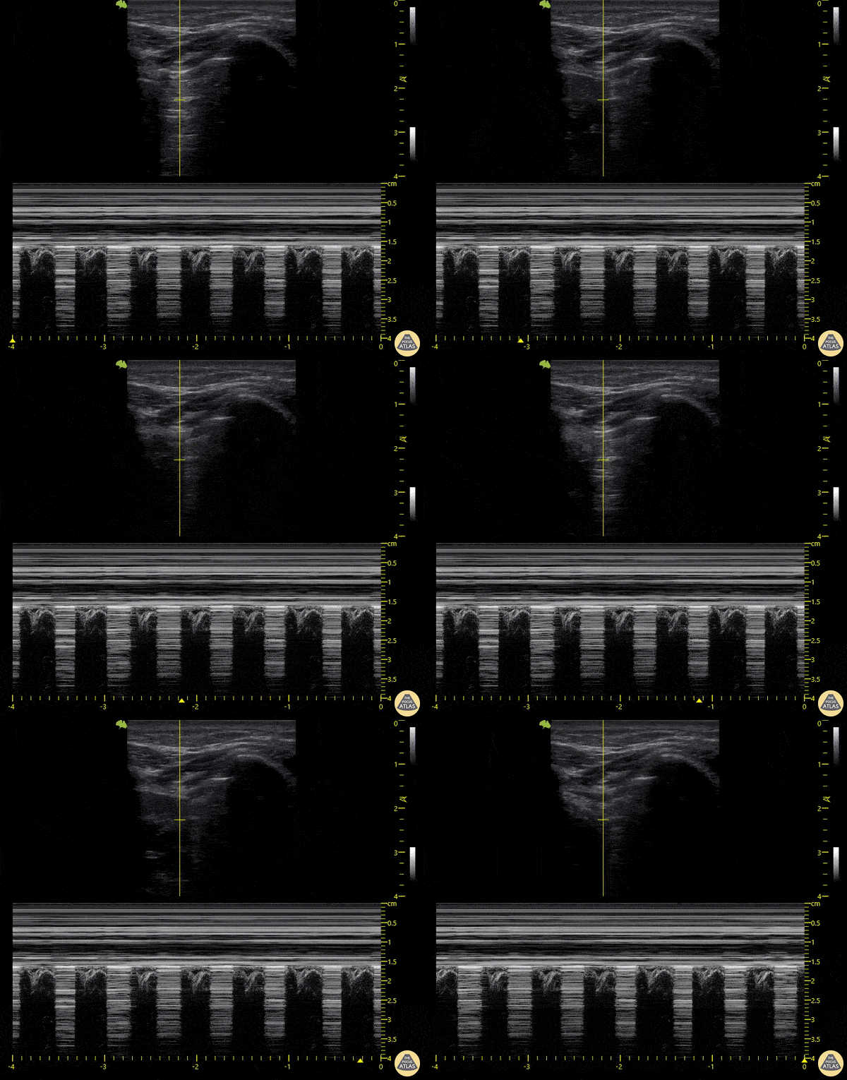
18-year-old patient who presented following a motorcycle accident in which he sustained closed chest trauma with bilateral hemopneumothoraces. In this sequence taken with a linear transducer in left pulmonary zone 1, the "pulmonary point" indicative of pneumothorax can be seen.
Libardo Valencia Chicue
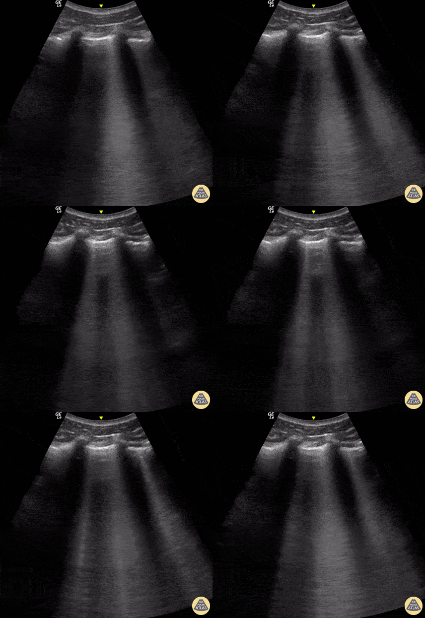
A 23-year-old male was admitted to the ED following a motorcycle accident. He subjectively reported dyspnea; objectively was hypoxic. POCUS seen here (obtained from L3 zone using curvilinear probe) reveals multifocal B-lines consistent with pulmonary contusion. This case illustrates that bedside US is useful beyond diagnosing pneumothorax and hemothorax in trauma patients.
Renato Tambelli, Emergency Physician Hospital das Clínicas de Marília, Brazil.
@R_Tambelli / @JediPocus
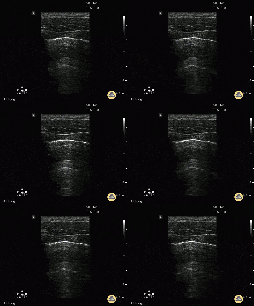
A young male presented to ED with stab wound to chest. Upright CXR was normal. POCUS showed double lung point indicative of small PTX. This was monitored without evidence of progression. No intervention was required.
Image courtesy of Robert Jones DO, FACEP @RJonesSonoEM
Director, Emergency Ultrasound; MetroHealth Medical Center; Professor, Case Western Reserve Medical School, Cleveland, OH
View his original post here
Other Abdominal Trauma
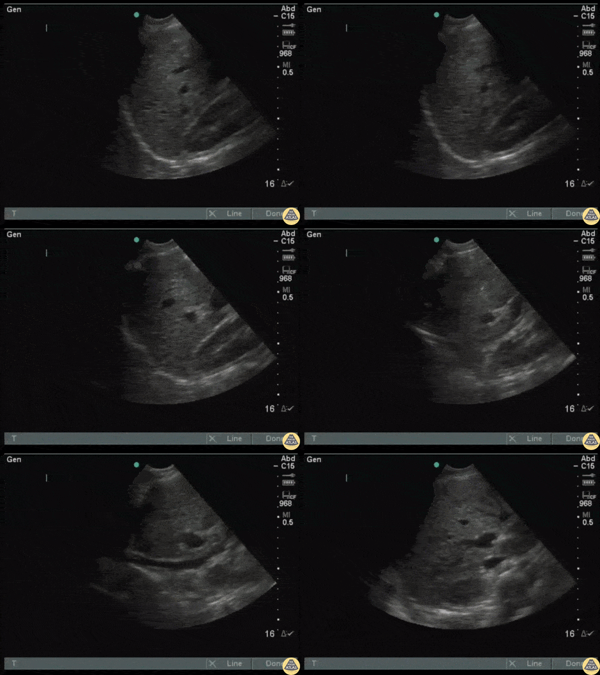
Seen in the superficial region of the liver is an easy to miss hepatic laceration. Ultrasound has a low specificity for detecting solid organ lacerations. Diagnosis was confirmed via CT.
Image courtesy of Robert Jones DO, FACEP @RJonesSonoEM
Director, Emergency Ultrasound; MetroHealth Medical Center; Professor, Case Western Reserve Medical School, Cleveland, OH
View his original post here
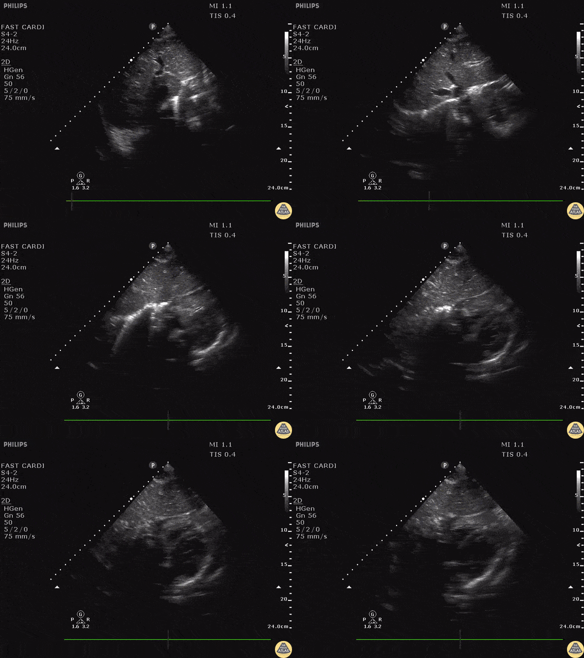
Subcostal window in a patient with a gunshot wound revealed a hyperechoic structure with strong comet tail reverberation artifact. These findings are consistent with a metallic foreign body, such as a retained bullet in this patient. Visit the original post for a labelled image.
Image courtesy of Robert Jones DO, FACEP @RJonesSonoEM
Director, Emergency Ultrasound; MetroHealth Medical Center; Professor, Case Western Reserve Medical School, Cleveland, OH
View his original post here
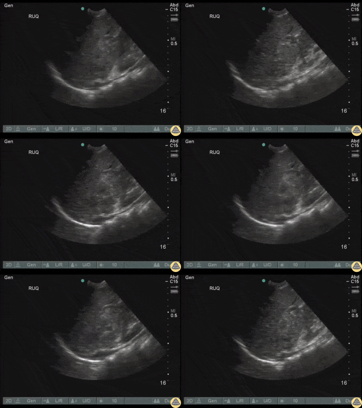
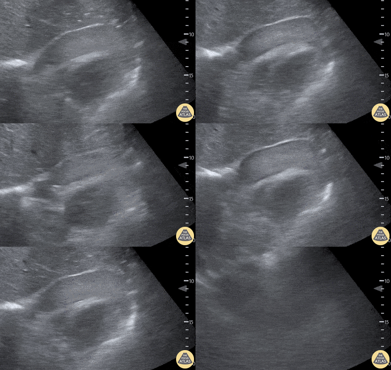
An adult male presented to the ED following a MVC with a fractured femur, right sided flank pain, and transient hypotension. FAST exam revealed an isoechoic, oval structure just cephalad to the right kidney indicative of an adrenal hemorrhage/hematoma.
Image courtesy of Robert Jones DO, FACEP @RJonesSonoEM
Director, Emergency Ultrasound; MetroHealth Medical Center; Professor, Case Western Reserve Medical School, Cleveland, OH
View his original post here
MSK Trauma
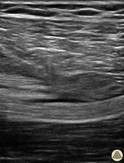
Patient sustained a gun shot wound through left mid thigh.
Image is long axis view of sciatic nerve mid thigh. Nerve is running distal to proximal from left to right at approximately 3cm depth. There is a hypoechoic fluid collection seen superficial to the nerve. The epineurium is intact and we can see smaller nerve fibers contained in it.
Patient had 0/5 plantar/dorsiflexion of ankle on admission, consistent with a sciatic nerve injury. The ultrasound exam on the sciatic nerve did not show any gross abnormality other than this fluid collection around the nerve. Within 7 days (4 days after this image) he regained strength in plantar/dorsiflexion in the ankle.
Mike Guju, MD @MichaelMGujuMD
Resident PM&R EVMS
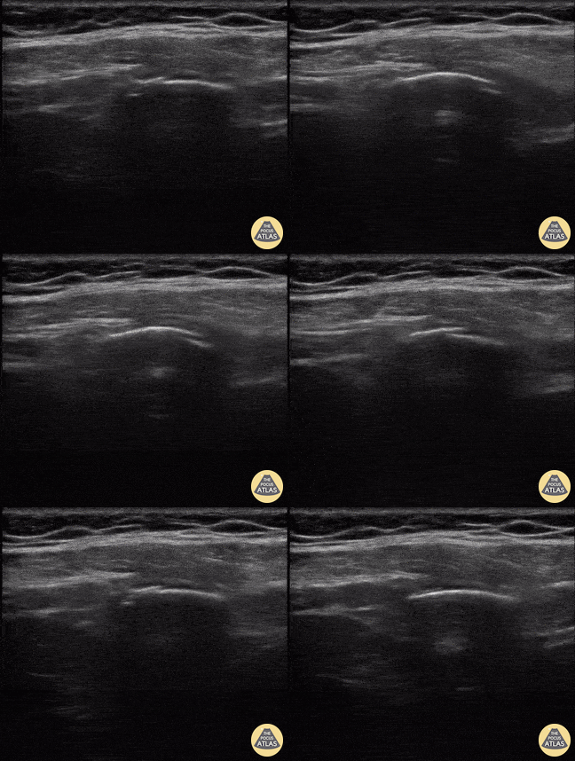
A middle aged man presented 1 week after sustaining a fall with direct injury to his left chest. He reported pain with inspiration and coughing; he localized pain to one specific area of his chest wall. Seen here is the image obtained when the linear probe was placed in the longitudinal plane to his area of point-tenderness. Notice the disruption of the hyperechoic cortex of the rib. Findings were confirmed in the transverse plan. The patient went on to have an anterior serratus nerve block for pain control related to his rib fracture.
Mandy Peach, MD @mandy_peach
Saint John Regional Hospital. NB, Canada
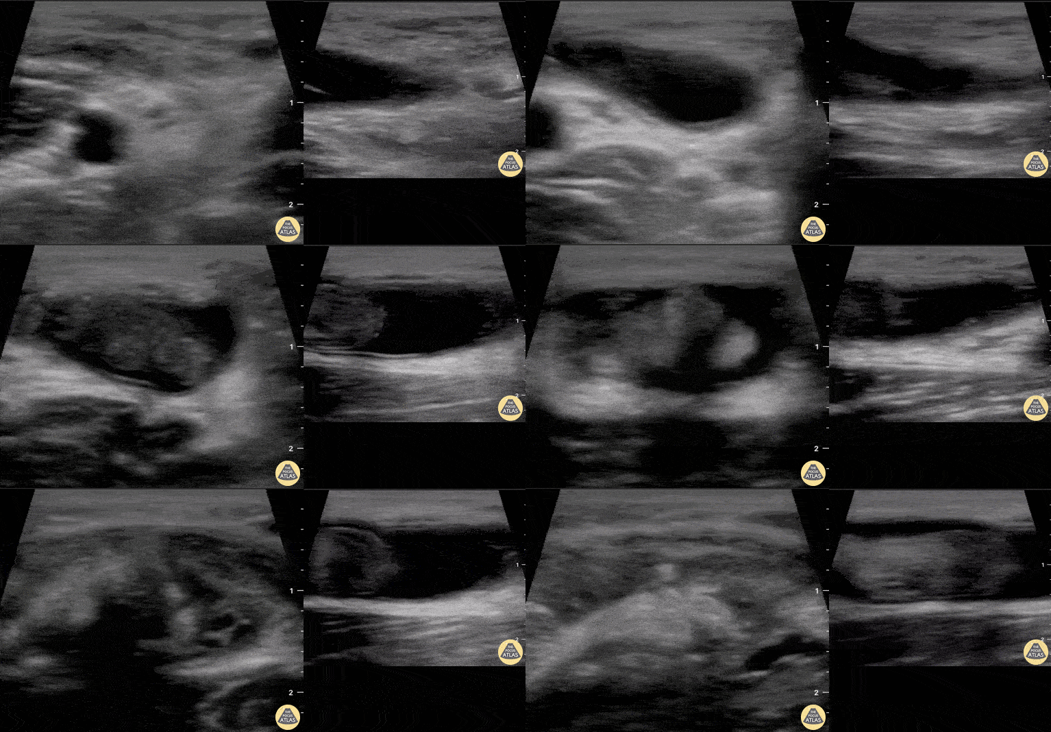
A 30-year-old male presented with acute-onset left anterior elbow pain after feeling a “pop” while weight-lifting. On exam, there was an obvious reverse popeye sign, swelling and mild ecchymosis. There was slight weakness in flexion and supination. Hook test was positive. A Butterfly IQ was used on an MSK soft tissue setting to assess the tendon in long and short axis via an anterior approach. Discontinuity of the tendon and surrounding anechoic fluid representing hematoma were noted. Orthopaedic surgery was consulted and reviewed ultrasound images. As a result, the patient had an urgent MRI and underwent expedited surgical repair.
Melanie Leclerc, MD CCFP(EM). @MelanieLecler19
David Lewis MB,BS FRCS FCEM CFEU PGDipSEM
Saint John Regional Hospital, New Brunswick, Canada
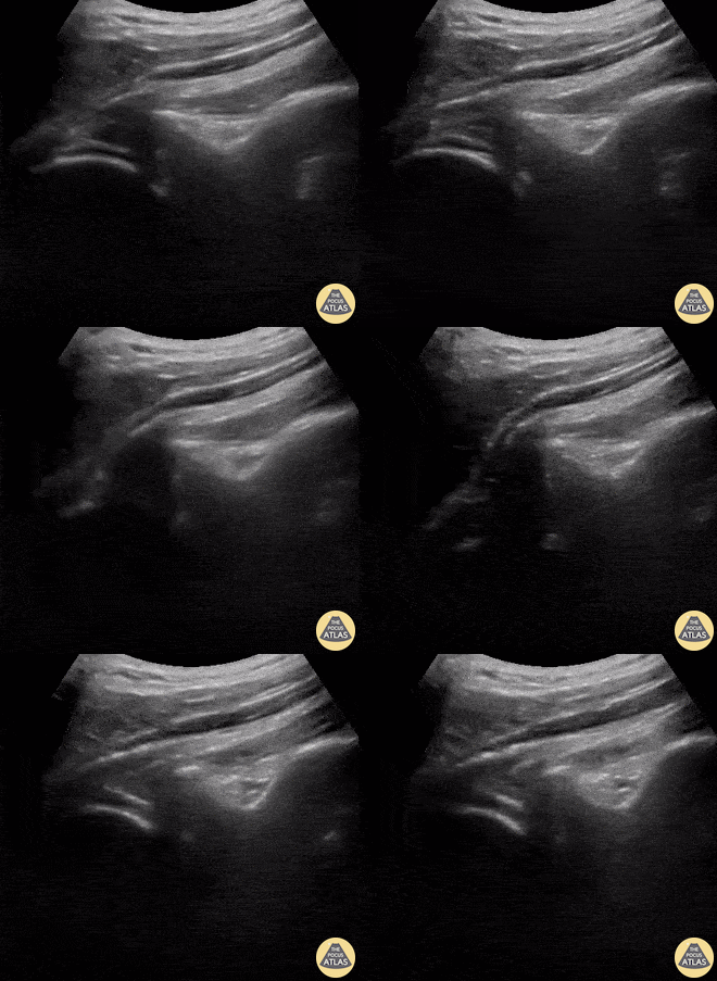
A 9-year-old female presented with left shoulder pain. She has a history of multiple dislocations and, as seen here on POCUS, is able to reduce the dislocation herself.
Julie Klensch, PEM Fellow & Paul Khalil, MD
University of Louisville/Norton Children's Hospital

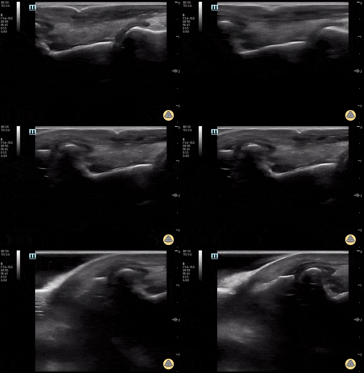
A 32-year-old male pedestrian struck by a jeep traveling at 30 mph was evaluated with POCUS for left knee injury. He sustained a left-sided rupture of ACL, LCL, and an avulsion fracture of his IT band. Pictured here is his left lateral knee US performed 10-days after the acute injury. You can appreciate the popliteus notch in the lateral femur, typically both the origin of the popliteus muscle and the plane the LCL traverses to the fibula; here only notable for diffuse edema.
Eben Alexander