
OB/Gyn
Obstetrics and Gynecology 👼🏽
Note: These search bar results are only for the OB/Gyn image archive. To search the entire site, click here.
Recently Added
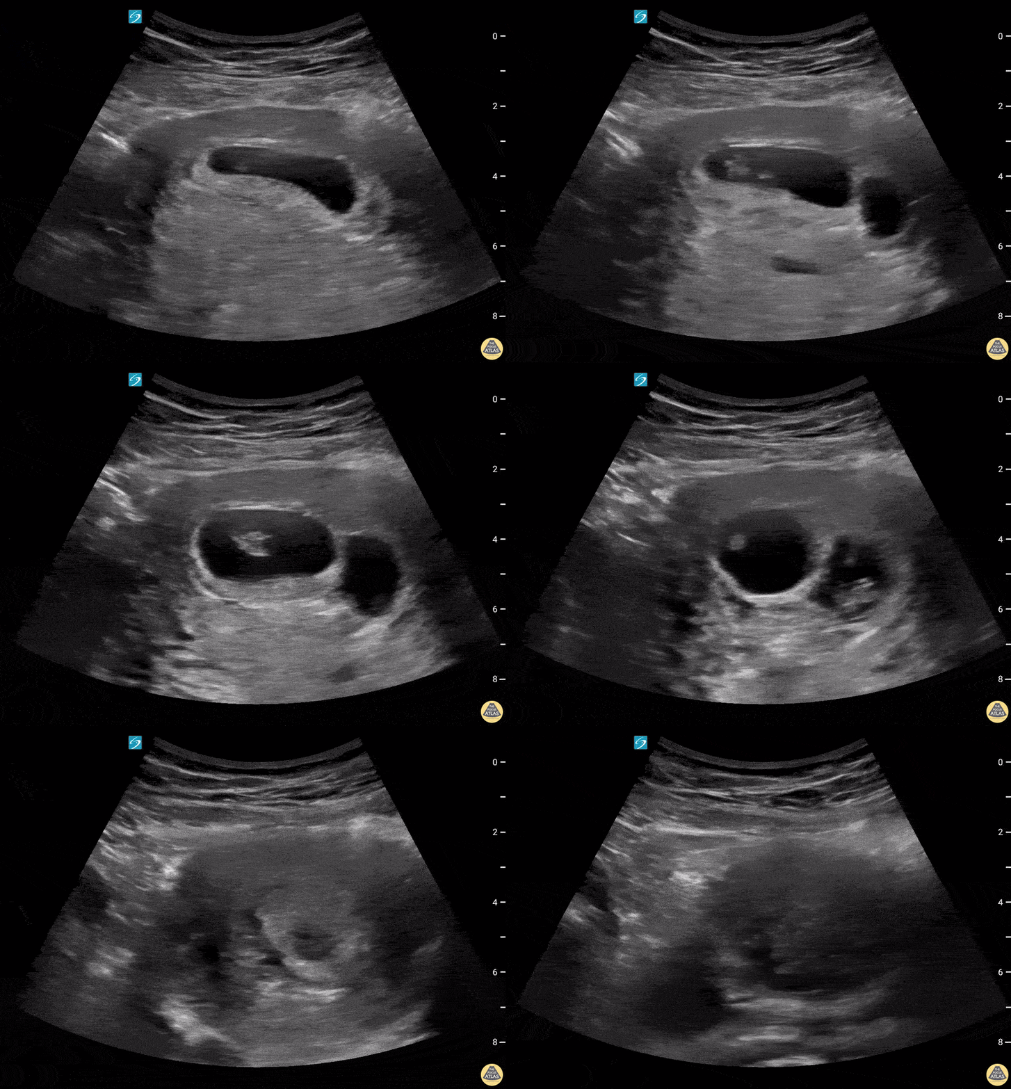
A 27-year-old female presented for evaluation of cough, congestion, decreased appetite, muscle aches, and fatigue. Patient was unsure of the date of her last menstrual period.
This image is a transabdominal ultrasound of the uterus demonstrating a dichorionic diamniotic twin intrauterine pregnancy. The pregnancy was dated at approximately 8 weeks and 2 days, with fetal heart rates of 167 bpm and 174 bpm.
Arthur Sieron BMBS, PGY-1 Emergency Medicine, Central Michigan University; Eric Spencer DO, PGY-3 Emergency Medicine, Central Michigan University; Halimah Hamidu, MS4 Central Michigan University College of Medicine.
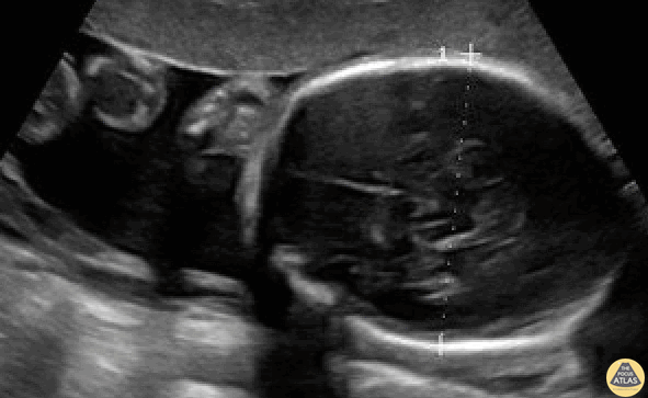
BPD should be measured in the axial plane, perpendicular to the falx cerebri, and from the outer edge of the near calvarial wall to the inner edge of the far calvarial wall.
Contributors:
Stephen Holihan (MD); Dillon Nerland (MD); Madison Waddell (MS4)
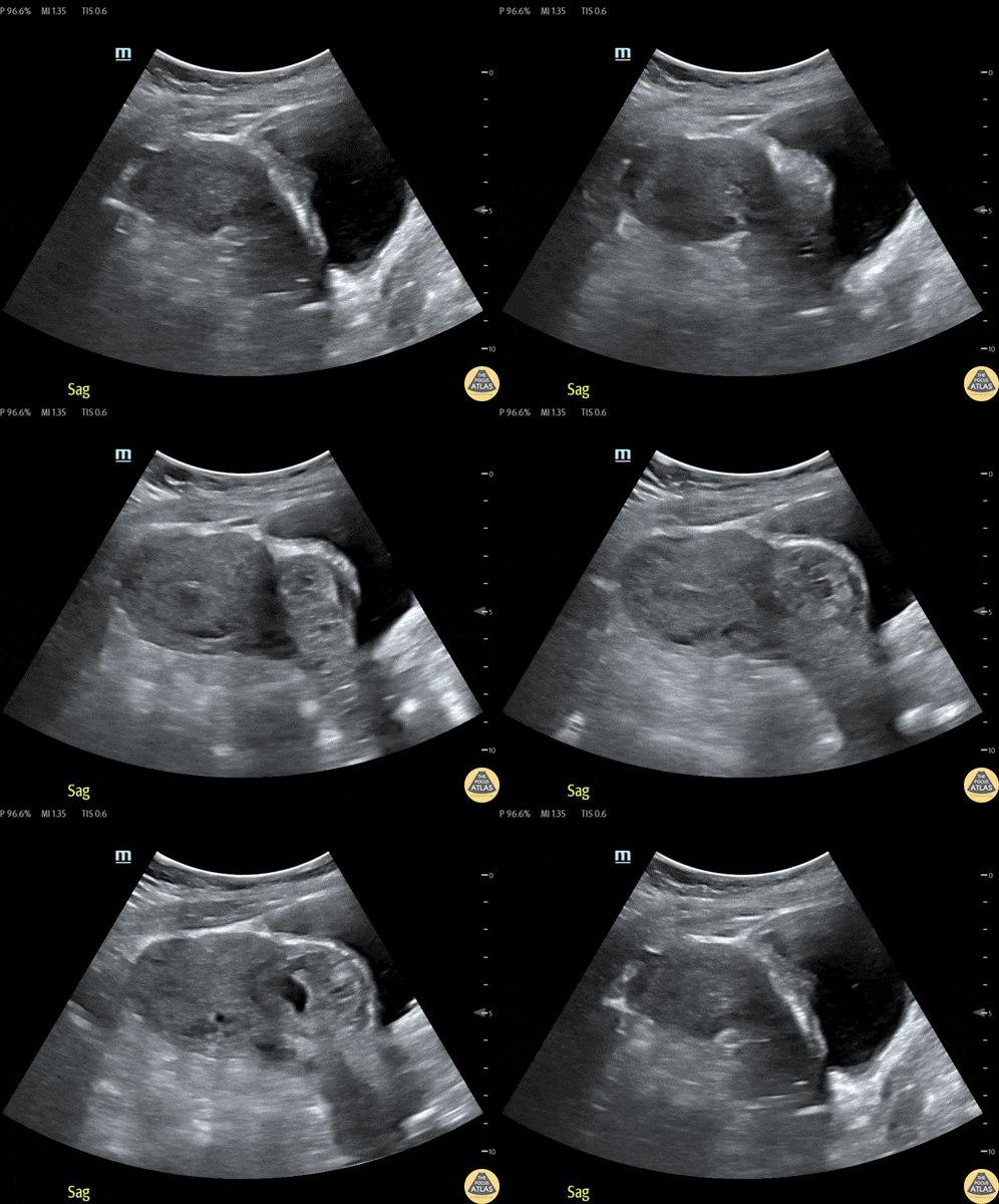
32 year old female at 8-9 weeks gestation by LMP referred to the ED for pregnancy of unknown location. She reported 1-2 weeks of mild pelvic cramping and vaginal spotting.
Bedside trans-abdominal imaging demonstrated a round, heterogenous structure without a clear yolk sack or fetal pole, but with several hallmarks of caesarean scar ectopic pregnancy: position in the anterior uterine wall; in the lower (closer to the cervix) half of the uterus; bulging into the neighboring bladder; with thin interposed myometrium. After confirmatory formal ultrasound, she was admitted for methotrexate termination, which proved successful.
Contributors: Nicholas Maurer, MD, MPH (PGY-1); Megan Chenworth (PGY-3)
Emergency Medicine, Northwestern McGaw Medical Center
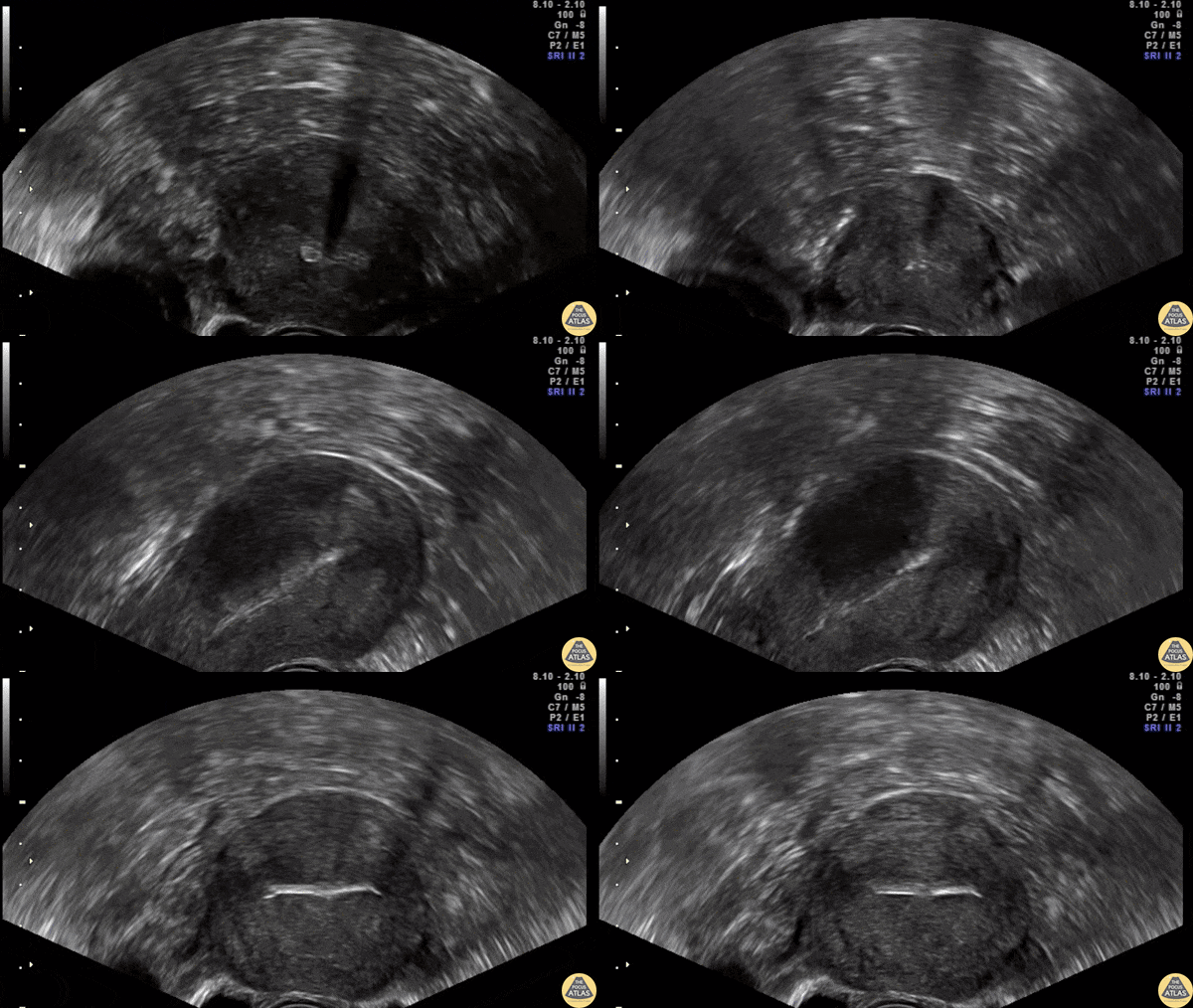
Transvaginal ultrasound showing the IUD correctly placed in both sagittal and transverse plane.
Contributor: Dr. Nicolay B. Werner
Akershus University Hospital, Norway
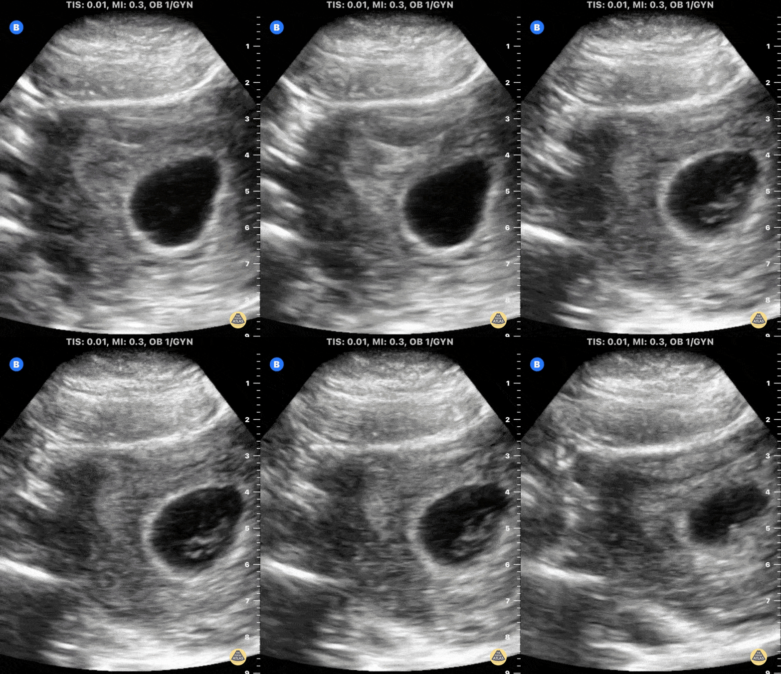
This is a transverse uterus view in early pregnancy demonstrating a a fetal pole with visualized cardiac activity.
Mike Macias, MD, Emergency Physician, @emedcurious
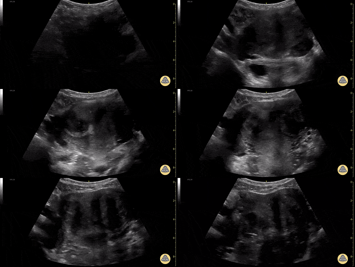
Heterogeneous uterus with multiple well defined hypoechoic masses consistent with leiomyomas (fibroids).
Dimitri Livshits DO, Ultrasound Fellow, Kings County/SUNY Downstate; Jane Belyavskaya MD, Ultrasound Fellow, Kings County/SUNY Downstate; Chris Hanuscin MD, Ultrasound Division Director, Kings County/SUNY Downstate;
Normal Anatomy
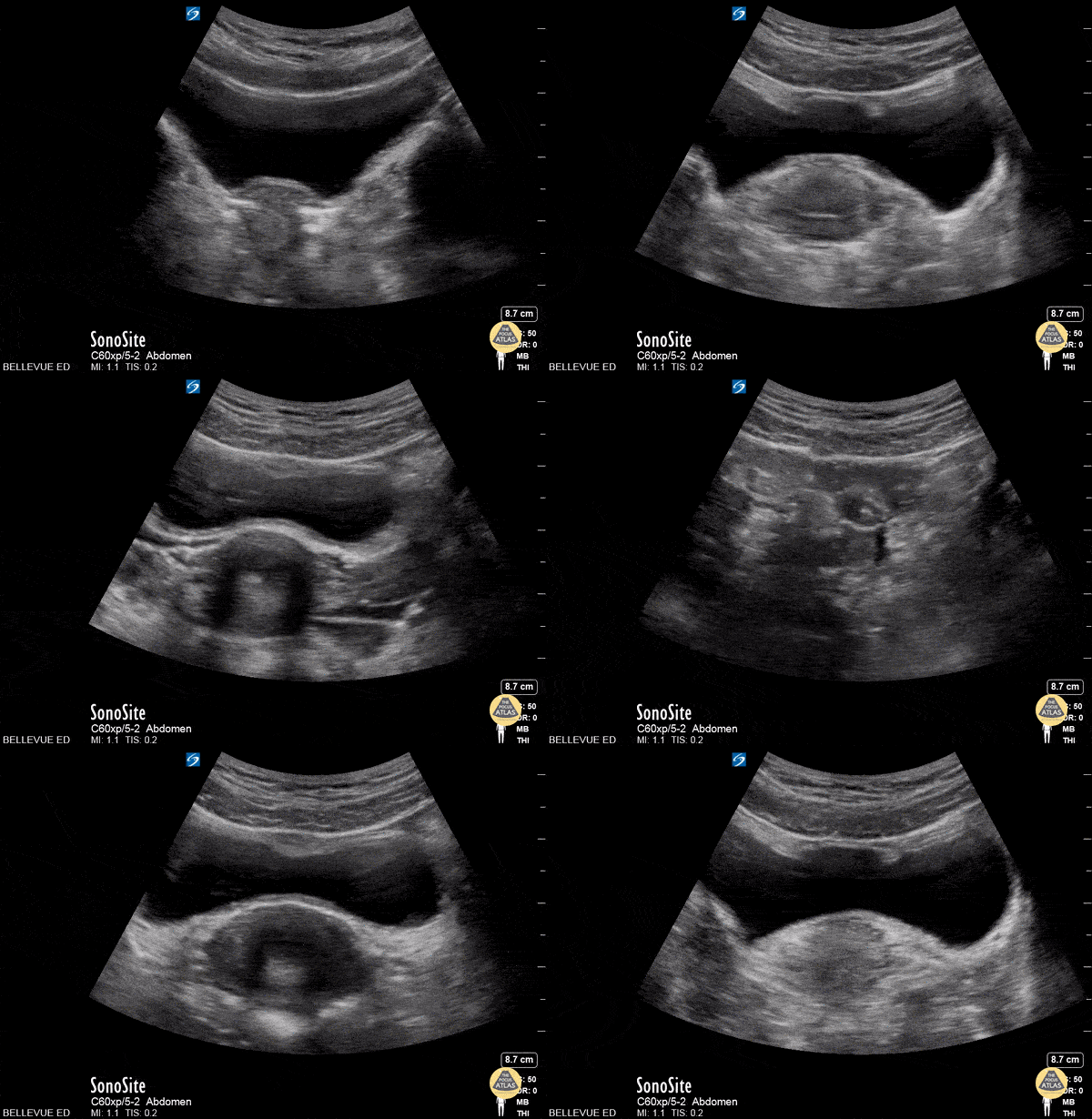
This is a clip of the uterus in transverse view using the transabdominal approach. The large anechoic structure in the center is the bladder in transverse view, and immediately deep to the bladder is the round uterine fundus. As the probe fans through the uterus we see some dark shadowing within it, likely due to artifact created by an IUD. The fallopian tubes are also faintly visible bilaterally branching off the uterus.
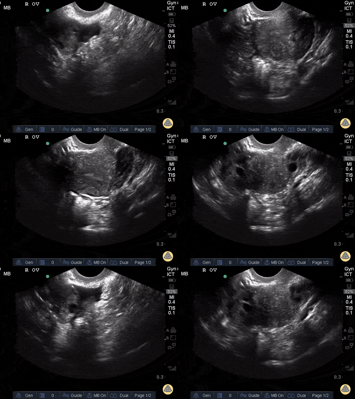
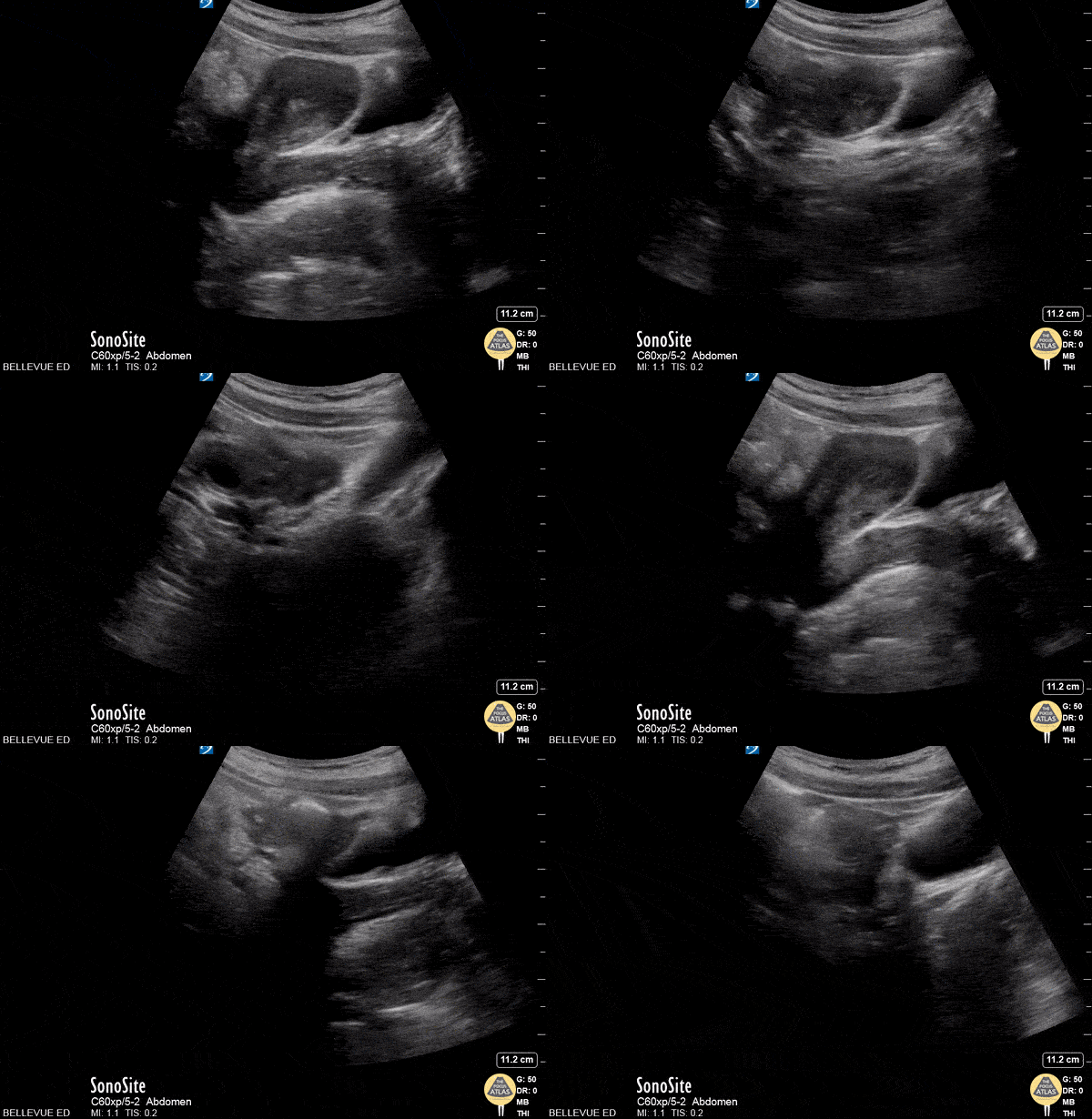
In this transabdominal view of the uterus in transverse, we see the left fallopian tube branching off the uterus leading to the left ovary in which multiple small hypoechoic follicles are visible, giving the ovary its typical “chocolate chip cookie” appearance. At the end of the clip, one of the left iliac vessels is seen in long axis just deep and lateral to the ovary. The bright hyperechoic lines with dark shadowing in the surrounding fields are bowel gas.
Hannah Kopinksi and Dr. Lindsay Davis - NYU Emergency Medicine
Colorized Normal Anatomy
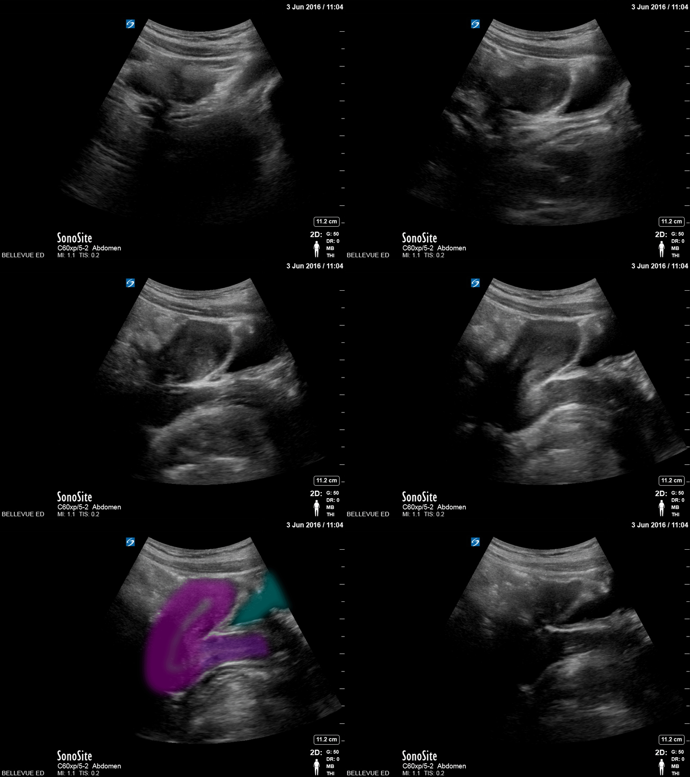
Transabdominal Uterus (sagittal view)
Pink: Uterus, Purple: Cervix, Teal: Bladder
Images: Dr. Lindsay Davis, Dr. Hannah Kopinski. Image Editing: Michael Amador and Dr. Matthew Riscinti
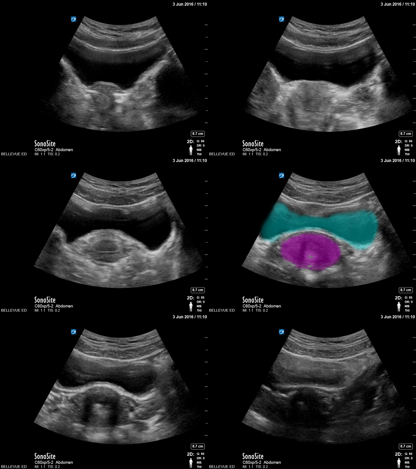
Transabdominal Uterus (transverse)
Teal: Bladder, Pink: Uterus
Images: Dr. Lindsay Davis, Dr. Hannah Kopinski. Image Editing: Michael Amador and Dr. Matthew Riscinti
Pregnancy
A 27-year-old female presented for evaluation of cough, congestion, decreased appetite, muscle aches, and fatigue. Patient was unsure of the date of her last menstrual period.
This image is a transabdominal ultrasound of the uterus demonstrating a dichorionic diamniotic twin intrauterine pregnancy. The pregnancy was dated at approximately 8 weeks and 2 days, with fetal heart rates of 167 bpm and 174 bpm.
Arthur Sieron BMBS, PGY-1 Emergency Medicine, Central Michigan University; Eric Spencer DO, PGY-3 Emergency Medicine, Central Michigan University; Halimah Hamidu, MS4 Central Michigan University College of Medicine.
BPD should be measured in the axial plane, perpendicular to the falx cerebri, and from the outer edge of the near calvarial wall to the inner edge of the far calvarial wall.
Contributors:
Stephen Holihan (MD); Dillon Nerland (MD); Madison Waddell (MS4)
This is a transverse uterus view in early pregnancy demonstrating a a fetal pole with visualized cardiac activity.
Mike Macias, MD, Emergency Physician, @emedcurious
Ectopic
32 year old female at 8-9 weeks gestation by LMP referred to the ED for pregnancy of unknown location. She reported 1-2 weeks of mild pelvic cramping and vaginal spotting.
Bedside trans-abdominal imaging demonstrated a round, heterogenous structure without a clear yolk sack or fetal pole, but with several hallmarks of caesarean scar ectopic pregnancy: position in the anterior uterine wall; in the lower (closer to the cervix) half of the uterus; bulging into the neighboring bladder; with thin interposed myometrium. After confirmatory formal ultrasound, she was admitted for methotrexate termination, which proved successful.
Contributors: Nicholas Maurer, MD, MPH (PGY-1); Megan Chenworth (PGY-3)
Emergency Medicine, Northwestern McGaw Medical Center
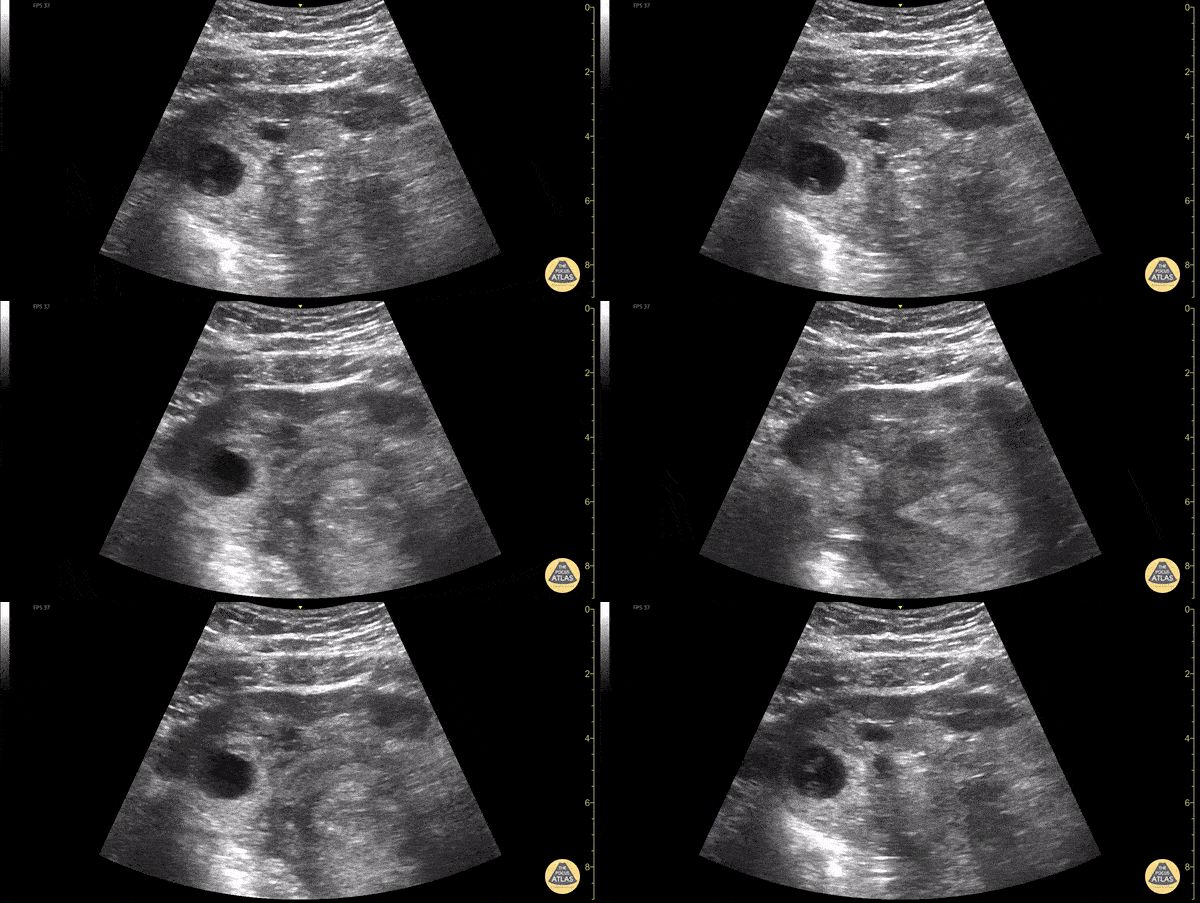
30s female currently 7 week pregnant with lower abdominal pain and tenderness to palpation to lower abdomen. POCUS showed a gestation sac containing a yolk sac and fetal pole outside of uterus. Also noted is a thickened endometrial stripe (to the right of the gestational sac). No free fluid was noted in the RUQ. The patient was taken to the OR for definitive management of ectopic pregnancy.
Dimitri Livshits DO, Ultrasound Fellow, Kings County/SUNY Downstate; Jane Belyavskaya MD, Ultrasound Fellow, Kings County/SUNY Downstate; Farnam Kazi MD, Ultrasound Faculty, Kings County/SUNY Downstate;
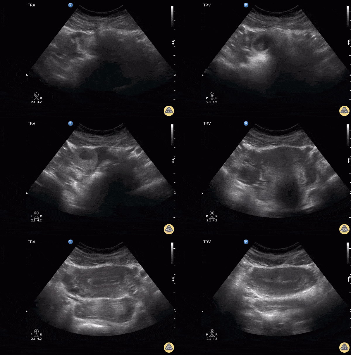
Notice to the left of the screen the presence of an adnexal mass separate to the ovaries which in this case indicates a right tubal ectopic pregnancy. Also noted here within the endometrium is an outer echogenic layer, middle hypoechoic layer and an inner hyperechoic stripe, which makes up the classic trilaminar pattern that is usually observable during the proliferative phase of menstruation.
Image courtesy of Robert Jones DO, FACEP @RJonesSonoEM
Director, Emergency Ultrasound; MetroHealth Medical Center; Professor, Case Western Reserve Medical School, Cleveland, OH
View his original post here
Ovarian Cysts
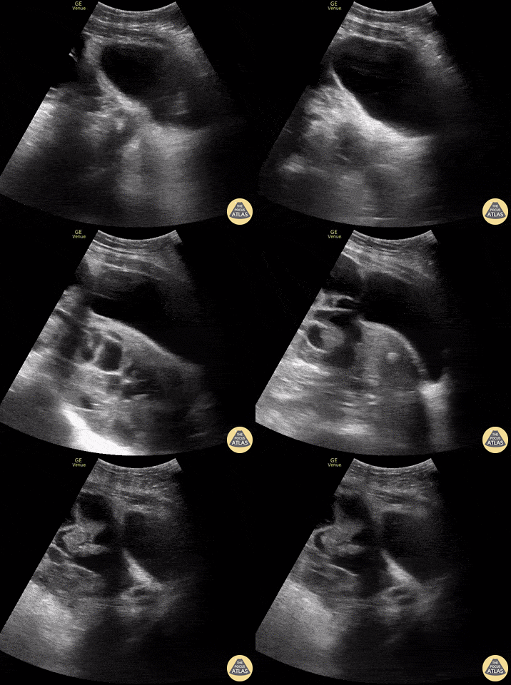
20 y/o female s/p egg retrieval for egg donation 3 days prior presents for diffuse abdominal pain and bloating. In the clip (bladder sagittal view), you can see massive enlargement of the ovary and its multiple follicles. FAST revealed free fluid throughout the abdomen likely secondary to leakage of fluid from these follicles, a process referred to as ovarian hyperstimulation syndrome (OHSS). The patient was admitted to GYN for possible drainage.
Jennifer Kaminsky, MD PGY-2; @jen_kaminskyMD
Pamela Santivanez, MD PGY-1
Sean Beckman, Rocky Vista University COM OMS-4
Joshua Greenstein, MD, Director of ED Ultrasound
Northwell Health - Staten Island University Hospital
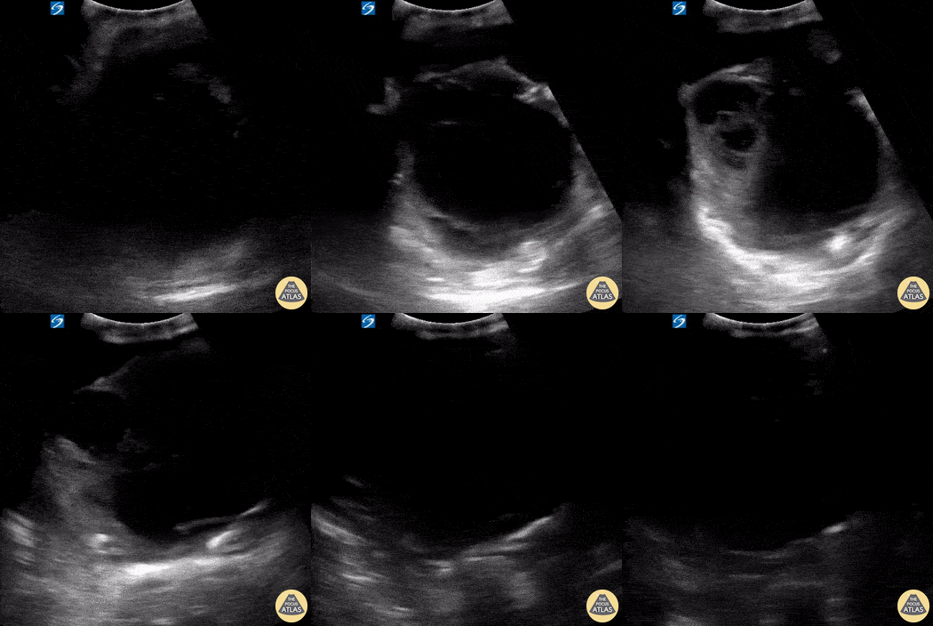
A middle-aged female with a history of colon cancer presented to the ED with constipation and abdominal pain. A recent colonoscopy had revealed rectosigmoid adenocarcinoma. Her physical exam was notable for a distended abdomen that was tender to palpation. POCUS revealed the presence of a large, complex, cystic structure within the pelvis. Subsequent CT confirmed a loculated fluid collection within the pelvis compressing the rectum and sigmoid colon; this mechanical obstruction was likely contributing to patient’s constipation. Differential diagnosis included loculated ascites versus cystic tumor.
Kyla Walworth, MS-4 & Matthew McDowell, PGY-1
Central Michigan University College of Medicine
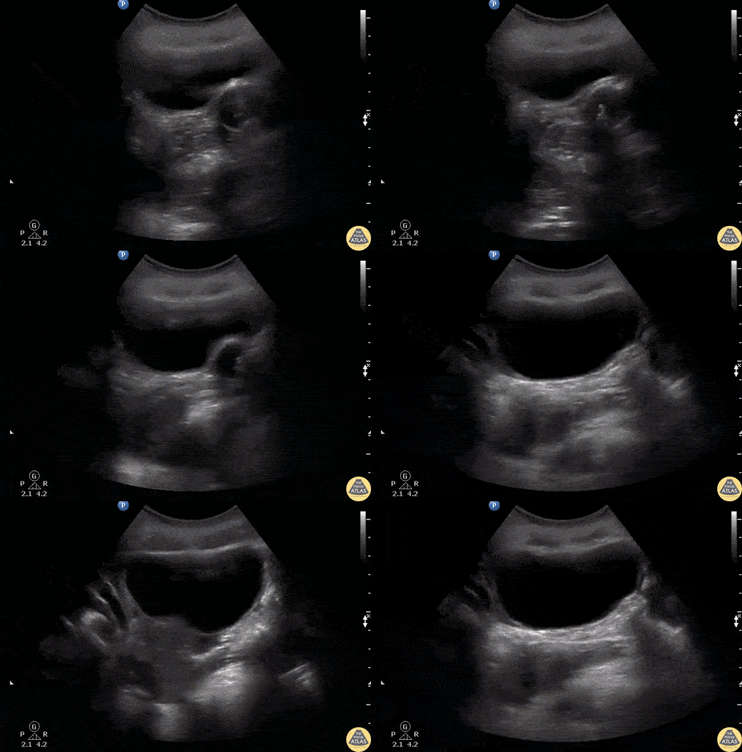
A young female presented to the ED with sudden pelvic pain. She has no PMH and a negative hCG. Bedside sagittal transabdominal ultrasound revealed a large right ovarian cyst mimicking the urinary bladder. Notice the bladder decompressed with a foley balloon.
Image courtesy of Robert Jones DO, FACEP @RJonesSonoEM
Director, Emergency Ultrasound; MetroHealth Medical Center; Professor, Case Western Reserve Medical School, Cleveland, OH
View his original post here
Other
Transvaginal ultrasound showing the IUD correctly placed in both sagittal and transverse plane.
Contributor: Dr. Nicolay B. Werner
Akershus University Hospital, Norway
Heterogeneous uterus with multiple well defined hypoechoic masses consistent with leiomyomas (fibroids).
Dimitri Livshits DO, Ultrasound Fellow, Kings County/SUNY Downstate; Jane Belyavskaya MD, Ultrasound Fellow, Kings County/SUNY Downstate; Chris Hanuscin MD, Ultrasound Division Director, Kings County/SUNY Downstate;
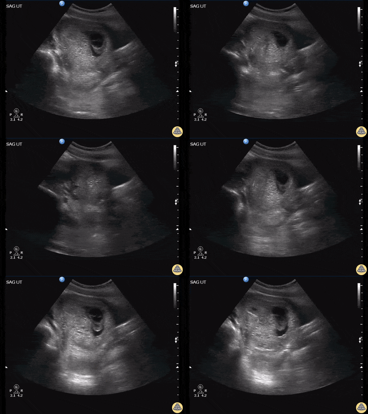
This is a saggital view of the uterus belonging to a patient who returned to the emergency department after persistent vomiting. An initial urine pregnancy test performed yielded a negative result however this patient’s ultrasound scan ultimately revealed a molar pregnancy. As Dr. Jones explains, this patient false-negative urine pregnancy test is explained by a phenomenon known as the High Dose Hook Effect.
Image courtesy of Robert Jones DO, FACEP @RJonesSonoEM
Director, Emergency Ultrasound; MetroHealth Medical Center; Professor, Case Western Reserve Medical School, Cleveland, OH
View his original post here