
Vascular
Vascular
Note: These search bar results are only for the Vascular image archive. To search the entire site, click here.
Recently Added
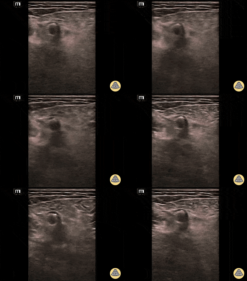
Femoral artery stent can be appreciated in this short axis view as a bright, hyperechoic structure within the femoral artery borders.
Contributed by: Brittany Garza, DO; Saleem Nasseh, MD; Sadie Ellerson, MS4
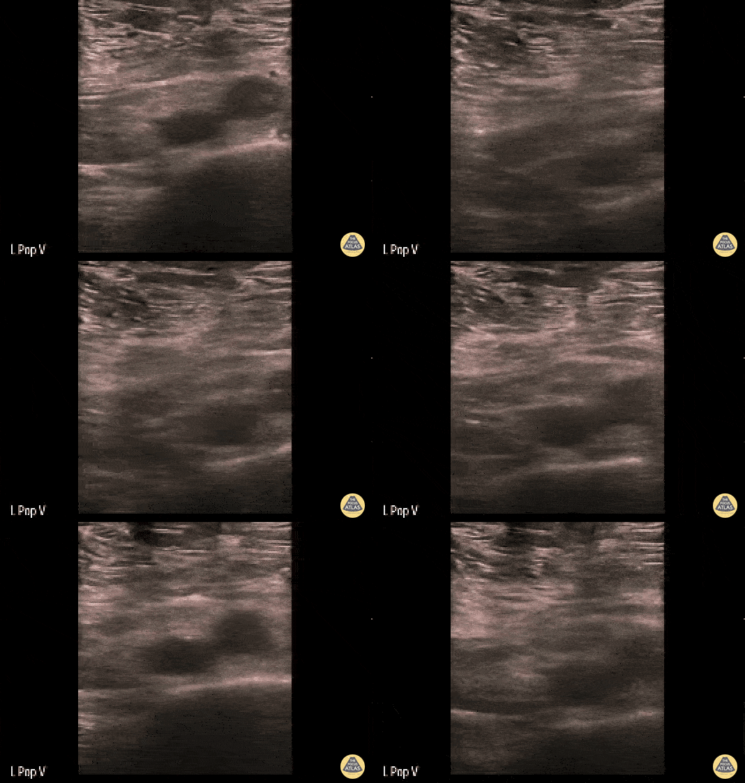
This clip demonstrates a non-compressible left popliteal vein consistent with deep venous thrombosis.
Contributed by: Brittany Garza, DO; Saleem Nasseh, MD; Sadie Ellerson, MS4
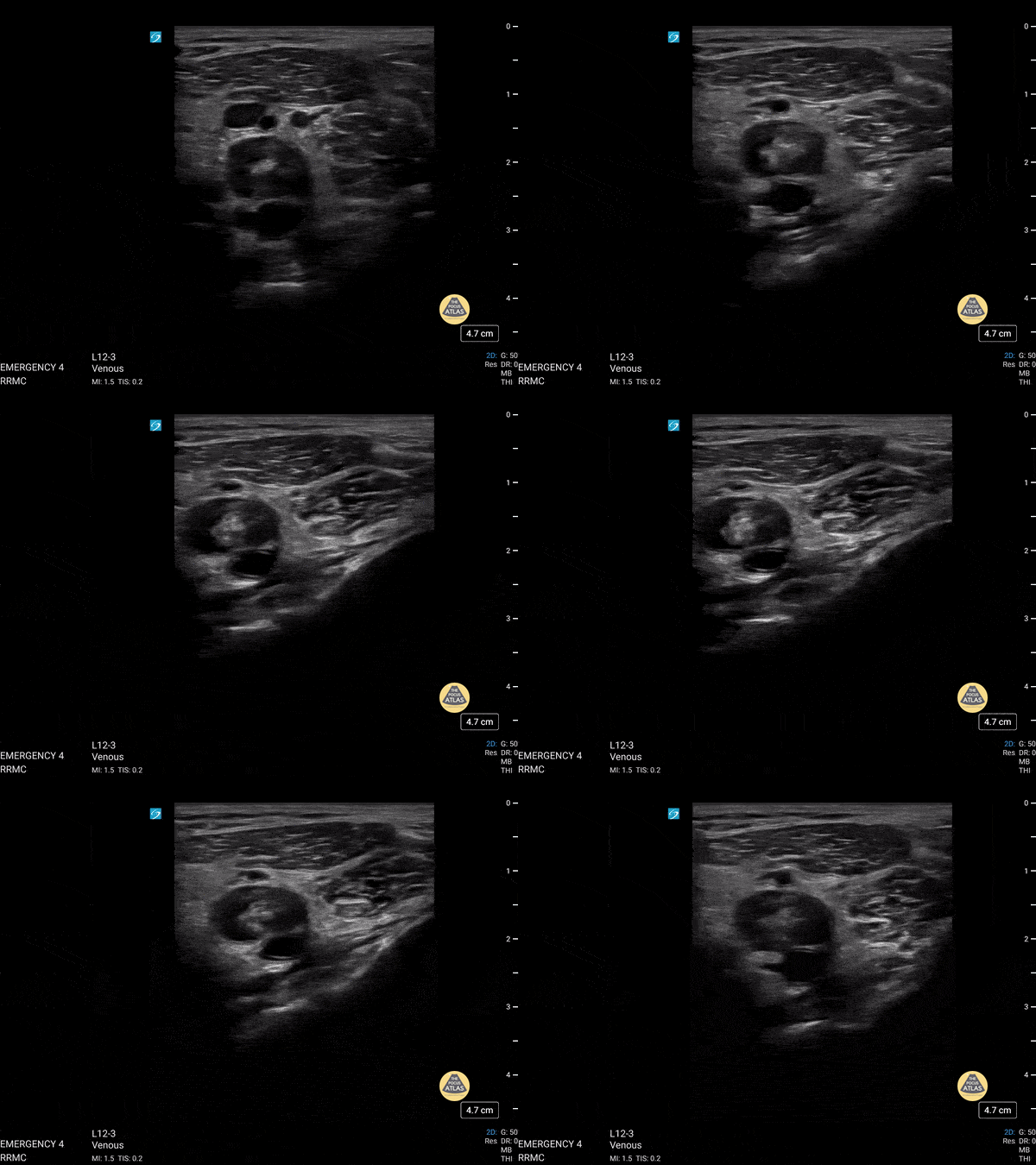
This is of a 52 year old male who presented to the emergency department complaining of leg pain and erythema while at work. He was evaluated prior to his arrival to the emergency department and was noted to have an elevated d-dimer level. Point of care ultrasound was performed starting from the great saphenous vein and common femoral vein, descending along his femoral vein. Initial compression did not reveal any thrombus until arriving at his popliteal vein which revealed a non-compressible vein due to a large thrombus.
Dr. Christopher Paulo, DO, PGY-1
Riverside Regional Medical Center Emergency Medicine Program (Newport News, VA)
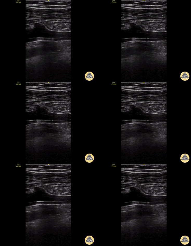
55 y/o man with septic shock and multiorgan failure due to SBP and liver failure. Picc line visualized within the R subclavian vein. longitudinal view.
Alex Steinberg
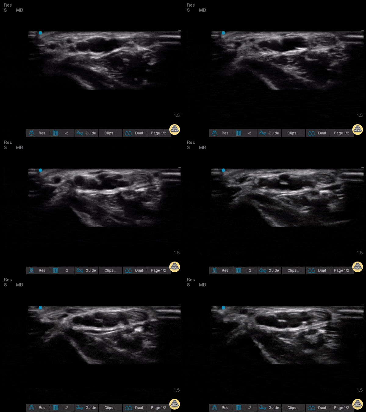
Very interesting find while performing a wrist ultrasound where we see the presence of two radial arteries
Image courtesy of Robert Jones DO, FACEP @RJonesSonoEM
Director, Emergency Ultrasound; MetroHealth Medical Center; Professor, Case Western Reserve Medical School, Cleveland, OH
View his original post here
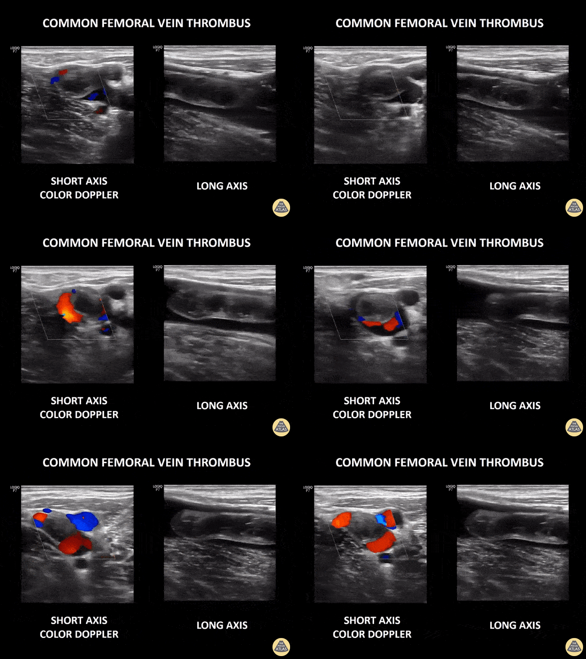
Elderly patient with a history of dyspnea and desaturation. POCUS of the common femoral vein in its transverse axes with color Doppler and longitudinal axes, showing partially mobile hyperechoic content in its interior associated with the absence of venous compressibility.
Paula Martins, @paula_smrtns
Medical Student at The Faculdade de Medicina de Marília
Normal Anatomy
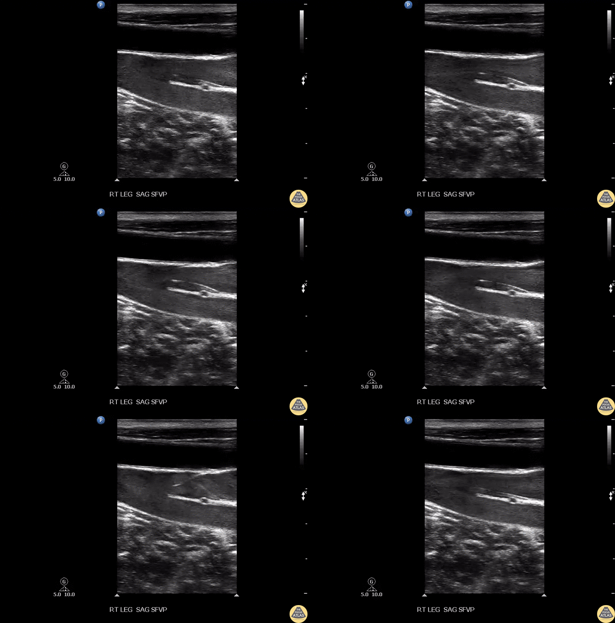
Seen here is healthy appearing venous system with effectively functioning valves. Important distinction between the arterial and venous systems.
Image courtesy of Robert Jones DO, FACEP @RJonesSonoEM
Director, Emergency Ultrasound; MetroHealth Medical Center; Professor, Case Western Reserve Medical School, Cleveland, OH
View his original post here
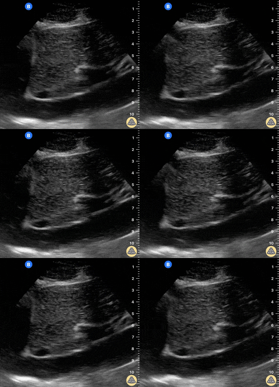
The IVC drains into the RA. A normal IVC frequently demonstrates transmitted pulsations from the RA. This clip is taken over the subxiphoid region, longitudinally to the body’s axis, and with the probe marker oriented to the patient’s head and RA. The hepatic vein can also be seen draining into the IVC.
Dr. Felipe Urriola P., Emergency Unit, Puerto Aysen Hospital, Chilean Patagonia.
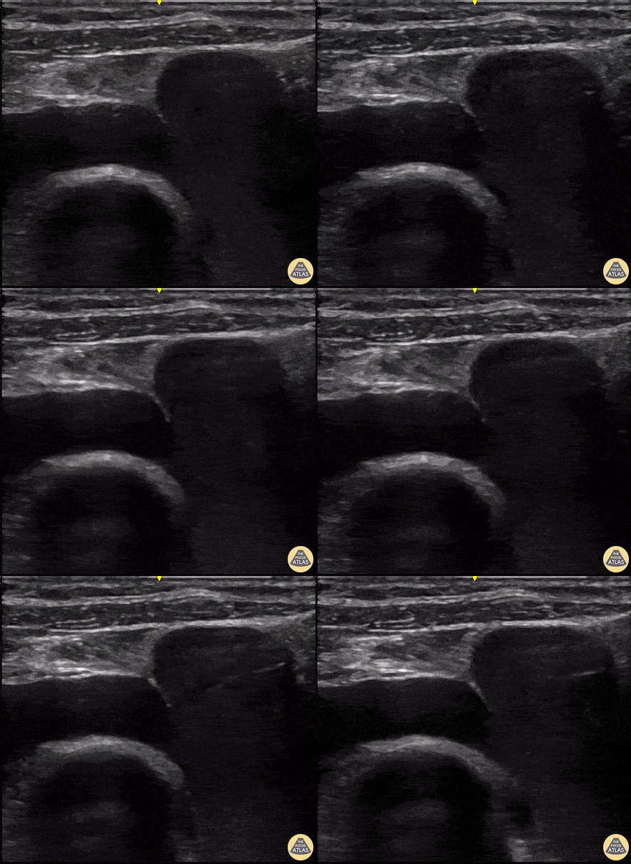
This POCUS revealss a supraclavicular long axis view of the subclavian vein. It is an ideal view for US-guided central venous access.
Renato Tambelli, mergency Physician Hospital das Clínicas de Marília, Brazil.
@R_Tambelli // @JediPocus
Colorized Normal Anatomy
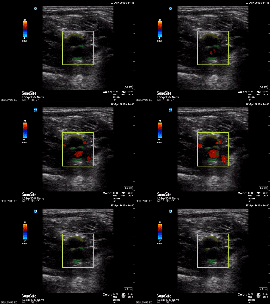
Popliteal vessels
Green: Popliteal artery, Yellow: Popliteal vein
Images: Dr. Lindsay Davis, Dr. Hannah Kopinski. Image Editing: Michael Amador and Dr. Matthew Riscinti
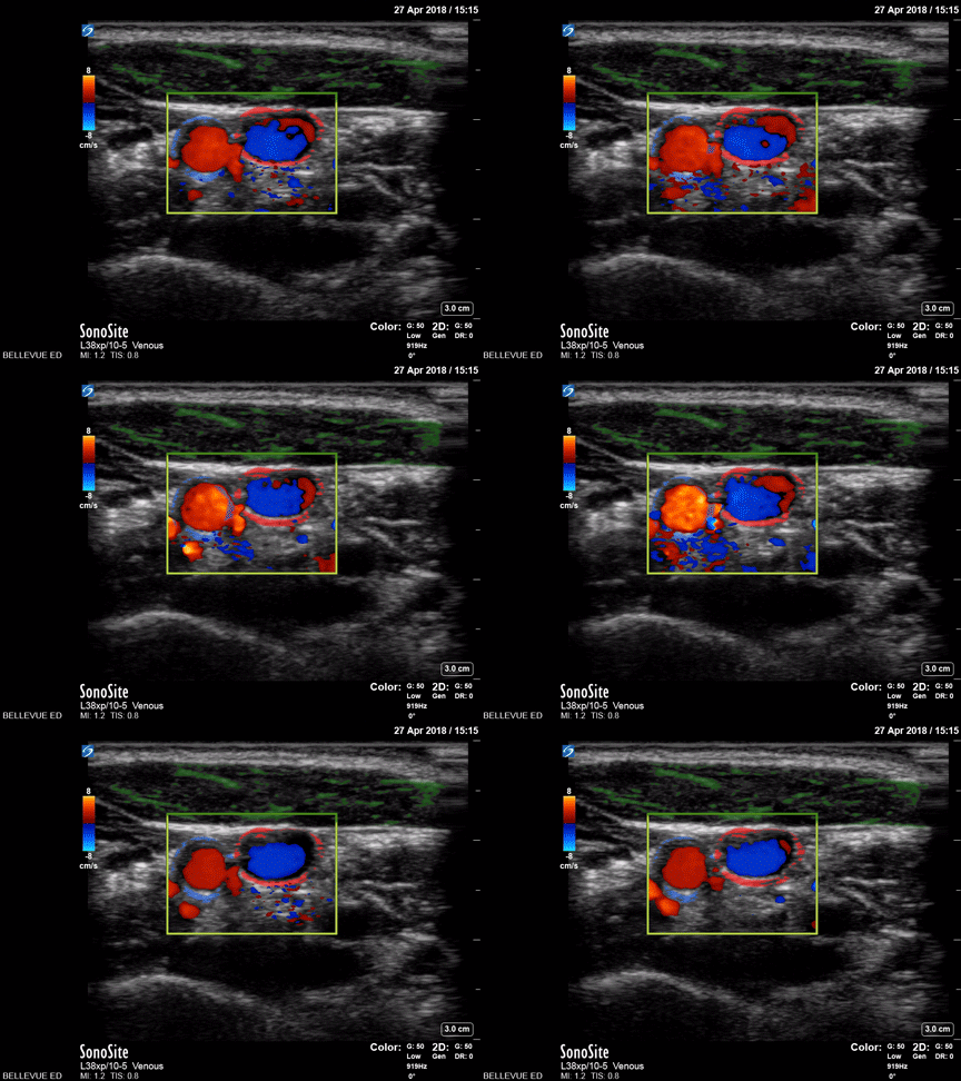
Carotid Artery and Internal Jugular Doppler
Green: Sternocleidomastoid, Blue outline: Common carotid artery, Red outline: Internal jugular vein
Images: Dr. Lindsay Davis, Dr. Hannah Kopinski. Image Editing: Michael Amador and Dr. Matthew Riscinti
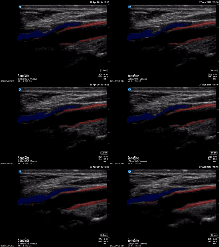
Carotid Bulb (long axis)
Red: Common carotid artery, Blue: Lumen of internal jugular vein
Images: Dr. Lindsay Davis, Dr. Hannah Kopinski. Image Editing: Michael Amador and Dr. Matthew Riscinti
Venous Thrombosis
This clip demonstrates a non-compressible left popliteal vein consistent with deep venous thrombosis.
Contributed by: Brittany Garza, DO; Saleem Nasseh, MD; Sadie Ellerson, MS4
This is of a 52 year old male who presented to the emergency department complaining of leg pain and erythema while at work. He was evaluated prior to his arrival to the emergency department and was noted to have an elevated d-dimer level. Point of care ultrasound was performed starting from the great saphenous vein and common femoral vein, descending along his femoral vein. Initial compression did not reveal any thrombus until arriving at his popliteal vein which revealed a non-compressible vein due to a large thrombus.
Dr. Christopher Paulo, DO, PGY-1
Riverside Regional Medical Center Emergency Medicine Program (Newport News, VA)
Elderly patient with a history of dyspnea and desaturation. POCUS of the common femoral vein in its transverse axes with color Doppler and longitudinal axes, showing partially mobile hyperechoic content in its interior associated with the absence of venous compressibility.
Paula Martins, @paula_smrtns
Medical Student at The Faculdade de Medicina de Marília
Other Pathology
Femoral artery stent can be appreciated in this short axis view as a bright, hyperechoic structure within the femoral artery borders.
Contributed by: Brittany Garza, DO; Saleem Nasseh, MD; Sadie Ellerson, MS4
55 y/o man with septic shock and multiorgan failure due to SBP and liver failure. Picc line visualized within the R subclavian vein. longitudinal view.
Alex Steinberg
Very interesting find while performing a wrist ultrasound where we see the presence of two radial arteries
Image courtesy of Robert Jones DO, FACEP @RJonesSonoEM
Director, Emergency Ultrasound; MetroHealth Medical Center; Professor, Case Western Reserve Medical School, Cleveland, OH
View his original post here