
Renal/GU
Renal/GU
Note: These search bar results are only for the Renal/GU image archive. To search the entire site, click here.
Recently Added
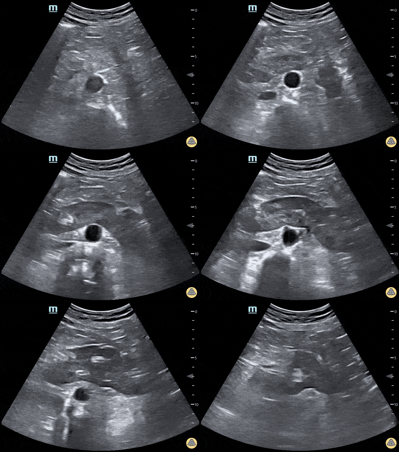
Horseshoe kidney. No kidney was identified in either flank area, this anterior abdominal view shows one large kidney that is anterior to the Aorta.
Contributed by: Marion Memmott, DO; Michael Bernard, DO; Central Michigan University Emergency Medicine Residency
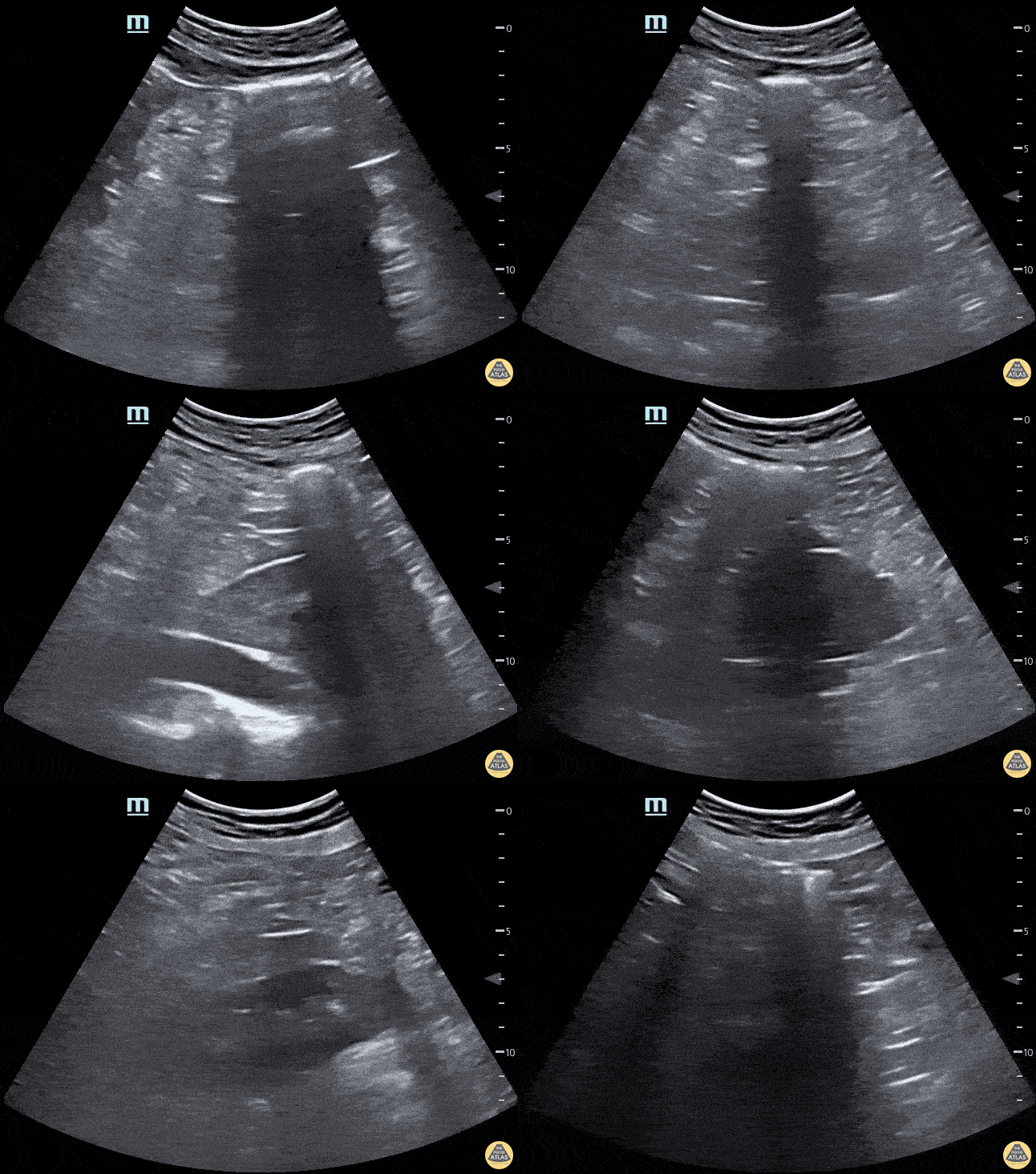
Horseshoe kidney. No kidney was identified in either flank area, this anterior abdominal view shows one large kidney that is anterior to the Aorta.
Marion Memmott, DO; Michael Bernard, DO; Central Michigan University Emergency Medicine Residency

Prominent prostate on ultrasound in a 72 year-old-male with known prostate carcinoma and recent PET scan with confirmation. No known masses or metastasis. This is seen in both transverse and sagittal views.
Lindsay Davis, DO, MPH, PGY3; Matthew Welch, MS4; Alex Schlangen, DO, PGY1
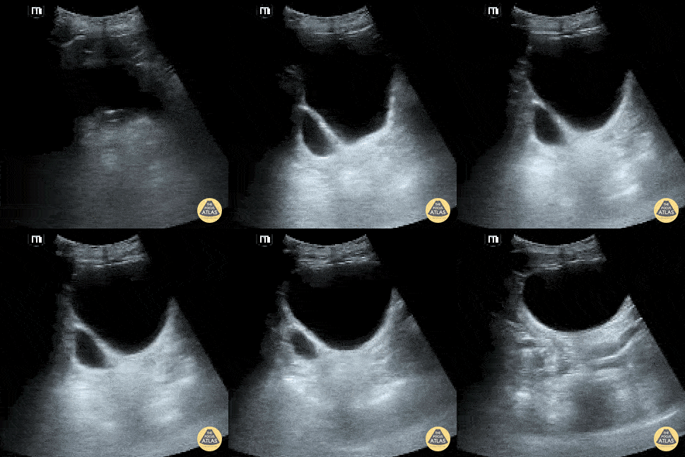
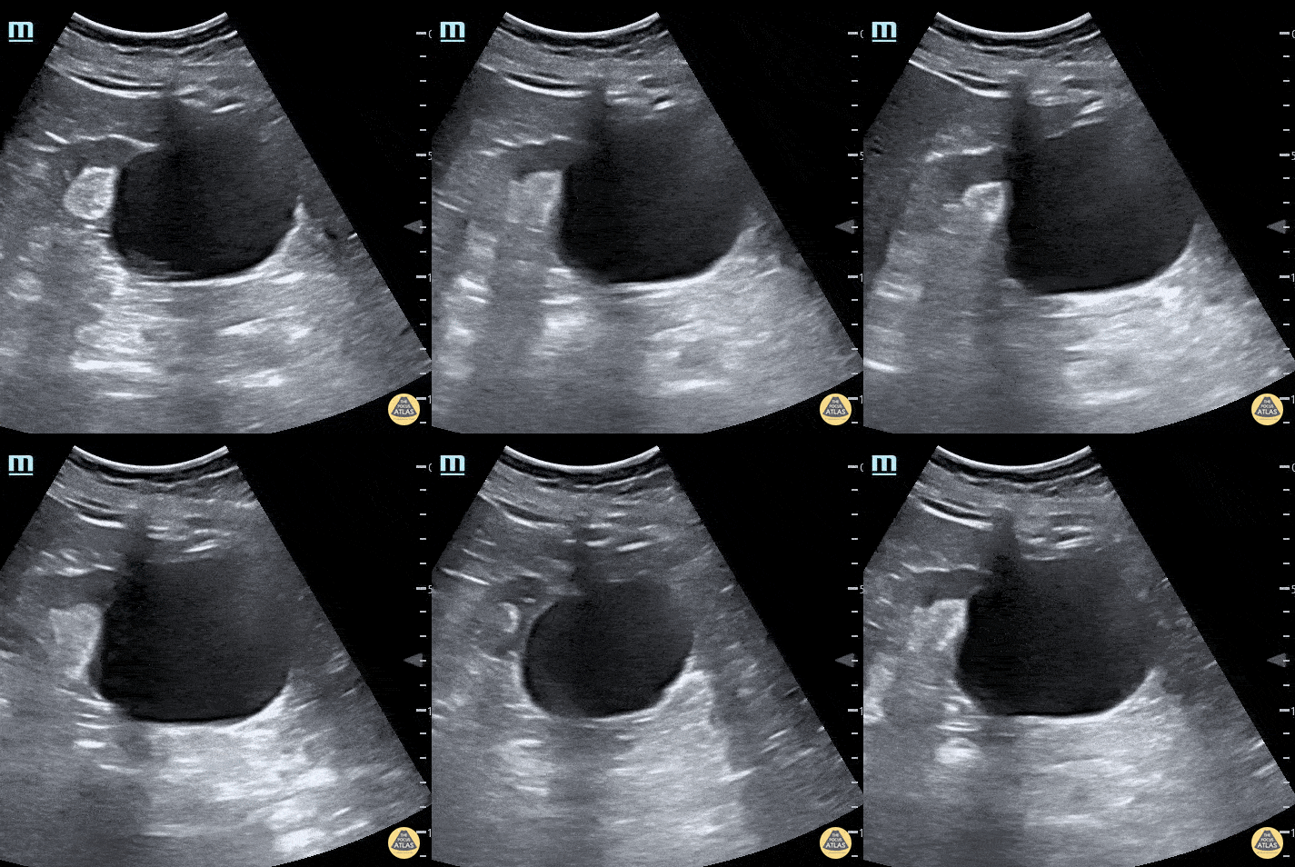
A bladder diverticulum is demonstrated deep to the bladder as the ultrasound probe is fanned.
Contributed by: Brittany Garza, DO; Saleem Nasseh, MD; Sadie Ellerson, MS4
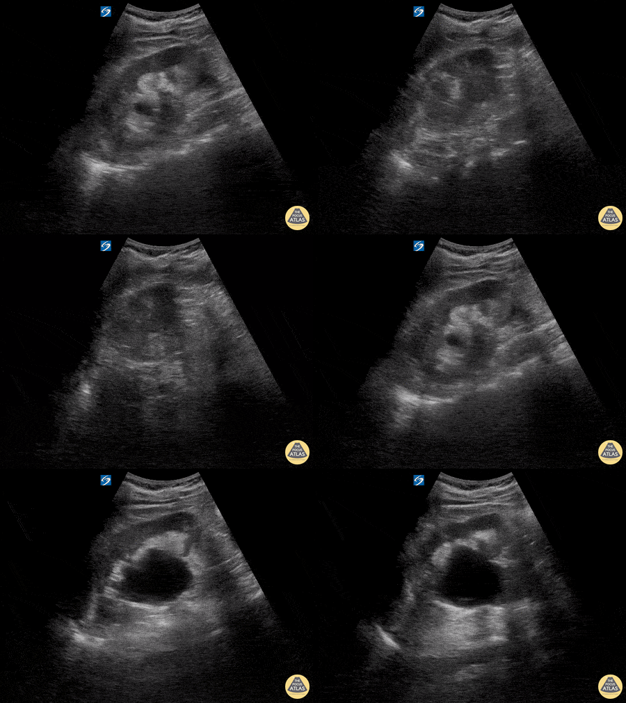
Here is an example of hydronephrosis in the presence of nephrolithasis. This patient presented to the emergency department complaining of new right flank pain after waking up in the morning. She was seen previously for urinary complaints, diagnosed with UTI and started on antibiotic regiment that had been altered twice due to concerns of sub-optimal coverage. She demonstrated right CVA tenderness on physical exam, raising suspicion for pyelonephritis as well as hydronephrosis. Subsequent point of care ultrasound revealed moderate hydronephrosis. CT was obtained to further evaluate and confirmed the presence of an obstructing kidney stone. Note presence of hydronephrosis as the probe is fanned/tilted through the kidney, highlighting the importance of scanning through the entire organ.
Dr. Carlo Zamora, DO, PGY-1
Riverside Regional Medical Center Emergency Medicine Residency (Newport News, VA)

A young female presented with acute RLQ abdominal pain and found to have a 5mm right ureter stone at the UVJ with ureteral dilation. The patient also had moderate hydronephrosis.
Matthew Petruso, DO; Harpreet Grewal, MD; Eric Versluis, MS4; Central Michigan University: Emergency Medicine Residency.
Normal Anatomy
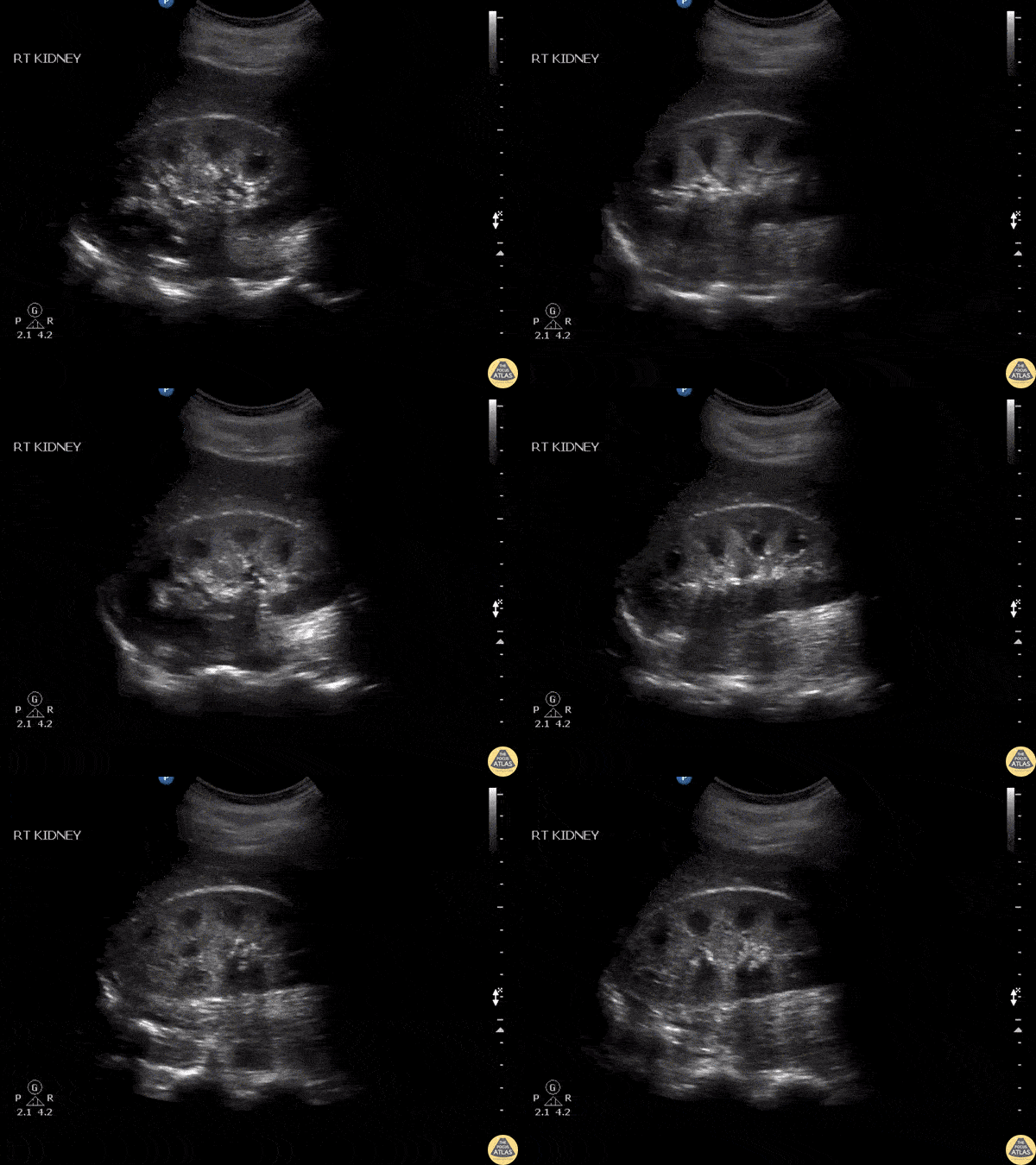
A patient was being evaluated for flank pain. This clip shows findings of normal but prominent medullary pyramids as indicated by how they taper down toward the renal pelvis.
Image courtesy of Robert Jones DO, FACEP @RJonesSonoEM
Director, Emergency Ultrasound; MetroHealth Medical Center; Professor, Case Western Reserve Medical School, Cleveland, OH
View his original post here
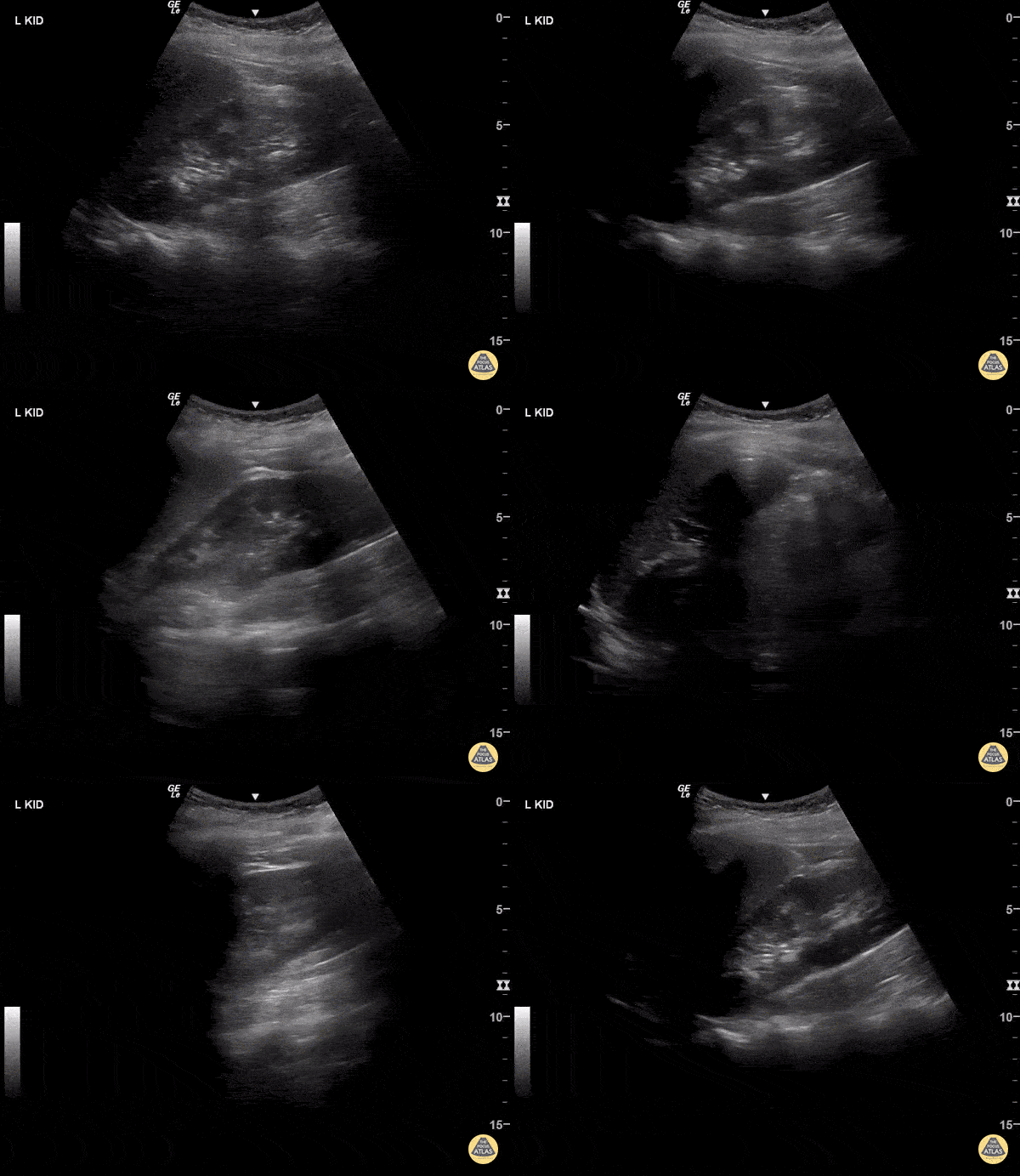

Normal kidney - No hydro.
One of the common pitfalls in identifying hydronephrosis is not using color flow. You must use color flow doppler to ensure that what you're looking at is actually hydronephrosis, not simply vasculature.
Dr. Bryan Jarret - Kings County Emergency Medicine
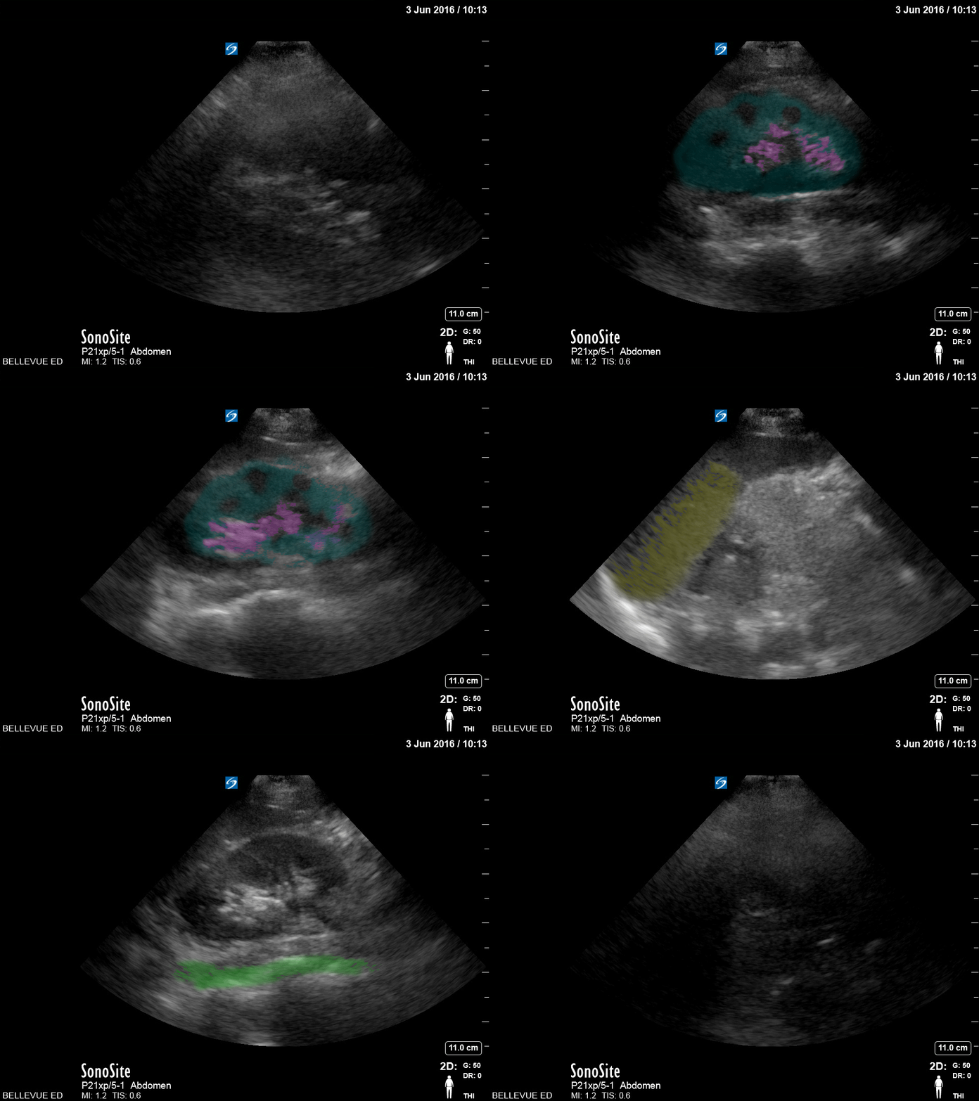
Colorized Normal Anatomy
Hydronephrosis
Here is an example of hydronephrosis in the presence of nephrolithasis. This patient presented to the emergency department complaining of new right flank pain after waking up in the morning. She was seen previously for urinary complaints, diagnosed with UTI and started on antibiotic regiment that had been altered twice due to concerns of sub-optimal coverage. She demonstrated right CVA tenderness on physical exam, raising suspicion for pyelonephritis as well as hydronephrosis. Subsequent point of care ultrasound revealed moderate hydronephrosis. CT was obtained to further evaluate and confirmed the presence of an obstructing kidney stone. Note presence of hydronephrosis as the probe is fanned/tilted through the kidney, highlighting the importance of scanning through the entire organ.
Dr. Carlo Zamora, DO, PGY-1
Riverside Regional Medical Center Emergency Medicine Residency (Newport News, VA)
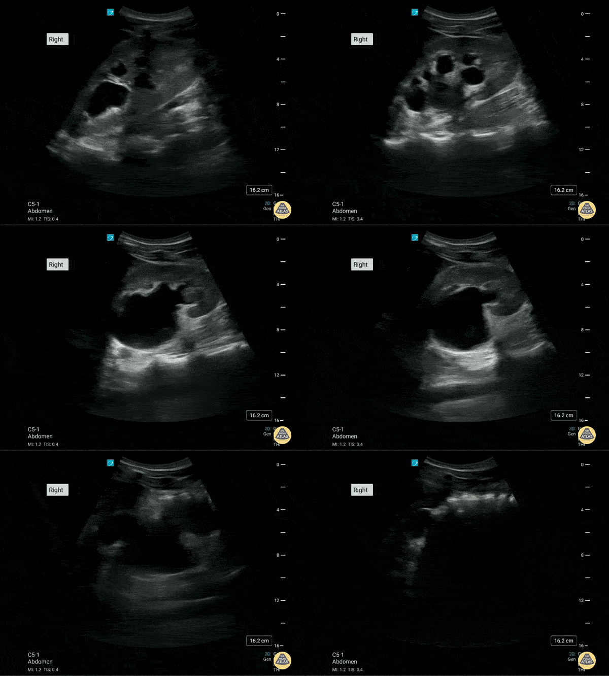
This is the right renal ultrasound of a 20 year old male with a past medical history of recurrent obstructing nephrolithiasis. There is a moderate to severe hydronephrosis present. with what appears to be some thinning of the renal cortex.
Brittany Ladson, DO; Sara Schumbach, MD
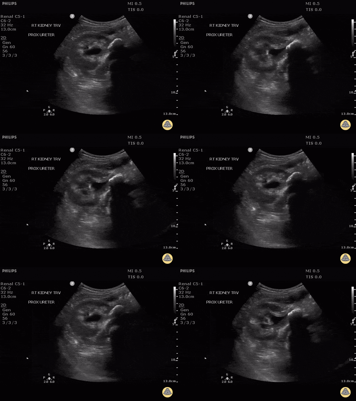
This patient presented with fever and flank pain with a known history of medullary nephrocalcinosis. Here we can see internal echoes within the dilated collecting system consistent with pyonephrosis. A large stone can be seen in the proximal ureter as a hyperechoic line with posterior shadowing.
Image courtesy of Robert Jones DO, FACEP @RJonesSonoEM
Director, Emergency Ultrasound; MetroHealth Medical Center; Professor, Case Western Reserve Medical School, Cleveland, OH
View his original post here
Urinary Tract Calculi
A young female presented with acute RLQ abdominal pain and found to have a 5mm right ureter stone at the UVJ with ureteral dilation. The patient also had moderate hydronephrosis.
Matthew Petruso, DO; Harpreet Grewal, MD; Eric Versluis, MS4; Central Michigan University: Emergency Medicine Residency.
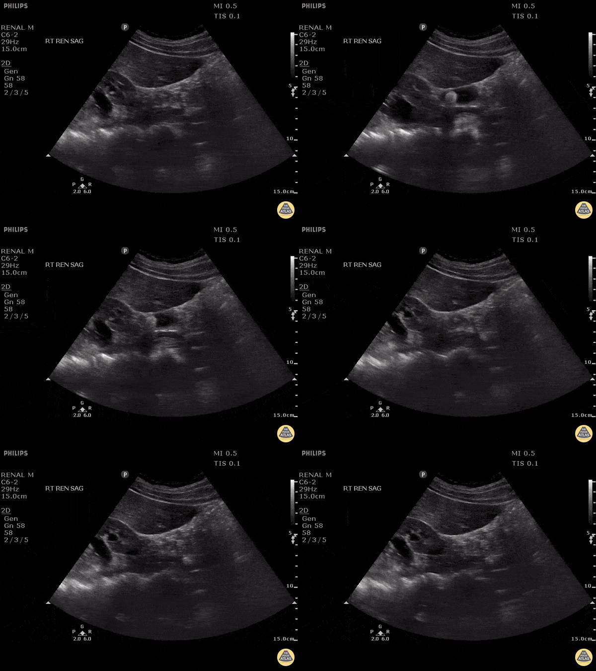
Ultrasound clip demonstrating both cholelithiasis as well as obstructing stone in the ureter!
Image courtesy of Robert Jones DO, FACEP @RJonesSonoEM
Director, Emergency Ultrasound; MetroHealth Medical Center; Professor, Case Western Reserve Medical School, Cleveland, OH
View his original post here.
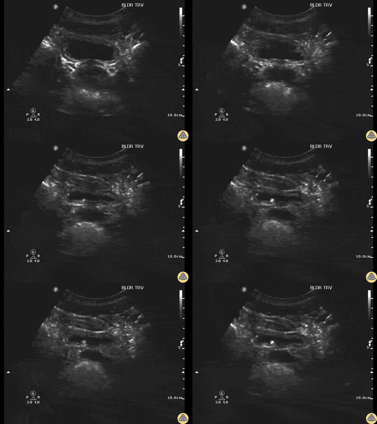
Patient with flank pain was found to have a 3mm unilateral obstructing ureteric stone without hydronephrosis.
Image courtesy of Robert Jones DO, FACEP @RJonesSonoEM
Director, Emergency Ultrasound; MetroHealth Medical Center; Professor, Case Western Reserve Medical School, Cleveland, OH
View his original post here
Renal Cysts
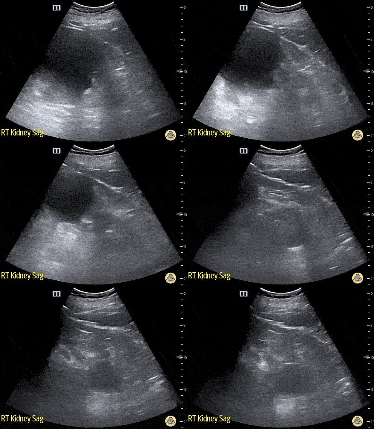
83 year old male presented to the ED for right flank pain.
The renal ultrasound showed two right renal cysts. The superior renal pole cyst measuring 12 x 13cm. The inferior pole cyst measuring 4 x 5cm.
Laura Schroeder, MS, MD; Syeda Zehra, MD; Conner Shea (Medical Student) Central Michigan University Emergency Medicine Residency
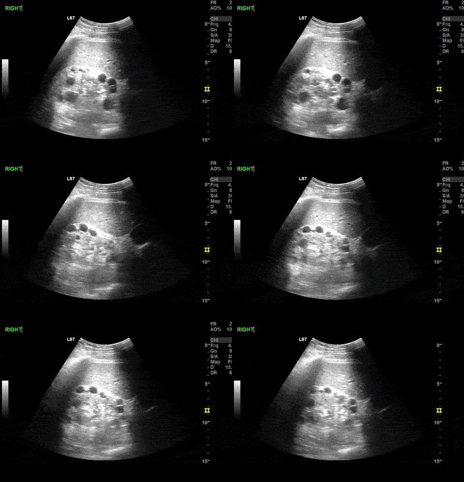
A 12 year old male presented after a MVC. A FAST exam was performed demonstrating incidental finding of Polycystic Kidney Disease (PCKD).
Paul Khalil, MD @Khalil3Paul
Assistant PEM POCUS director at University of Louisville/Norton Children’s
Bladder Pathology
A bladder diverticulum is demonstrated deep to the bladder as the ultrasound probe is fanned.
Contributed by: Brittany Garza, DO; Saleem Nasseh, MD; Sadie Ellerson, MS4
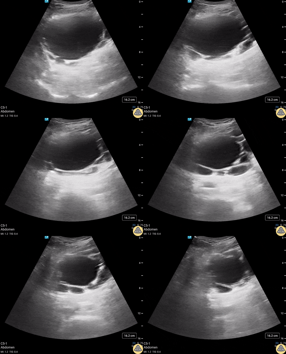
82 year old male with history of subarachnoid bleed presented for evaluation after a fall. During a EFAST exam, bladder US revealed multiple, anechoic, cystic-appearing structures consistent with bladder diverticula (confirmed by CT).
A bladder diverticulum is a protrusion of the mucosa through the muscular wall of the bladder and can be congenital or acquired. Acquired diverticula are more common, with bladder outlet obstruction being the most common cause. Urology follow up is recommended to confirm the cause and screen for other concerning etiologies or complications including carcinoma, recurrent UTIs and bladder calculi.
Contributors: Michael Huber, 4th year Medical Student; Chad Bambrick, MD; Ana Camagay, MD
Central Michigan University College of Medicine
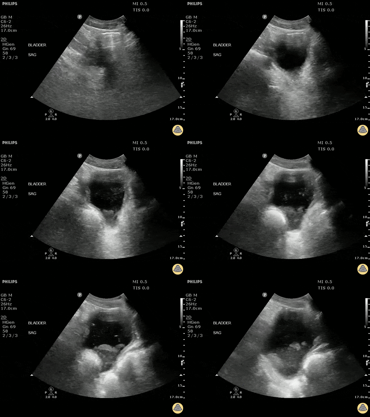
This sagittal image of the bladder demonstrates air within the bladder. This could be from emphysematous cystitis but could also be iatrogenic due to recent foley placement. Also note the presence of layered echoes in the bladder due to an infectious process.
Image courtesy of Robert Jones DO, FACEP @RJonesSonoEM
Director, Emergency Ultrasound; MetroHealth Medical Center; Professor, Case Western Reserve Medical School, Cleveland, OH
View his original post here
Scrotal Pathology
Other Pathology
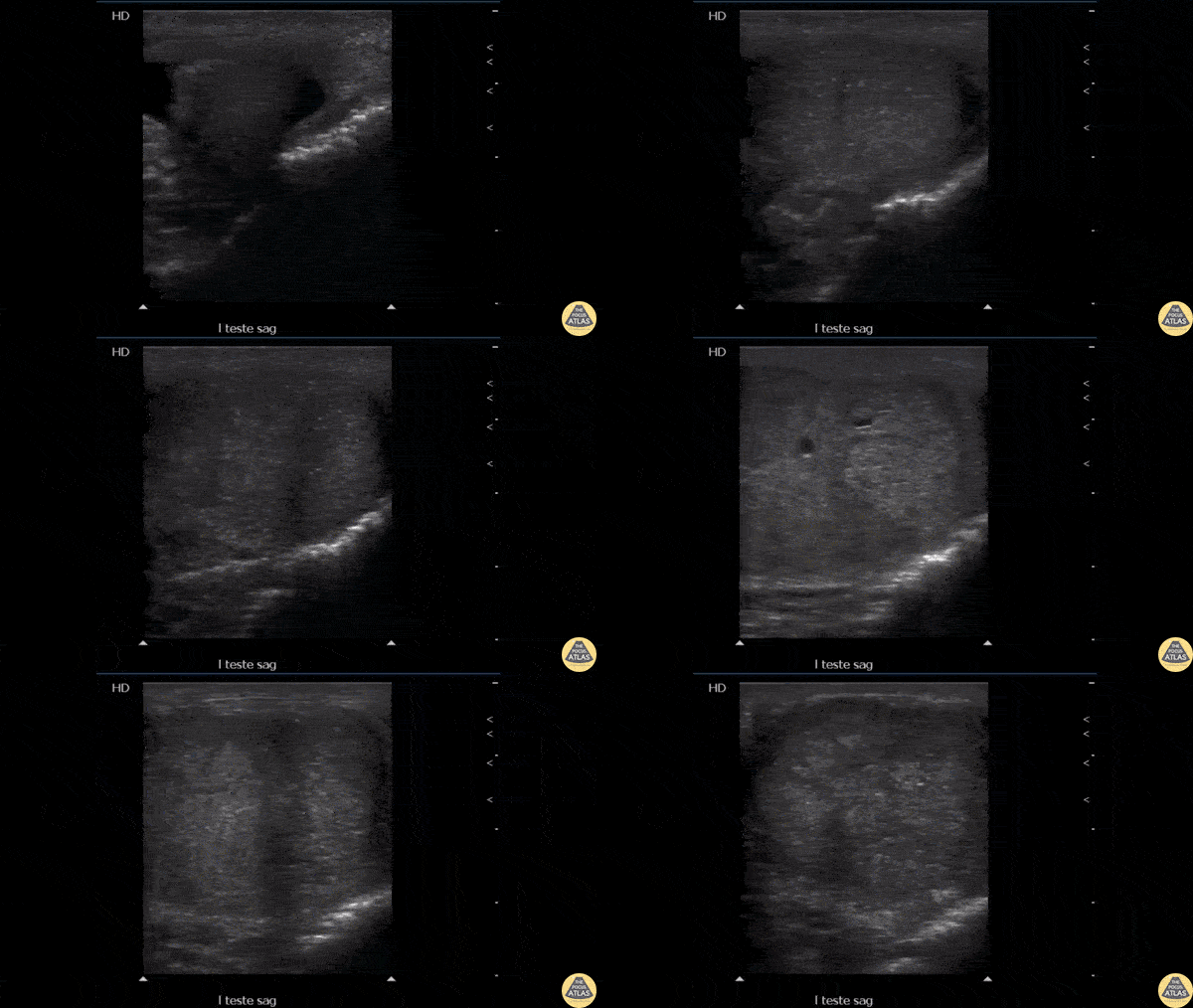
Testicular ultrasound revealed microcalcifications diffusely within the testes presenting with testicular pain. These findings can be associated with germ cell tumors.
Image courtesy of Robert Jones DO, FACEP @RJonesSonoEM
Director, Emergency Ultrasound; MetroHealth Medical Center; Professor, Case Western Reserve Medical School, Cleveland, OH
View his original post here
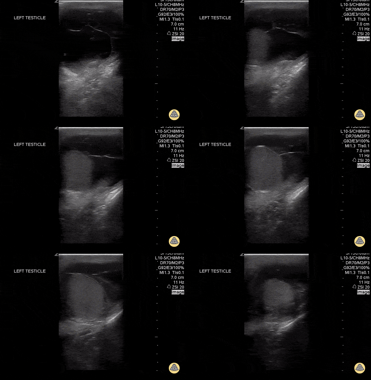
A 30 year old sexually active male with no previous medical history presented to the emergency department with testicular pain. POC US demonstrates a septated hydrocele.
Mario Corro, MD, PGY-3 Staten Island University Hospital
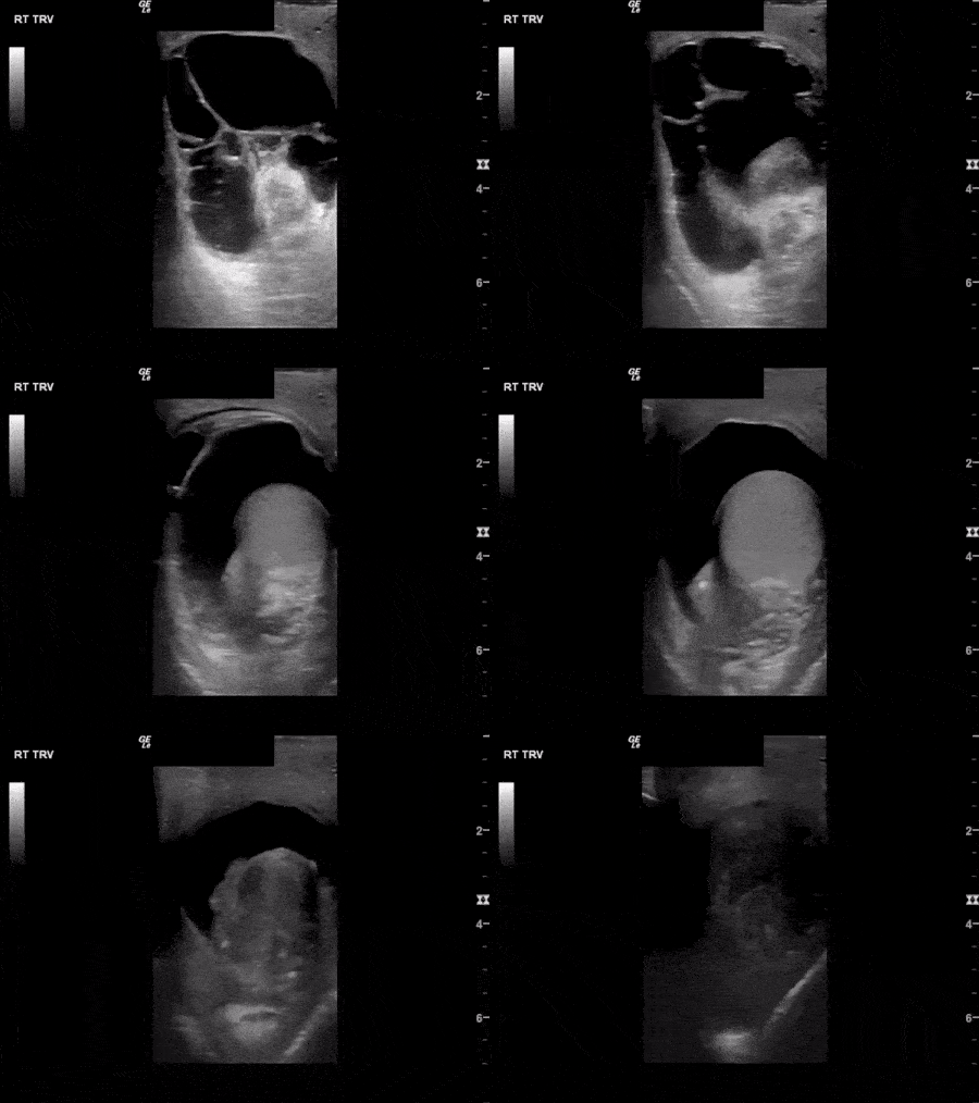
This image demonstrates fluid filling the scrotal sac with multiple thin septations consistent with a complex hydrocele. In the setting of epididymitis, a pyocele should be considered.
Image courtesy of Aventura Ultrasound
See Original Post Via their Twiiter: @AventuraEUS
Horseshoe kidney. No kidney was identified in either flank area, this anterior abdominal view shows one large kidney that is anterior to the Aorta.
Contributed by: Marion Memmott, DO; Michael Bernard, DO; Central Michigan University Emergency Medicine Residency
Horseshoe kidney. No kidney was identified in either flank area, this anterior abdominal view shows one large kidney that is anterior to the Aorta.
Marion Memmott, DO; Michael Bernard, DO; Central Michigan University Emergency Medicine Residency
Prominent prostate on ultrasound in a 72 year-old-male with known prostate carcinoma and recent PET scan with confirmation. No known masses or metastasis. This is seen in both transverse and sagittal views.
Lindsay Davis, DO, MPH, PGY3; Matthew Welch, MS4; Alex Schlangen, DO, PGY1