
Soft Tissue
Soft Tissue
Note: These search bar results are only for the Soft Tissue image archive. To search the entire site, click here.
Recently Added
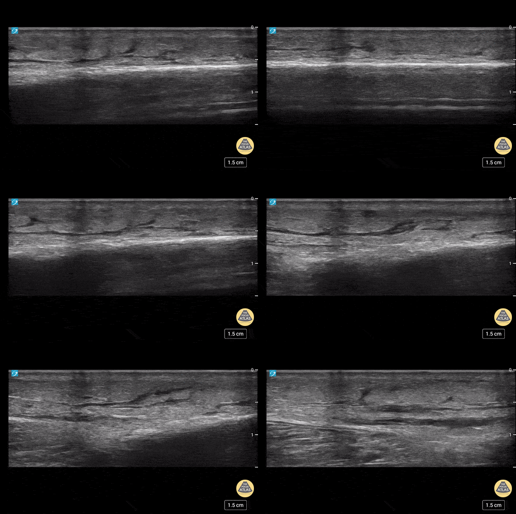
This is an example of “cobblestoning”. This patient initially presented to the emergency department with lower anterior leg pain, especially with walking. Physical exam revealed minor erythema but also exquisite tenderness to palpation as well as with plantar flexion. Although cellulitis is clinically diagnosed, point of care ultrasound can be used to support it. The cobblestone presence is a result of edema between subcutaneous fat.
Dr. Carlo Zamora, DO, PGY-1
Riverside Regional Medical Center Emergency Medicine Residency (Newport News, VA)
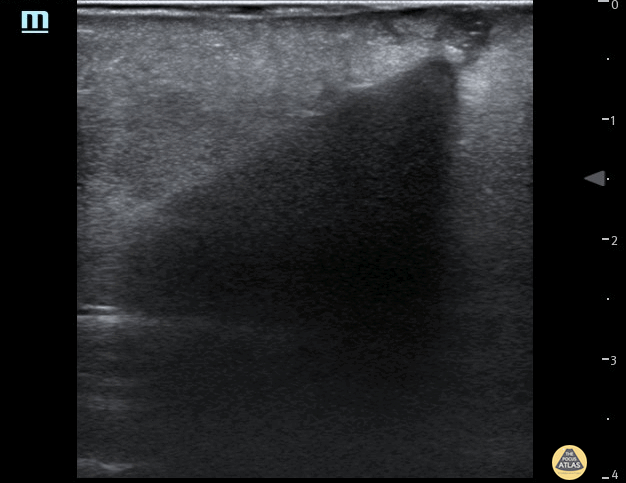
35 year female presented to the Emergncy Department with a self harm retained foreign body in the left forearm. The foreign body was a wooden cocktail stick not seen on x-ray. POCUS easily identified the cocktail stick by it's acoustic shadowing which allowed removal under local anaesthetic. Repeat POCUS scan showed no other foreign bodies.
Dr Pete Hulme @Dr_Pete_EmMed
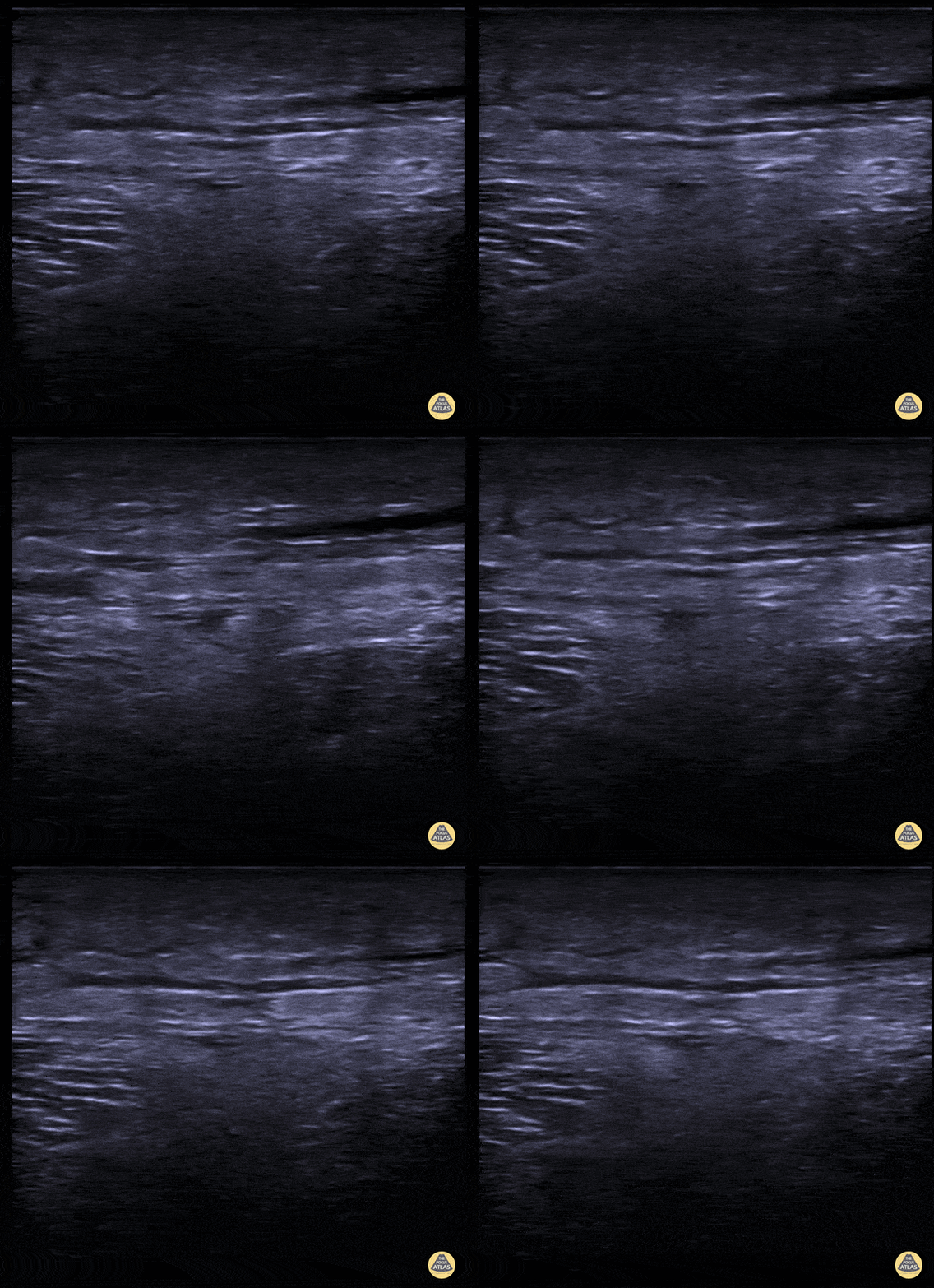
A middle aged male presented to the ED for left leg pain after sustaining puncture wound to the foot 4 hours prior arrival. The ultrasound image demonstrates free air causing dirty shadowing seen deep to the fascial plane obscuring the normal muscle architecture in the middle of the screen.
Nicmarie Maldonado, MD PGY-2; Eddie G. Rodriguez, MD; Miguel Agrait, MD; Michelle Surillo, MD - Division of Emergency Ultrasound, St. Luke's Medical Center/PHSU, Ponce, PR
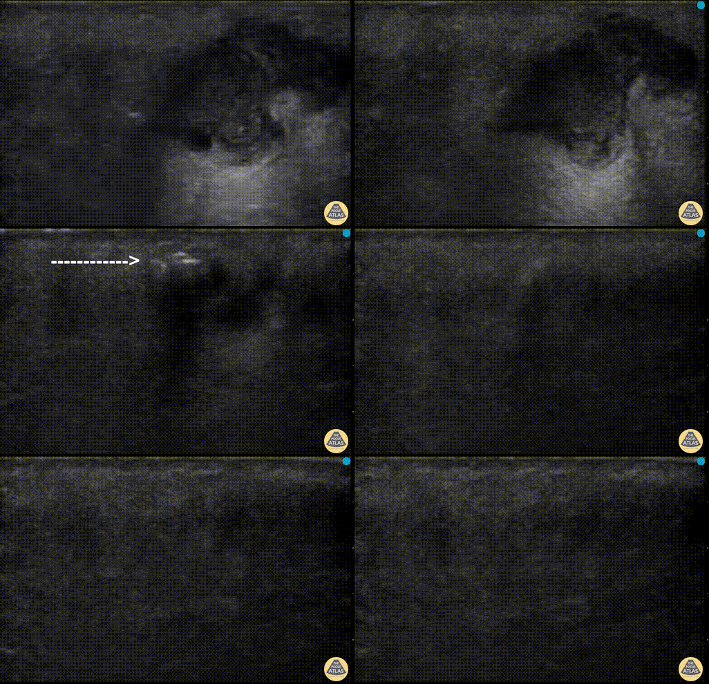
POCUS can help distinguish simple abscess from necrotizing fasciitis. “Dirty air shadowing” visualized in the clip suggests a necrotizing soft tissue infection.
Dr. Farnam Kazi Ultrasound Faculty; Dr. Dimitri Livshits Ultrasound Fellow; Dr. Jane Belyavskaya Ultrasound Fellow (All at Kings County Hospital/SUNY Downstate)
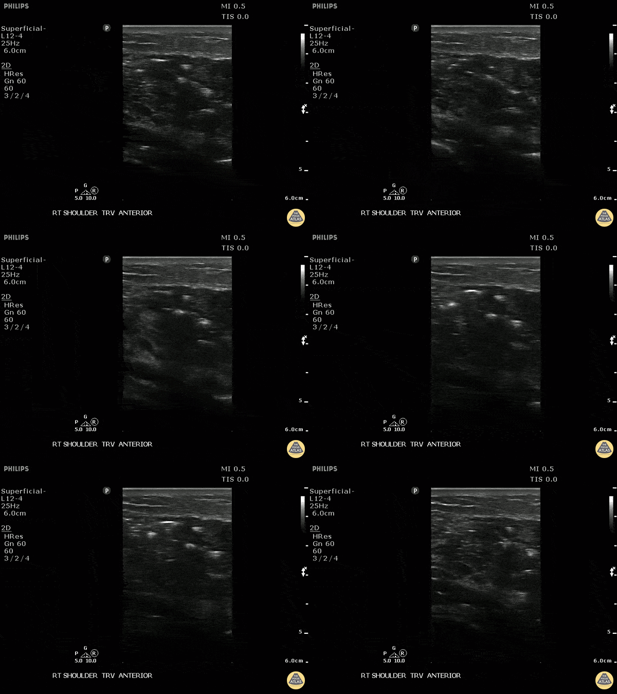
This is a transverse view of the anterior shoulder of a patient who presented to the emergency department with a fever and shoulder pain causing limited mobility. Looking closer, we can observe hyperechoic densities caused by air within the tissues, otherwise no signs of septic joint or bursitis.
Image courtesy of Robert Jones DO, FACEP @RJonesSonoEM
Director, Emergency Ultrasound; MetroHealth Medical Center; Professor, Case Western Reserve Medical School, Cleveland, OH
View his original post here
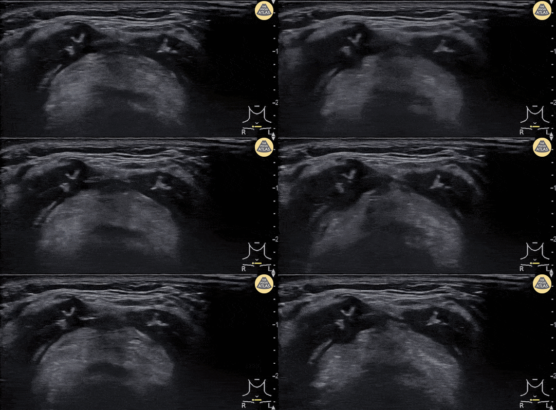
A 30 year-old female presented to the emergency department with high fever, severe sore throat and shortness of breath. POCUS of the neck was performed and revealed a swollen epiglottis, especially in the right side. Further X-ray of the neck revealed thumb sign and nasopharyngoscopy showed epiglottitis with swelling of right tongue base and arytenoid.
Although the normal range of the epiglottis thickness measured by ultrasound varied from 2 to 3 mm in previous studies, significant swelling and thickening of the epiglottis on ultrasound may indicate epiglottitis.
Chi-Hsin Chen
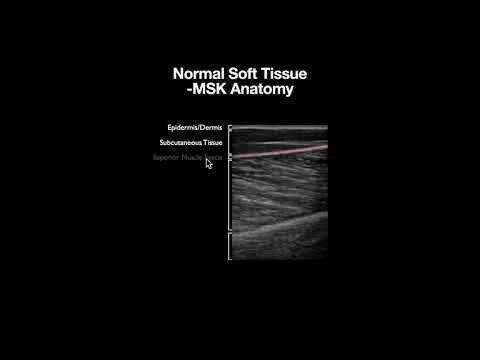
Normal Soft Tissue Anatomy

Abscess

This is an example of a large soft tissue abscess. Note the presence of a large well circumscribed fluid collection taking up the screen. Particulate can be seen throughout the fluid collection signifying increased density from purulent material.
Michael Macias, MD
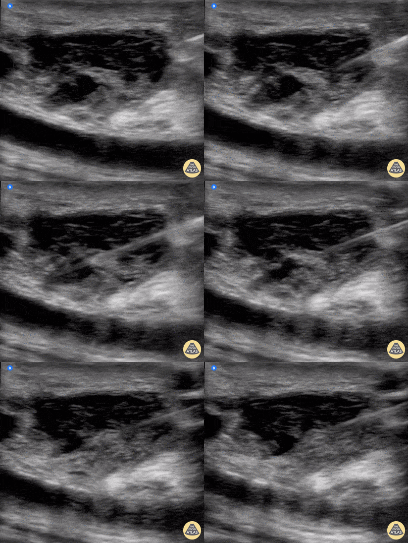
Needle aspiration of a fluid collection superficial to a femoropopliteal bypass.
Wolfgang Geisser, @fentanyl05
Bayern, Deutschland
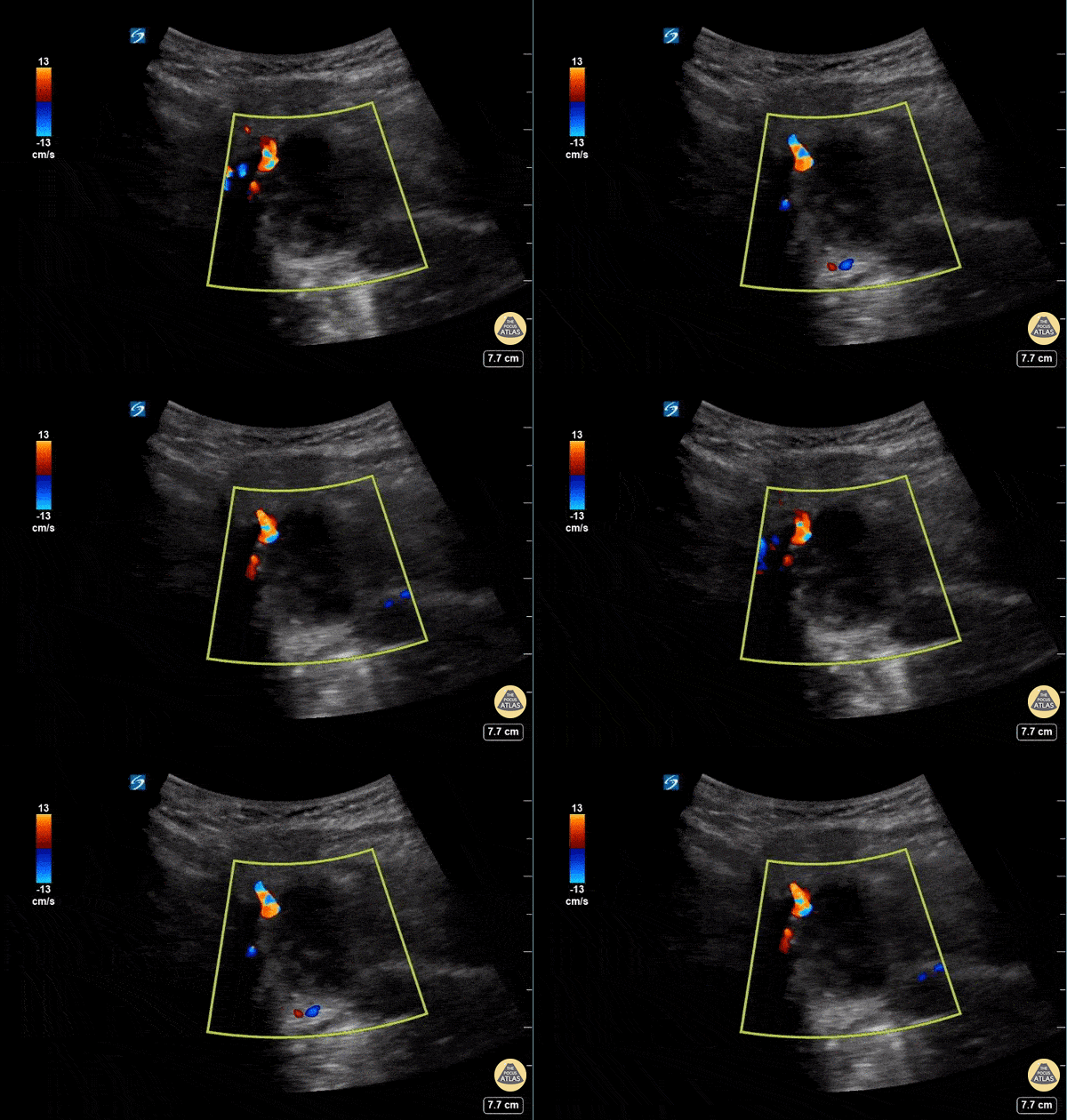
This is a submandibular view of a peritonsillar abscess. Color doppler is used to visualize blood vessels immediately deep to the abscess. By convention, the left side of the screen is posterior and the right side of the screen is anterior.
Garrett Ghent, Resuscitationist/Diagnostician; Norfolk, VA
@garrettghentMD
Cellulitis
This is an example of “cobblestoning”. This patient initially presented to the emergency department with lower anterior leg pain, especially with walking. Physical exam revealed minor erythema but also exquisite tenderness to palpation as well as with plantar flexion. Although cellulitis is clinically diagnosed, point of care ultrasound can be used to support it. The cobblestone presence is a result of edema between subcutaneous fat.
Dr. Carlo Zamora, DO, PGY-1
Riverside Regional Medical Center Emergency Medicine Residency (Newport News, VA)
A middle aged male presented to the ED for left leg pain after sustaining puncture wound to the foot 4 hours prior arrival. The ultrasound image demonstrates free air causing dirty shadowing seen deep to the fascial plane obscuring the normal muscle architecture in the middle of the screen.
Nicmarie Maldonado, MD PGY-2; Eddie G. Rodriguez, MD; Miguel Agrait, MD; Michelle Surillo, MD - Division of Emergency Ultrasound, St. Luke's Medical Center/PHSU, Ponce, PR
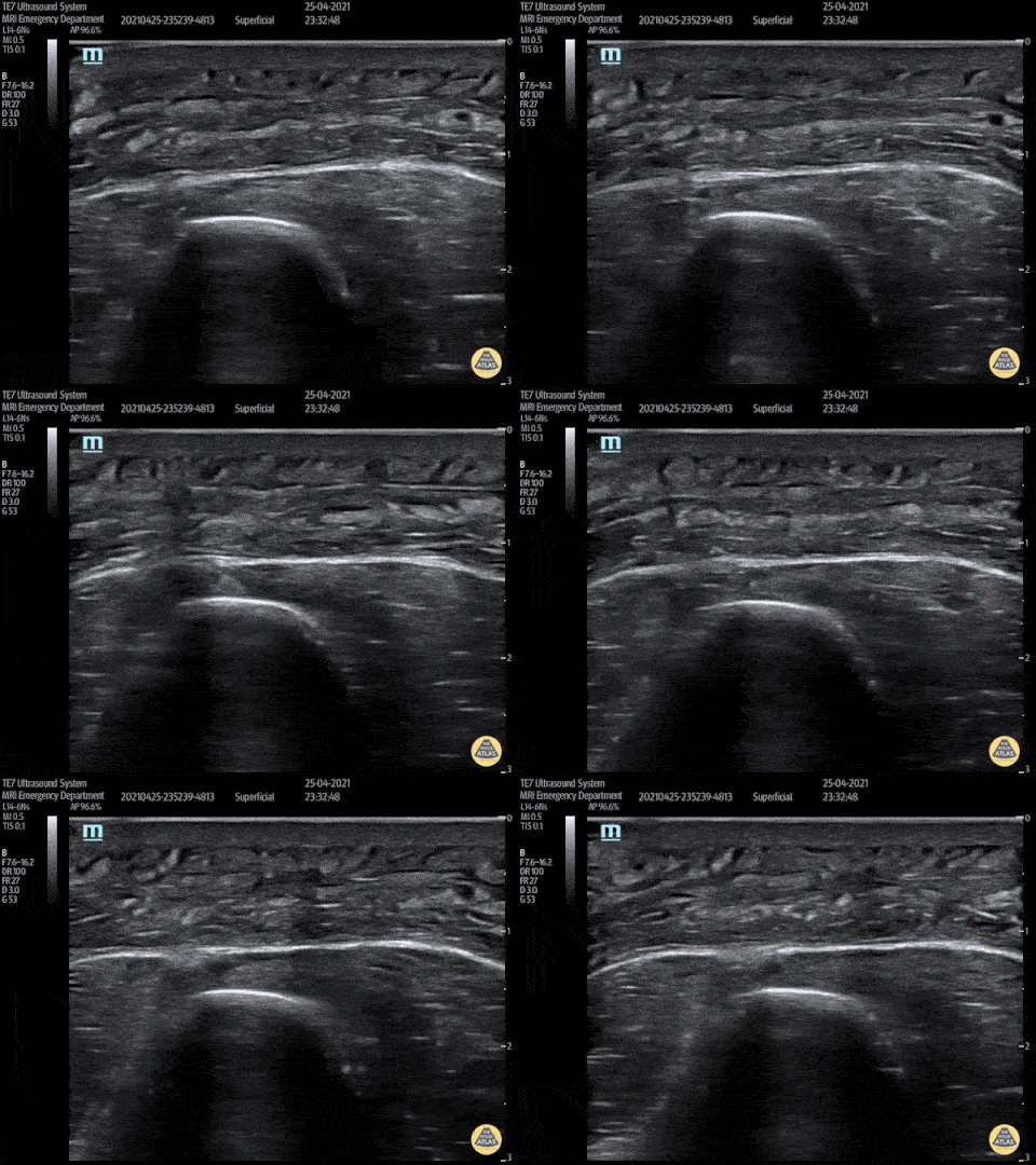
This video shows a young man who presented 3 days after being stabbed to the arm. His wound had been sutured at the time but he had represented with erythema, pain and swelling to his forearm. POCUS showed cobblestoning consisent with cellulitis and excluded abscess and a foreign body.
Peter Hulme
Twitter: @Dr_Pete_EmMed
Foreign Body
35 year female presented to the Emergncy Department with a self harm retained foreign body in the left forearm. The foreign body was a wooden cocktail stick not seen on x-ray. POCUS easily identified the cocktail stick by it's acoustic shadowing which allowed removal under local anaesthetic. Repeat POCUS scan showed no other foreign bodies.
Dr Pete Hulme @Dr_Pete_EmMed
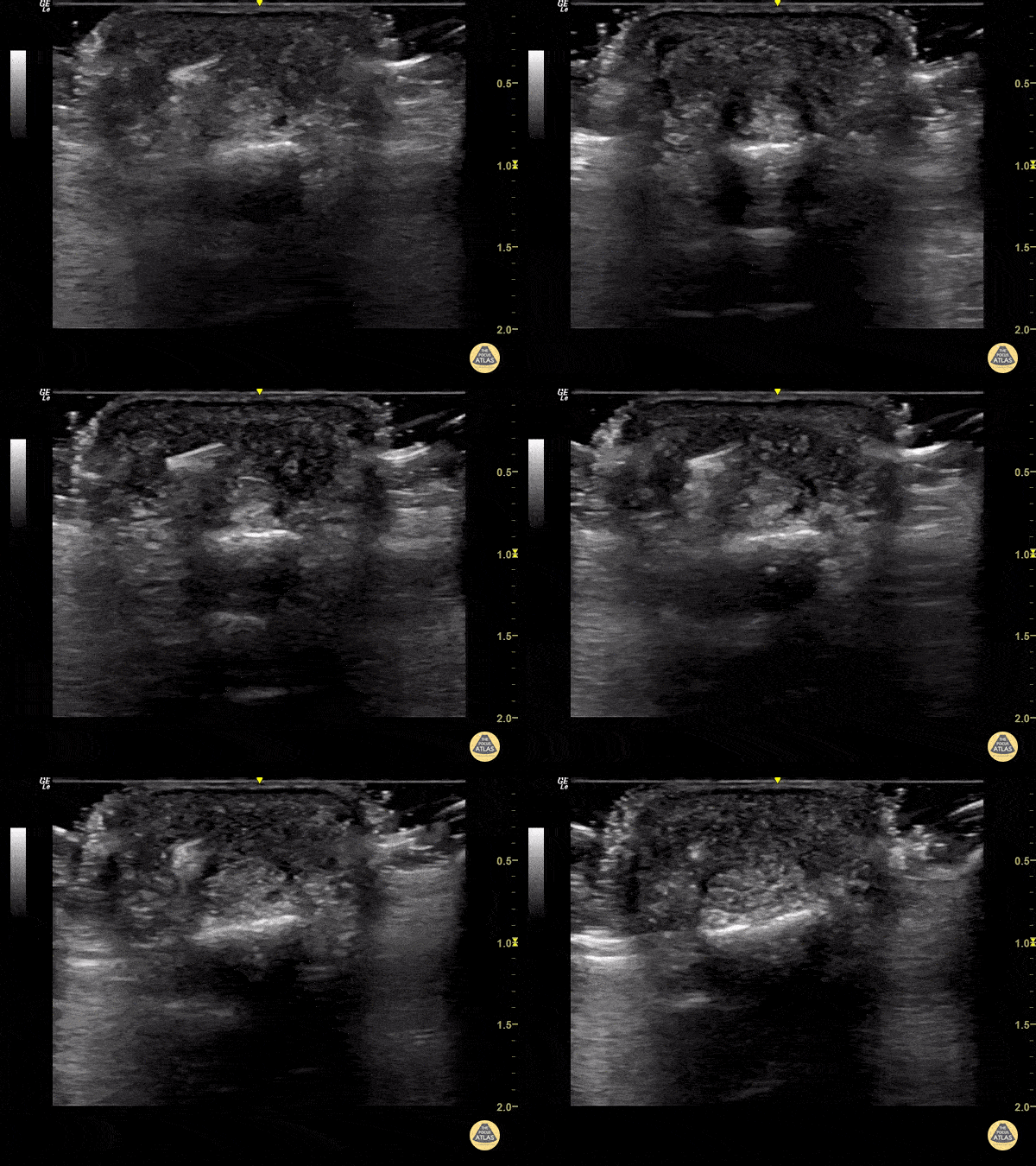
65 year old female with pain and swelling over 4th phalanx, no history of trauma. Hyperechoic density located above flexor tendon sheath. Patient sent to Ortho for surgical exploration.
Dr. Magner
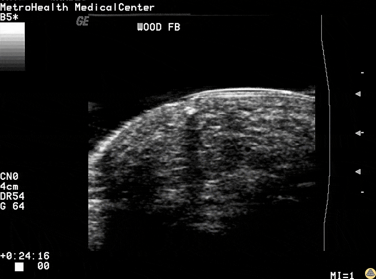
Wooden splinter in the fingertip of a patient. Ultrasound revealed the foreign body with posterior acoustic shadowing present.
Image courtesy of Robert Jones DO, FACEP @RJonesSonoEM
Director, Emergency Ultrasound; MetroHealth Medical Center; Professor, Case Western Reserve Medical School, Cleveland, OH
View his original post here
Other Pathology
A middle aged male presented to the ED for left leg pain after sustaining puncture wound to the foot 4 hours prior arrival. The ultrasound image demonstrates free air causing dirty shadowing seen deep to the fascial plane obscuring the normal muscle architecture in the middle of the screen.
Nicmarie Maldonado, MD PGY-2; Eddie G. Rodriguez, MD; Miguel Agrait, MD; Michelle Surillo, MD - Division of Emergency Ultrasound, St. Luke's Medical Center/PHSU, Ponce, PR
POCUS can help distinguish simple abscess from necrotizing fasciitis. “Dirty air shadowing” visualized in the clip suggests a necrotizing soft tissue infection.
Dr. Farnam Kazi Ultrasound Faculty; Dr. Dimitri Livshits Ultrasound Fellow; Dr. Jane Belyavskaya Ultrasound Fellow (All at Kings County Hospital/SUNY Downstate)
This is a transverse view of the anterior shoulder of a patient who presented to the emergency department with a fever and shoulder pain causing limited mobility. Looking closer, we can observe hyperechoic densities caused by air within the tissues, otherwise no signs of septic joint or bursitis.
Image courtesy of Robert Jones DO, FACEP @RJonesSonoEM
Director, Emergency Ultrasound; MetroHealth Medical Center; Professor, Case Western Reserve Medical School, Cleveland, OH
View his original post here