
Musculoskeletal Jr
Musculoskeletal Jr
Note: These search bar results are only for the Musculoskeletal Jr image archive. To search the entire site, click here.
Recently Added
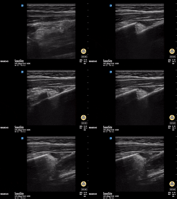
11y female with L both bone forearm fx from fall during gym class. Long axis view with clear discontinuity of proximal and distal components of the shaft. Transverse view with transition from single bony cortex to overlap of fracture ends.
Matthew Moake, MD PhD
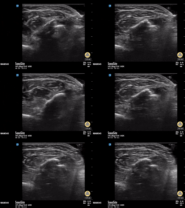
11y female with L BB forearm fx from fall during gym class. Long axis view with clear discontinuity of proximal and distal components of the shaft. Transverse view with transition from single bony cortex to overlap of fracture ends.
Matthew Moake, MD PhD
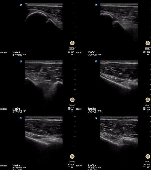
12y male presenting with RUE pain after bike injury. POCUS demonstrating proximal humeral fracture. Long axis and short axis views of fracture.
Matthew Moake, MD PhD
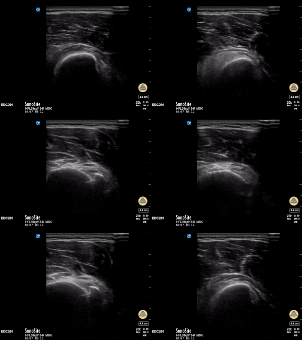
12y male presenting with RUE pain after bike injury. POCUS demonstrating proximal humeral fracture. Long axis and short axis views of fracture.
Matthew Moake, MD PhD
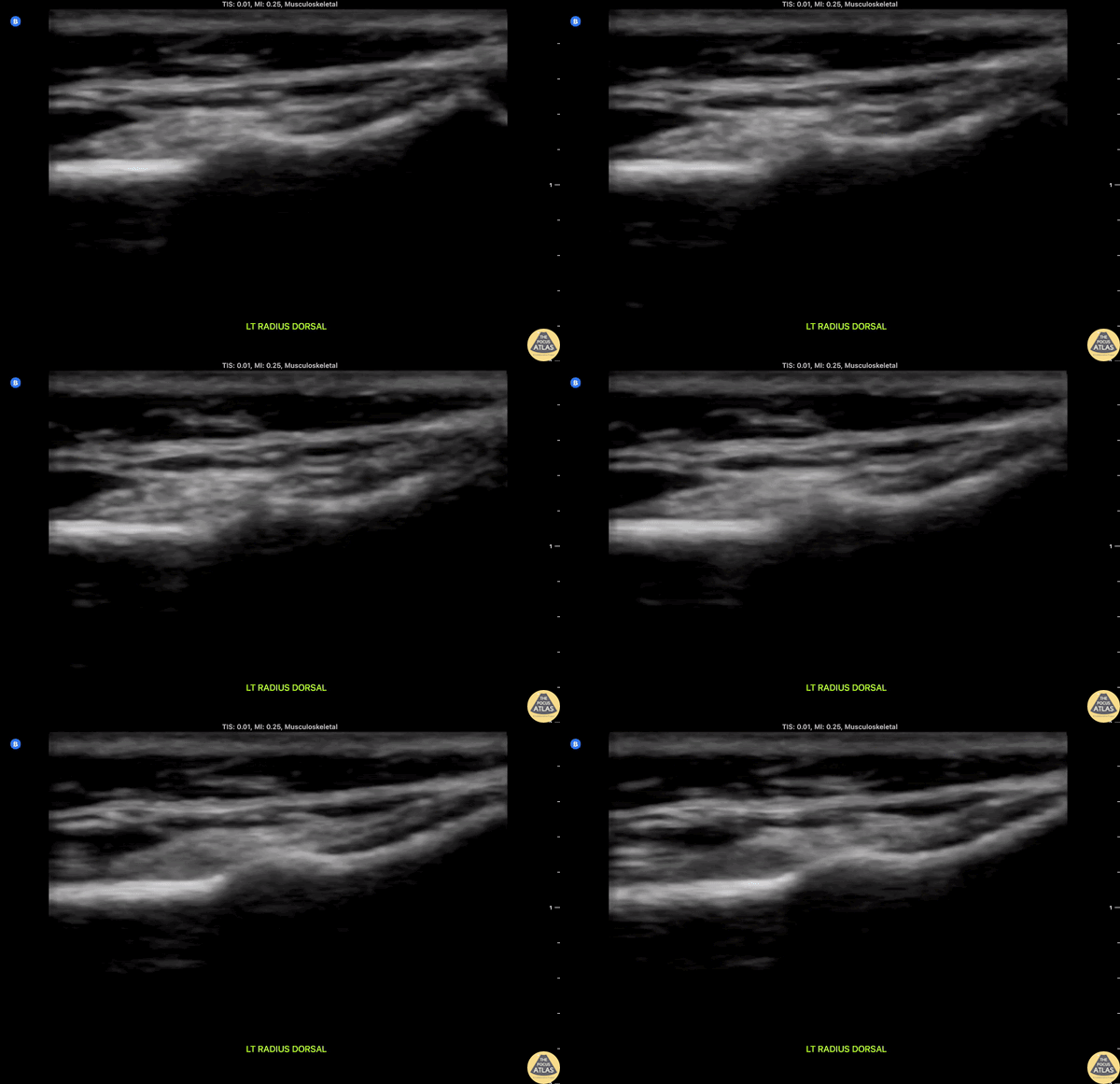
Clip shows longitudinal dorsal view of the radius with a "bump" in area of tenderness consistent with a torus or "buckle" fracture in a pediatric patient evaluated following a fall.
Miguel Agrait MD CAQ-SM, Eddie Rodriguez MD FPD-AEMUS
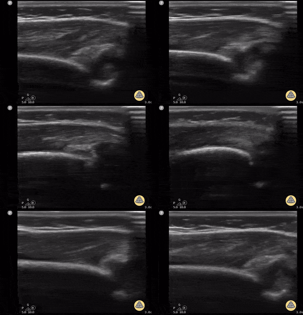
8 month old with arm swelling and fever. Longitudinal view of elbow with fluid collection displacing the posterior fat pad. Final diagnosis was septic arthritis of the elbow.
Contributor: Antonio Riera, MD, Yale University School of Medicine
Normal Anatomy
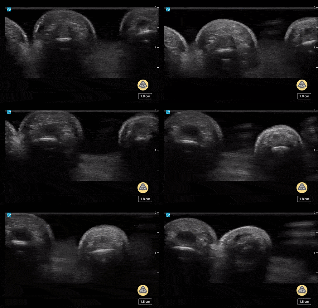
5 yo with normal finger anatomy in a waterbath. Case series 1/2
Contributor: Paul Khalil, MD Nicklaus Children's Hospital @khalil3paul
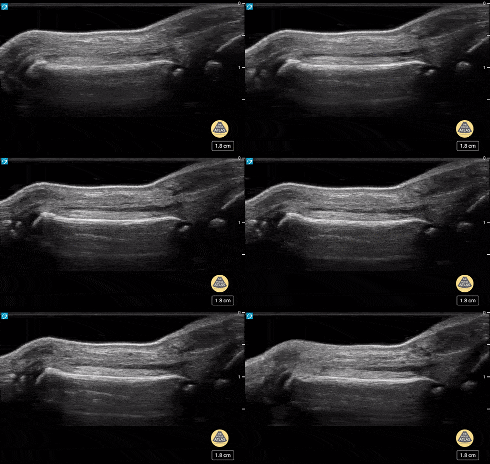
5 yo with normal finger anatomy in a waterbath. Case series 2 of 2
Contributor: Paul Khalil, MD Nicklaus Children's Hospital @khalil3paul
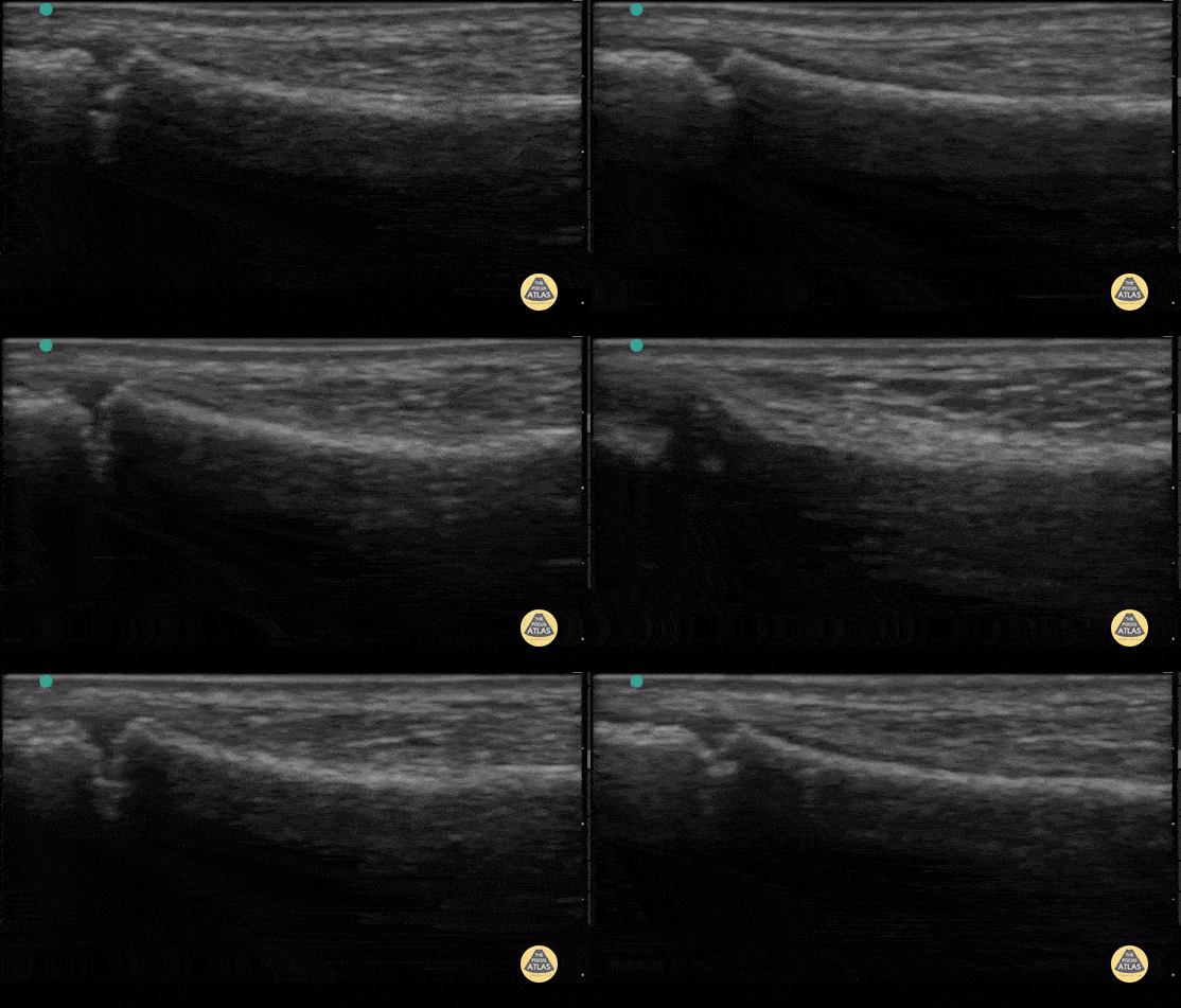
7yo right fibular ultrasound with notable physis on left hand side of screen.
Contributor: Antonio Riera, MD
Fractures
11y female with L both bone forearm fx from fall during gym class. Long axis view with clear discontinuity of proximal and distal components of the shaft. Transverse view with transition from single bony cortex to overlap of fracture ends.
Matthew Moake, MD PhD
11y female with L BB forearm fx from fall during gym class. Long axis view with clear discontinuity of proximal and distal components of the shaft. Transverse view with transition from single bony cortex to overlap of fracture ends.
Matthew Moake, MD PhD
12y male presenting with RUE pain after bike injury. POCUS demonstrating proximal humeral fracture. Long axis and short axis views of fracture.
Matthew Moake, MD PhD
Joint Effusions
8 month old with arm swelling and fever. Longitudinal view of elbow with fluid collection displacing the posterior fat pad. Final diagnosis was septic arthritis of the elbow.
Contributor: Antonio Riera, MD, Yale University School of Medicine
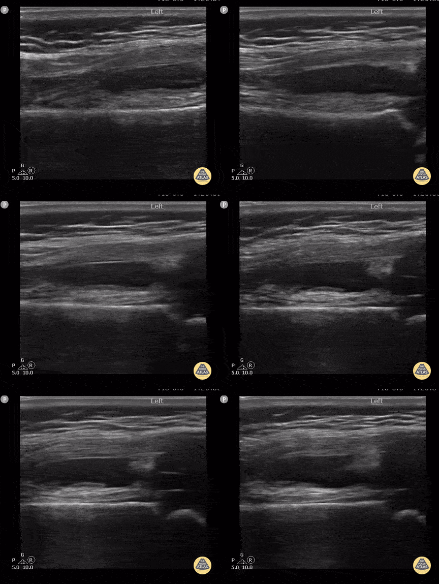
17 month old with knee effusion due to septic arthritis. Knee radiography was normal. Note the distal femur, physic and epiphysis seen on the lower part of the screen below the effusion and the hypoechoic non-ossified patella cartilage typically seen in this age.
Contributor: Antonio Riera, MD, Yale University School of Medicine
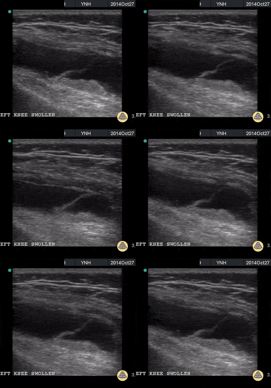
11 year old with large knee effusion in the suprapatellar bursa. Note the visible synovial plica membrane
Contributor: Antonio Riera, MD Yale University School of Medicine
Skull Fractures
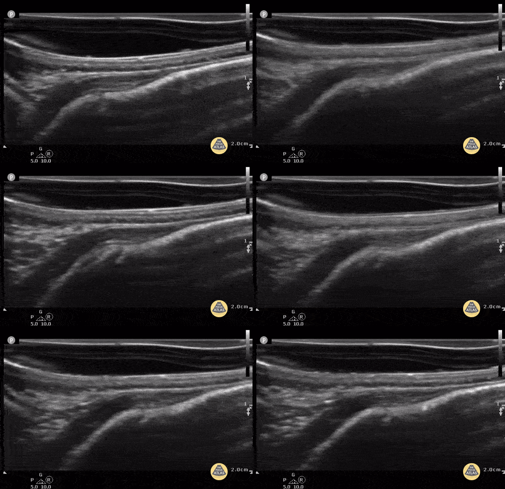
4 week old with non displaced parietal fracture using water filled glove as stand off. (Case Series 1/2)
Contributor: Antonio Riera, MD
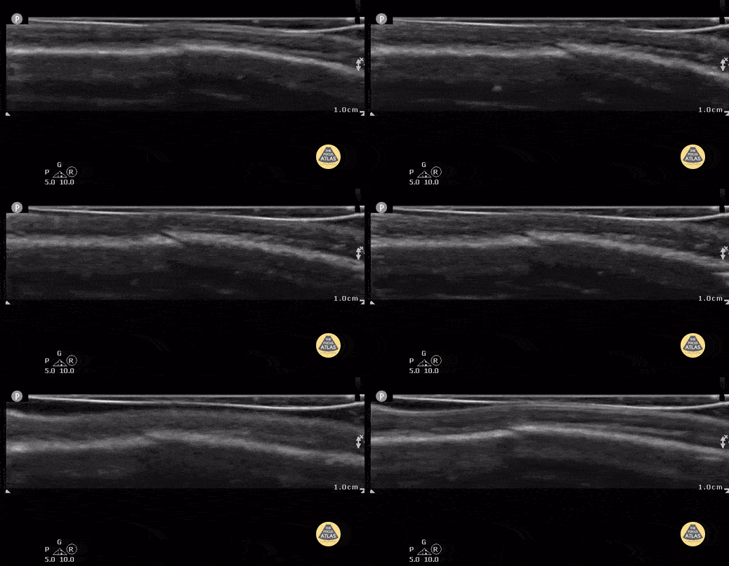
same 4 week old with non-displaced parietal fracture after compression and displacement of fluid from the water filled glove. (Case Series 2/2)
Contributor: Antonio Riera, MD
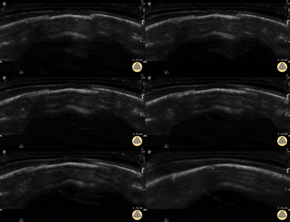
7 week old with left parietal skull fracture. Note irregular edges on both sides and slightly depressed bony cortex.
Contributor: Antonio Riera, MD
Tendon/Ligamentous Trauma
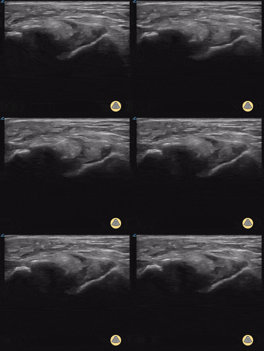
17 yo male inverted his ankle playing basketball. POCUS shows partial tear of the ATFL.
Contributor: Paul Khalil, MD Nicklaus Children's Hospital @khalil3paul
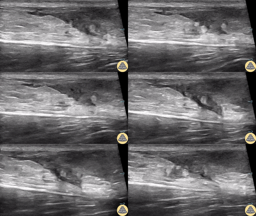
16-year-old male presented with acute onset sharp pain to his LE and inability to bare weight after having landed oddly while playing basketball. POCUS revealed a near-complete disruption of his Achilles Tendon.
Paul Khalil, MD. Assistant PEM POCUS director at University of Louisville/Norton Children’s
@Khalil3Paul
Dislocations
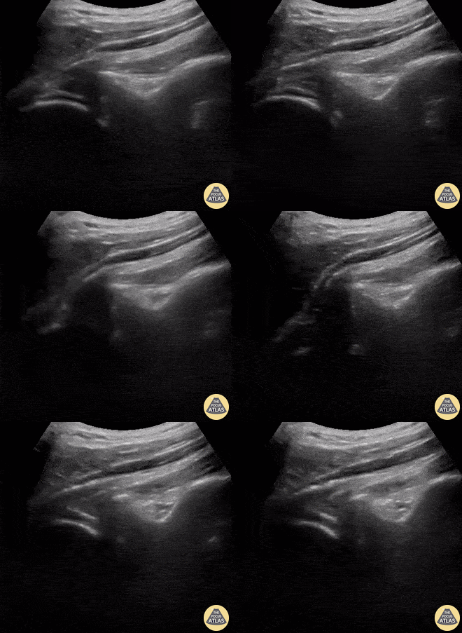
A 9-year-old female presented with left shoulder pain. She has a history of multiple dislocations and, as seen here on POCUS, is able to reduce the dislocation herself.
Julie Klensch, PEM Fellow & Paul Khalil, MD
University of Louisville/Norton Children's Hospital
Other Pathology
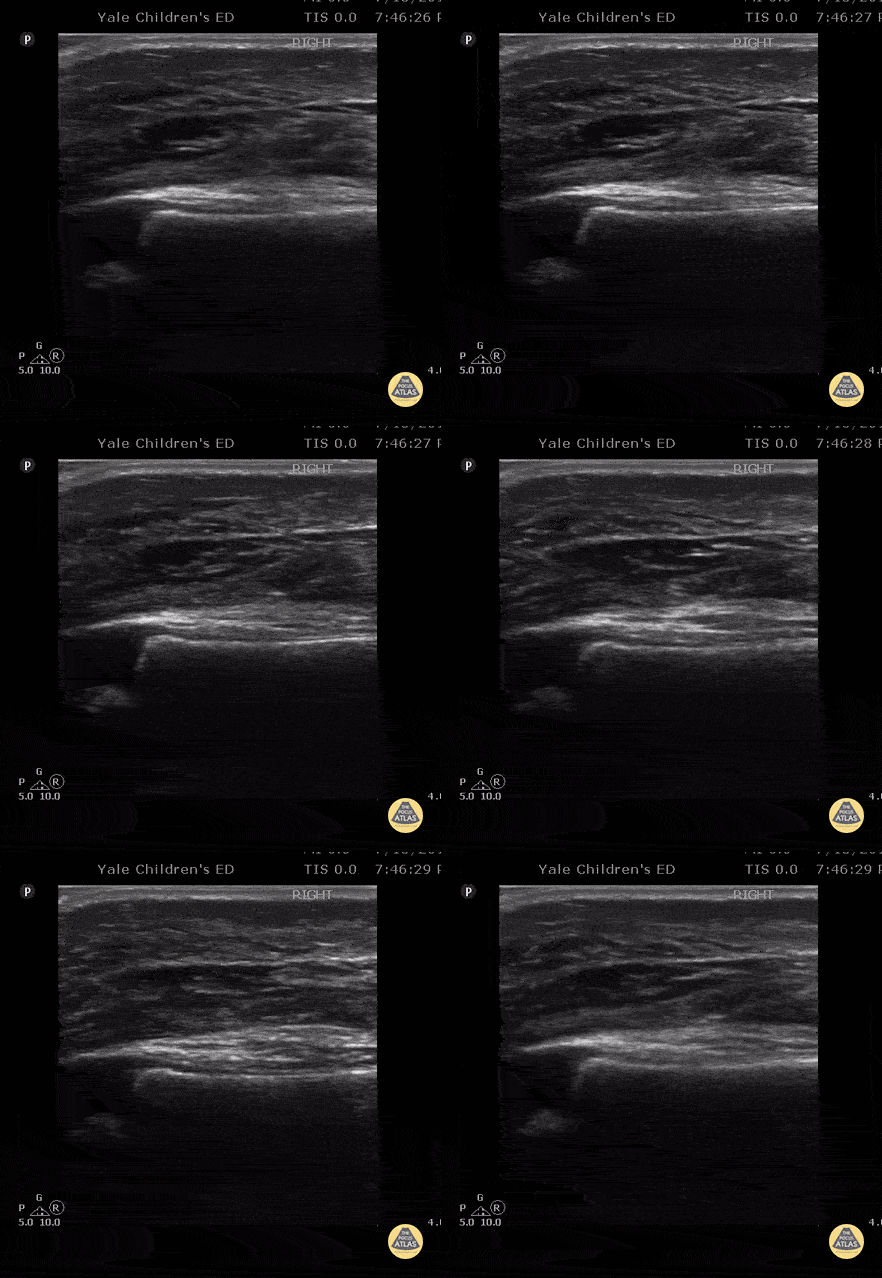
Longitudinal suprapatellar evaluation with linear probe in a 23 month old with swelling to knee / lower leg after vaccination given in thigh. Note the impressive soft tissue edema. There is no cobblestoning pattern and no fluid collection layering under the quadriceps tendon / no signs of suprapatellar effusion. The patient had a dry arthrocentesis performed under procedural sedation by a consulting service.
Contributor: Antonio Riera, MD, Yale University School of Medicine
Nursemaid's pre and post reduction.
Nathan Jia, Orthopedic Resident