
Aorta

Saccular AAA with Dissection Flap
Saccular AAA with Dissection Flap.
Contributor: Daniel Ostapowicz, MD

Sagittal View of Abdominal Aortic Aneurysm
Sagittal view of large abdominal aortic aneurysm incidentally found after accidental fall.
Image courtesy of Robert Jones DO, FACEP @RJonesSonoEM
Director, Emergency Ultrasound; MetroHealth Medical Center; Professor, Case Western Reserve Medical School, Cleveland, OH
View his original post here

Abdominal Aortic Aneurysm Presented As Cough
Here is another example of an abdominal aortic aneurysm that measured approximately 10.6 cm. The patient originally presented to the clinic complaining of a cough and was eventually admitted for surgical operation.
Image courtesy of Robert Jones DO, FACEP @RJonesSonoEM
Director, Emergency Ultrasound; MetroHealth Medical Center; Professor, Case Western Reserve Medical School, Cleveland, OH
View his original post here

AAA with Intramural Thrombus
The patient presented to the ED with a known abdominal aortic aneurysm (AAA). A point-of-care ultrasound (POCUS) examination of the abdominal aorta was performed using a curvilinear probe (longitudinal view shown). An infrarenal AAA was noted adjacent and superior to the bifurcation and measured a maximum diameter of approximately 5cm. Heterogeneous echogenic material was seen within the lumen of the aneurysm in keeping with intramural thrombosis, along with a hyperechoic line representing a chronic dissection.
This demonstrates the utility of POCUS in the rapid diagnosis of AAA and evaluation of size and intraluminal features such as intramural thrombus and dissection.
Andrew Namespetra, MB BCh BAO MSc
PGY-1 Emergency Medicine Resident, Central Michigan University (CMU)
Chad Bambrick, MD
PGY-2 Emergency Medicine Resident, CMU
Ryan Davis
MS4, CMU College of Medicine

AAA
Emergency response was called for an 88-year-old who experienced sudden onset thoracic and back pain. PMH significant for AAA (previously measuring 5.8 cm diameter on surveillance imaging 1 year ago). Mid-abdominal US view was obtained (shown here) and revealed AAA now measuring 7.2 cm diameter.
Wolfgang Geisser
@fentanyl05

Long Axis View of Large Abdominal Aortic Aneurysm
Long axis view of large abdominal aortic aneurysm containing intramural thrombus without evidence of the iliac arteries involvement.
Image courtesy of Giovanni Battista Fonsi

Smokin' AAA
A 70-year-old male presented as a trauma alert and was found to have this 10 cm abdominal aortic aneurysm (AAA) with associated echocardiographic smoke! Echogenic smoke is produced by the intersection of RBC’s and plasma proteins at low shear rate conditions.
Brian Toston, Internist. Adventura, FL
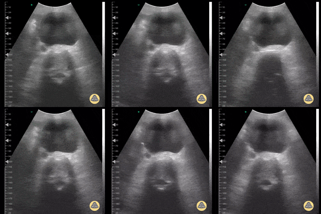
Abdominal Aortic Aneurysm
This image was taken from a pocket wireless device in the mesogastric region. We can see the short-axis aorta with its increased diameter and neighboring anatomical references such as the dorsal spine and inferior vena cava laterally.
Image courtesy of Dr. Renato Tambelli

Incidental AAA
Routine abdominal evaluation using a Sonosite X-Porte curvilinear ultrasound probe revealed this 6.0cm abdominal aortic aneurysm (without rupture or dissection) in a patient with risk factors for the same. This enabled our middle-aged diabetic male with 80 pack/yr smoking history to receive timely evaluation by vascular surgery and subsequent appropriate serial monitoring and follow-up.
Siang-Chean Kua MS-4
Central Michigan University College of Medicine
Additional contributors: Ryan Shelby, MD, Thomas Ferreri, MD, Therese Mead, DO, RDMS, FACEP
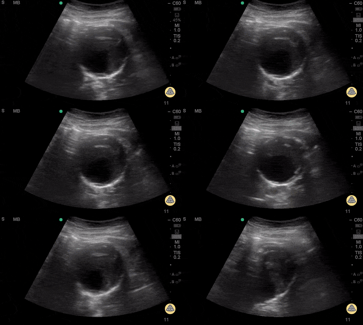
Mural Thrombus in AAA
74 y/o F hx stage 4 lung cancer, AAA, presented with chest pain and SOB x 1 day. POCUS shows the aorta is larger than the normal diameter of 3 cm, representing an abdominal aortic aneurysm, and measuring approximately 5.6 cm at its largest point. On cross section you are able to see a large mural thrombus with decreased diameter of blood flow through the center.
Juliana Jaramillo MD - Kings County Emergency Medicine
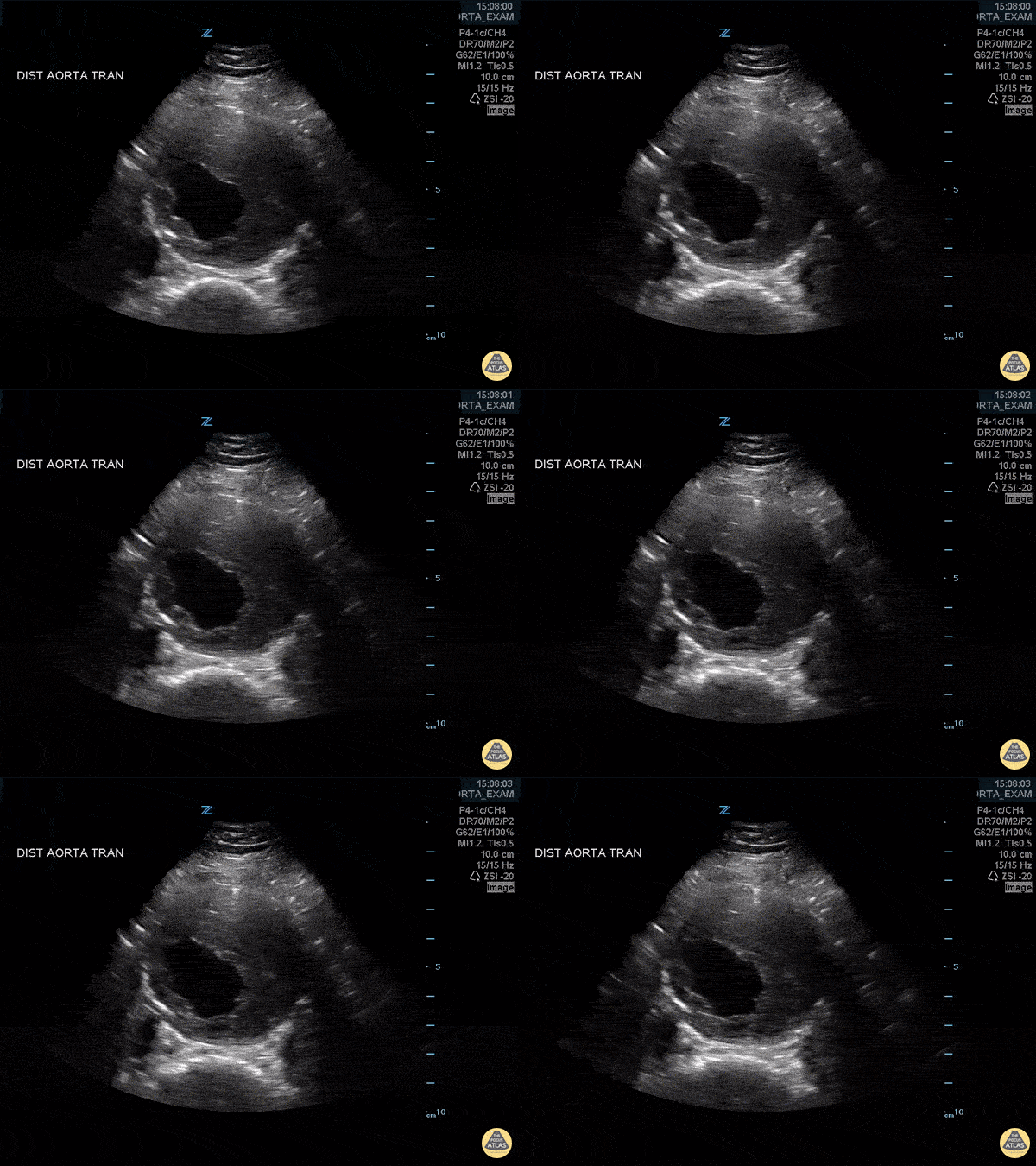
Abdominal Aortic Aneurysm with Thrombus
Approximately 6 cm abdominal aortic aneurysm with intramural thrombus.
Frances Russell, MD, RDMS
Assistant Professor of Emergency Medicine Division Chief, Ultrasound Fellowship Director, Ultrasound

Mural Thrombus in AAA - Doppler
74 y/o F hx stage 4 lung cancer, AAA, presented with chest pain and SOB x 1 day. POCUS shows the aorta is larger than the normal diameter of 3 cm, representing an abdominal aortic aneurysm, and measuring approximately 5.6 cm at its largest point. On cross section you are able to see a large mural thrombus with decreased diameter of blood flow through the center.
Juliana Jaramillo MD - Kings County Emergency Medicine
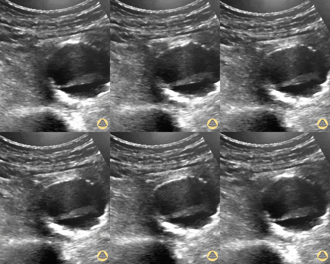
Pulsating Aortic Aneurysm
You may think this a dissection but it's actually an aortic aneurysm filled with thrombus, "with holes" making it a very "happy flappy." CT Confirmed.
Dr. Vincent Rietveld - Amsterdam, The Netherlands
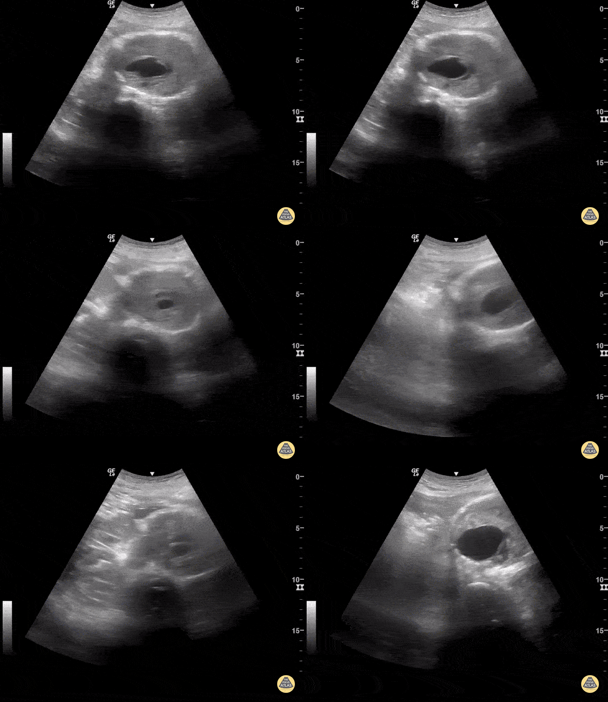
Abdominal Aortic Aneurysm
AAA is defined as a localized balloon-like dilatation of the abdominal aorta greater than 3cm. Risk factors include male sex, increased age, and tobacco use. AAAs should be closely monitored for changes in size. Due to the risk of rupture, elective surgery is recommended when the dilatation is greater than 5-5.5cm, or it is growing in size by greater than 1cm/year. The classic triad of a ruptured AAA include pulsatile abdominal mass, hypotension and pain.
This AAA has an intramural thrombus. Some studies have claimed that POCUS has a ~100% sensitive for increased diameter. 3cm from outer wall to outer wall defines an aneurysm. Slow, graded compression is key to move the bowel out of the way in any abdominal study.
Sukh Singh, MD
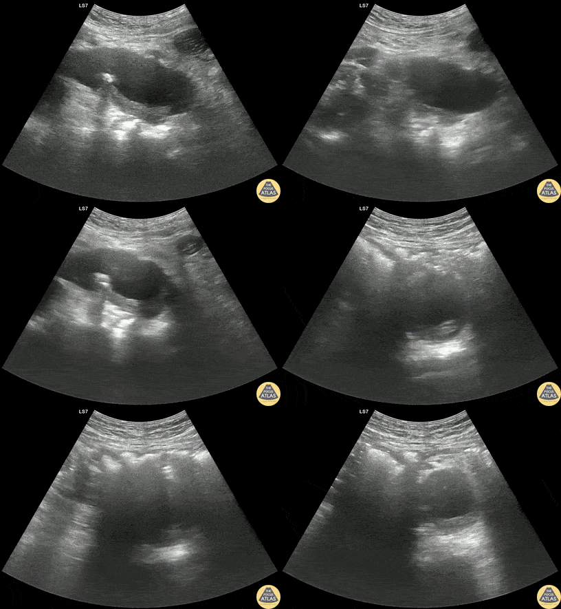
Thrombosed Type B Aortic Dissection
60s M PMH HTN, CKD stage 3 presented with bilateral flank pain x2 days. CT of the abdomen/pelvis with IV contrast showed a 3.8 cm x 4.6 cm infrarenal AAA with a 3.1 cm L internal iliac aneurysm. POCUS of the aorta was obtained re-demonstrating the AAA. Dedicated CTA showed that there actually demonstrating aneurysmal dilation of the descending thoracic aorta with a thrombosed type B dissection. The patient was managed with aggressive blood pressure and heart rate control with esmolol and was admitted to the ICU for medical management.
Dr. Henrik Galust, PGY4, Denver Health Residency in Emergency Medicine
Dr. Nimish Bhatt, Fellow, Denver Health Ultrasound Fellowship


