
Aorta

AAA in Undifferentiated Shock
70s-year-old male brought in for altered mental status and hypotension.
As part of the initial exam, POCUS was performed. This clip, obtained using the curvilinear probe in transverse orientation over the mid-epigastric region, revealed a AAA measuring 8 cm in AP diameter at the largest point with heterogeneous echogenic material consistent with thrombus. CT abdomen/pelvis with IV contrast confirmed ruptured AAA with thrombosis and only minor extravasation.
In this case, POCUS played a critical role in the rapid diagnosis of a patient with undifferentiated shock, particularly in a patient who was unable to provide any history.
Katherine Spencer, MD
Guy Youngblood, MD, FACEP, FAAEM

Large AAA Rupture
This is a clip demonstrating a large abdominal aortic aneurysm with significant intramural thrombus. Signs of rupture can be appreciated near the right lower portion of the screen as there is disruption of the aorta wall. Heterogeneity within the intramural thrombus can also be an indicator of rupture.
Michael Macias, MD

Ruptured AAA
Sagittal view of an abdominal aortic aneurysm with a hemorrhage anterior to the aorta. Be sure to distinguish a AAA from a dissection by assessing the abnormal wall motion seen at the site of rupture.
Image courtesy of Robert Jones DO, FACEP @RJonesSonoEM
Director, Emergency Ultrasound; MetroHealth Medical Center; Professor, Case Western Reserve Medical School, Cleveland, OH
View his original post here
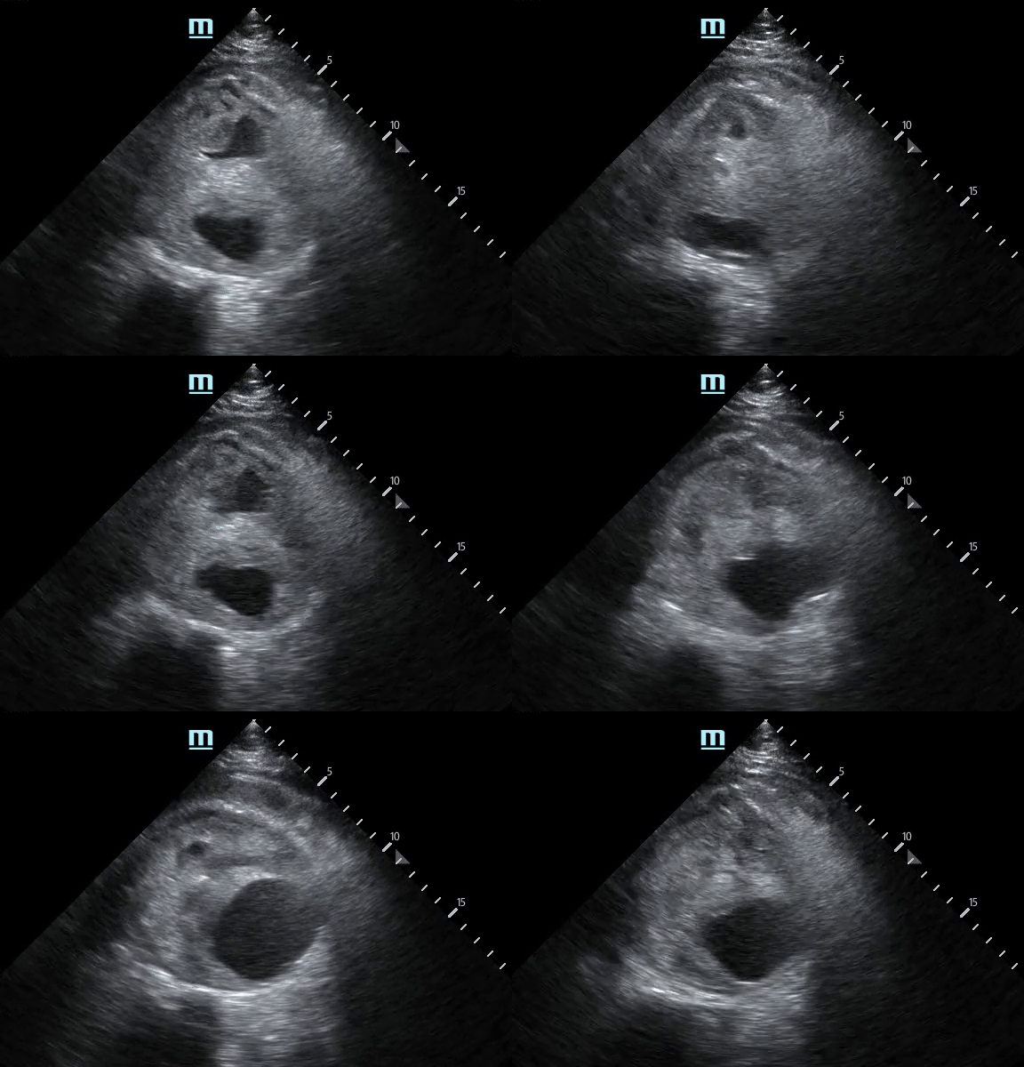
AAA Rupture: Not Your Typical Flank Pain
A 61-year-old male was brought in via EMS after a syncopal episode. He was diaphoretic and hypotensive, complaining of severe right flank pain.
While IV access was being obtained a bedside ultrasound was performed demonstrating a large abdominal aortic aneurysm with significant heterogeneous intraluminal clot. There is also appreciable focal hypoechoic disruption of the wall of the aneurysm consistent with rupture.
The patient was resuscitated in the ED and taken emergently to the OR to surgical repair.
Michael Macias, MD
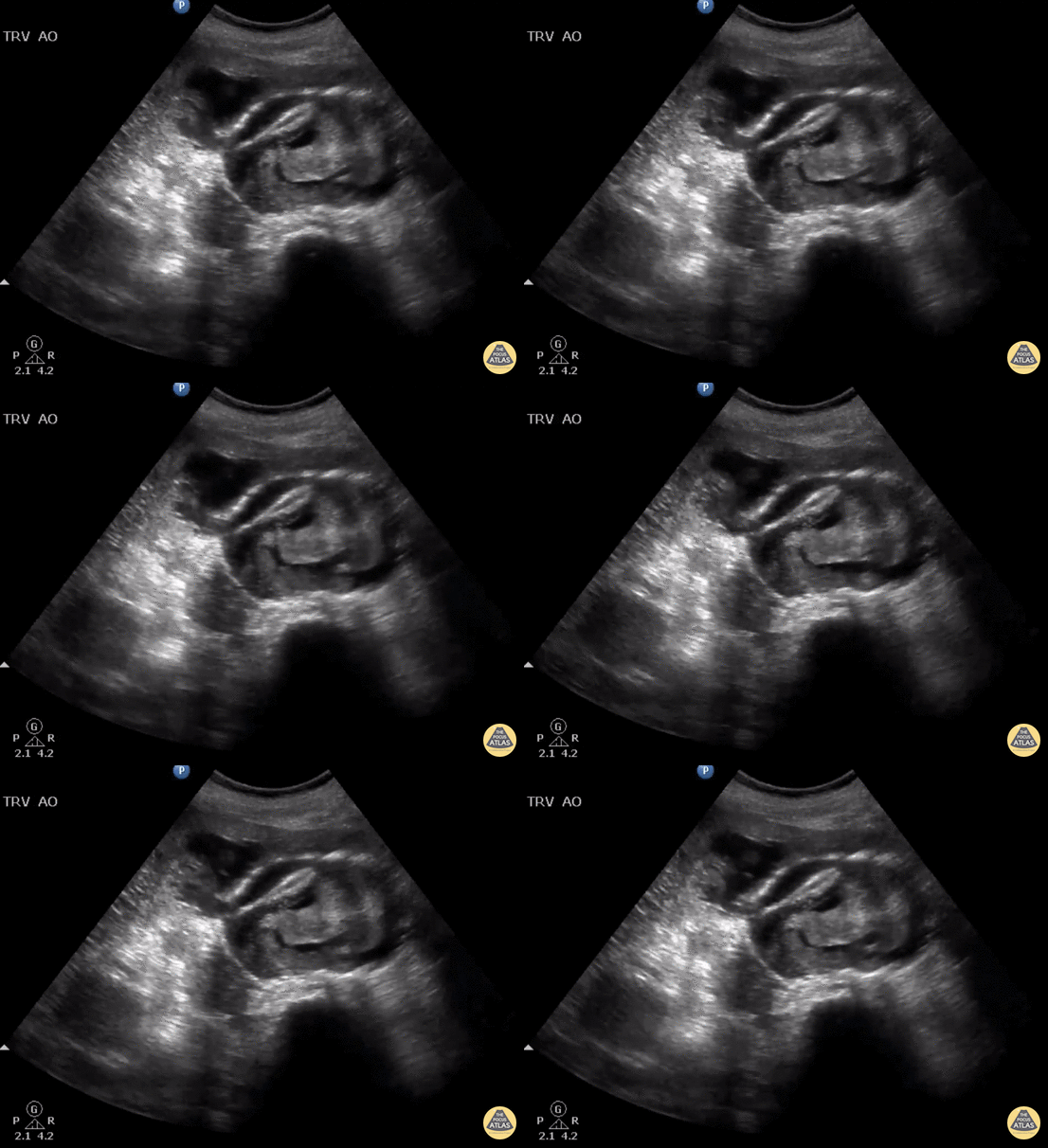
Ruptured AAA with Active Hemorrhage
Elderly female presents to ED with back pain and hypotension. Bedside US shows a ruptured AAA with active hemorrhage in the 9 o’clock position.
Image courtesy of Robert Jones DO, FACEP @RJonesSonoEM
Director, Emergency Ultrasound; MetroHealth Medical Center; Professor, Case Western Reserve Medical School, Cleveland, OH
View his original post here
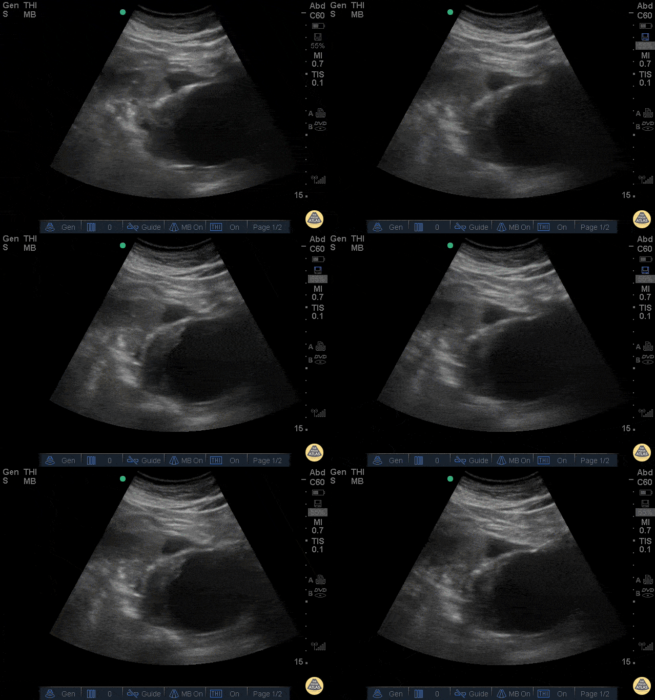
Ruptured AAA
The clip above is from a patient who presented with abdominal pain and syncope. His vitals were notable for tachycardia and hypotension. The image demonstrates a very large abdominal aortic aneurysm with anterior free fluid suggestive of rupture. The patient was taken emergently to the operating room for endograft repair and did well.
Jason Tanguay, DO
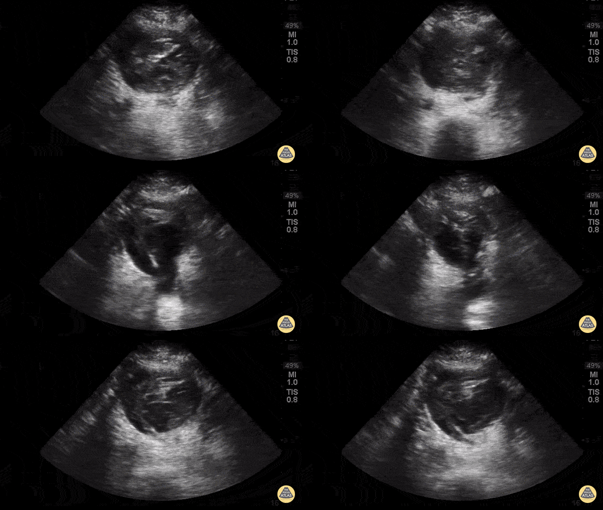
Aorta Posterior Wall Rupture
63yoM witnessed collapsed. Arrived VSA with PEA rhythm. A fast look abdominal aorta POCUS showed a large abdominal aortic aneurysm with internal thrombus and rupture through the posterior wall. Note the AAA is measured from echogenic outer wall to outer wall, not the hypoechoic internal lumen that is seen pulsating. The patient was resuscitated aggressively with blood and initially survived operative repair. Unfortunately he died in the ICU several days later from multiorgan failure.
Dr. Joey Newbigging
POCUS Utility in Suspected AAA Rupture
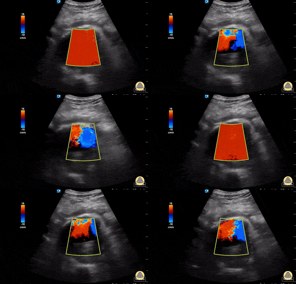
A 76 year old male presented to the ED for abdominal pain and syncope. He was tachycardic but normotensive. US performed with Sonosite 5-2MHz C60 probe with patient in supine position and probe held in transverse orientation revealed a AAA measuring ~9cm in AP diameter at its largest point. The AAA had an anechoic center with echogenic material in the posterior lumen, representing cholesterol deposit. Free fluid was seen anterior to the AAA suggestive of rupture which was confirmed by STAT CTA Aortagram. Patient was admitted to the OR for surgery. In this scenario, POCUS was a safe, non-invasive study that played a critical role in diagnosis of a ruptured AAA and accelerated surgical intervention.
Susan Dhamala, MS4, Drexel University College of Medicine
Brenton Elliot, MD, Crozer Chester Medical Center
Matthew Cully, DO, Nemours/Alfred I. duPont Hospital for Children
Kevin Conor Welch, DO, Crozer Chester Medical Center
Max Cooper, MD, RDMS, Director of Emergency Ultrasonography at Crozer Chester Medical Center


