1
2
3
4


Aortic Dissection
A chest pain received an echo and showed evidence of a dissection in the PLAX, PSAX, and AP4C views within the descending aorta.
Image courtesy of Robert Jones DO, FACEP @RJonesSonoEM
Director, Emergency Ultrasound; MetroHealth Medical Center; Professor, Case Western Reserve Medical School, Cleveland, OH
View his original post here
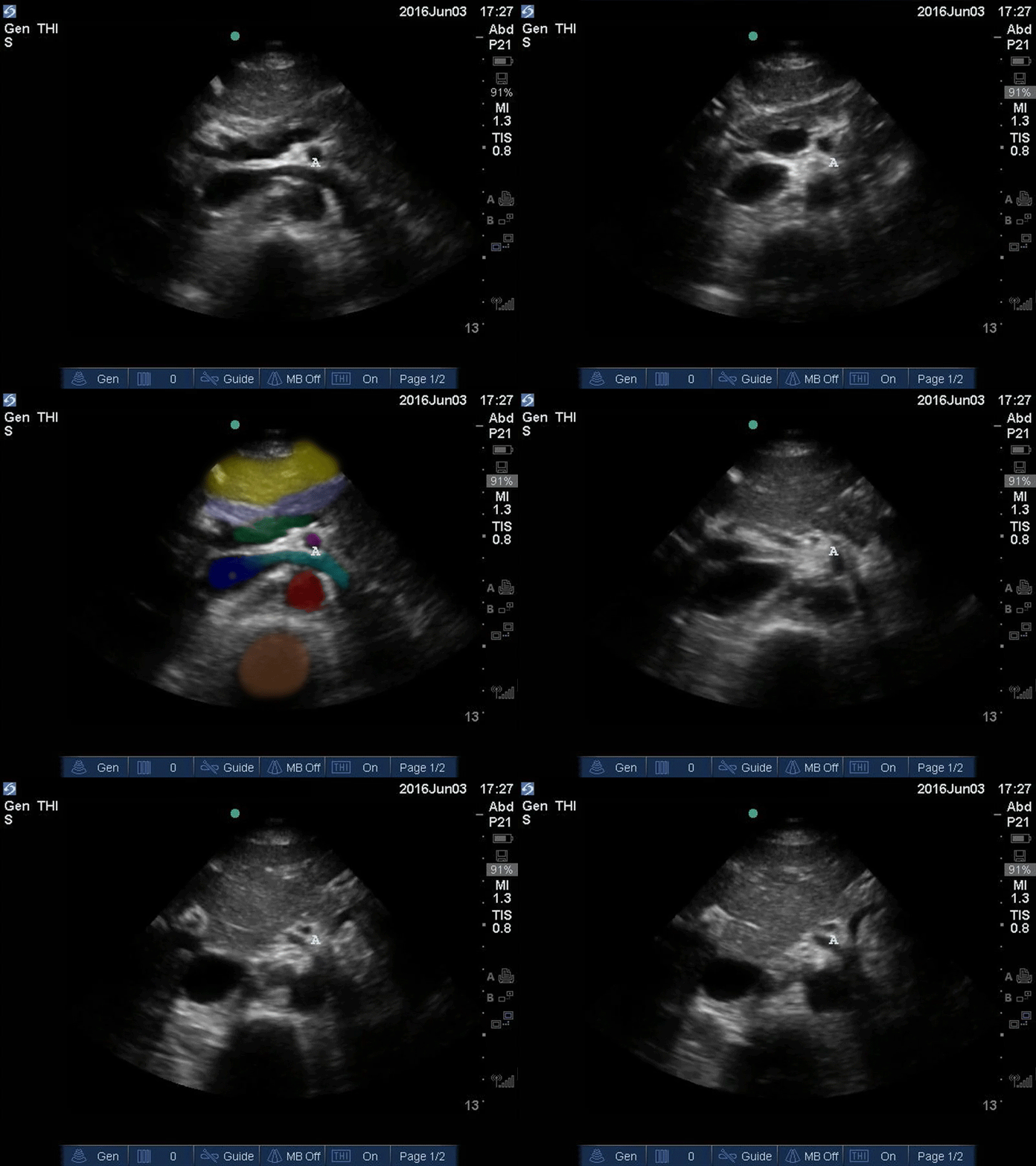
Upper Aorta - Colorized - The POCUS Atlas
Orange: Yellow: Liver, Light blue: Pancreas, Aqua: splenic vein/portal confluence, Blue: IVC with left renal vein, Purple: SMA, Red: Aorta, Orange: Spine
Images: Dr. Lindsay Davis, Dr. Hannah Kopinski. Image Editing: Michael Amador and Dr. Matthew Riscinti
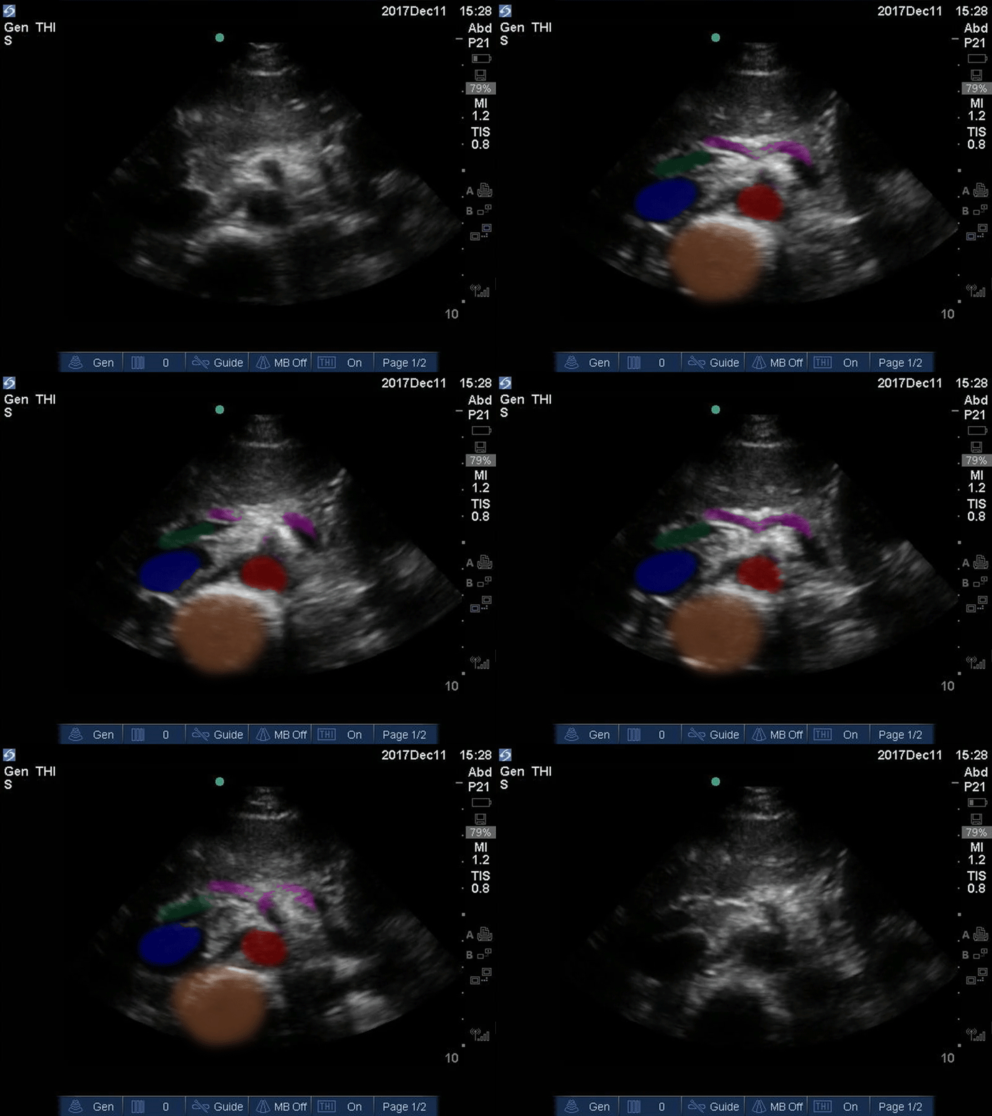
Seagull Sign - Colorized
Good Seagull Sign
Orange: Spine, Red: Aorta, Blue: IVC, Green: Portal venous confluence, Pink: “Seagull sign” aka celiac trunk
Images: Dr. Lindsay Davis, Dr. Hannah Kopinski. Image Editing: Michael Amador and Dr. Matthew Riscinti
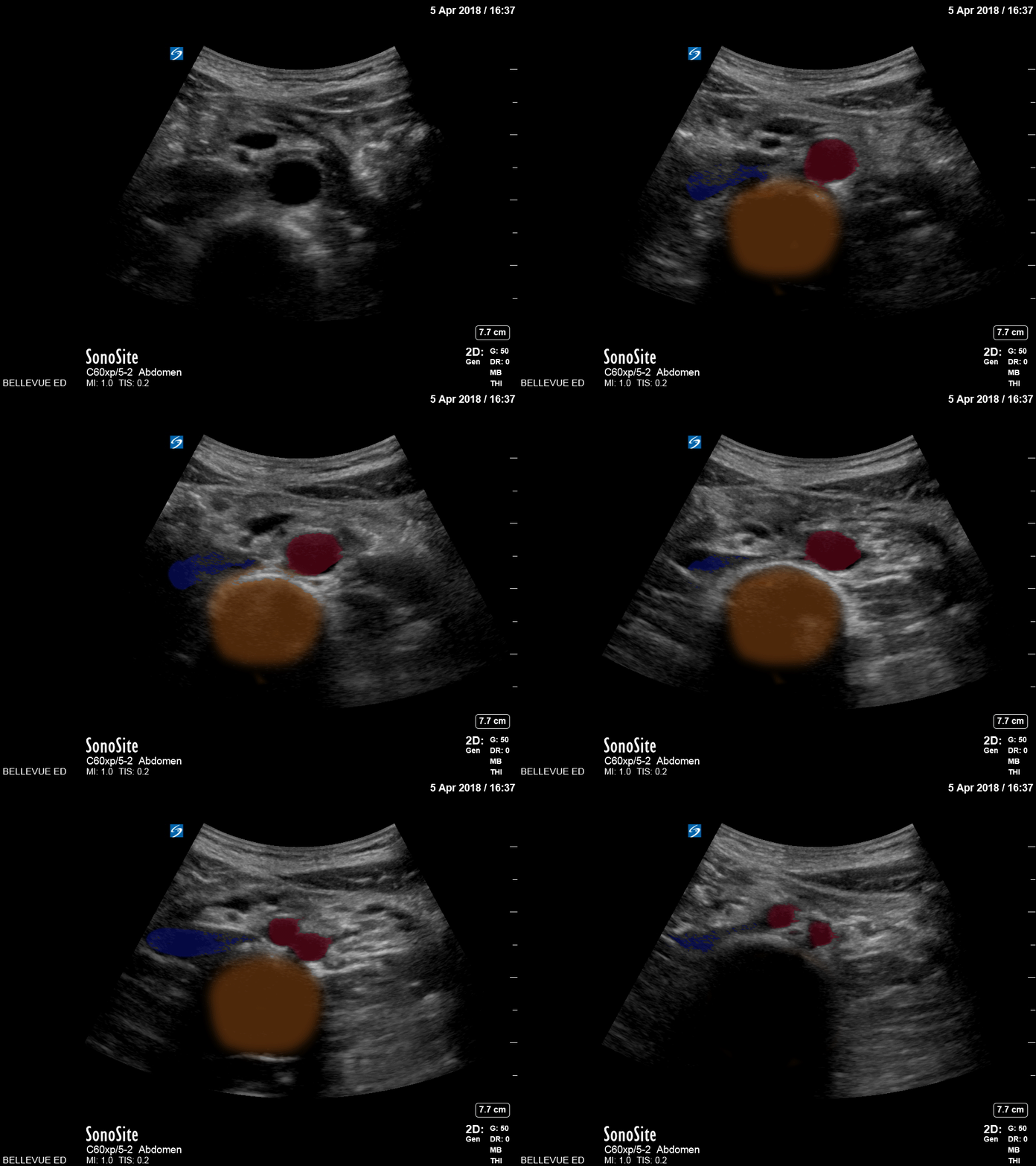
Distal Aorta - Colorized
Distal Aorta
Orange: Spine, Red: Aorta and Iliacs, Blue: IVC, Green: Portal venous confluence, Pink: “Seagull sign” aka celiac trunk
Images: Dr. Lindsay Davis, Dr. Hannah Kopinski. Image Editing: Michael Amador and Dr. Matthew Riscinti