Aorta

Suprasternal view of the aorta
Suprasternal notch view of the aortic arch with brachiocephalic artery (BCA), left common carotid artery (LCA), and left subclavian artery (LSA) branching off. Right pulmonary artery is also visible adjacent to the ascending aorta.
Charles Jang, EM PGY-3

Suprasternal view of the aorta with labels
Suprasternal notch view of the aortic arch with brachiocephalic artery (BCA), left common carotid artery (LCA), and left subclavian artery (LSA) branching off. Right pulmonary artery is also visible adjacent to the ascending aorta.
Charles Jang, EM PGY-3

Normal Aortic Arch - Suprasternal View
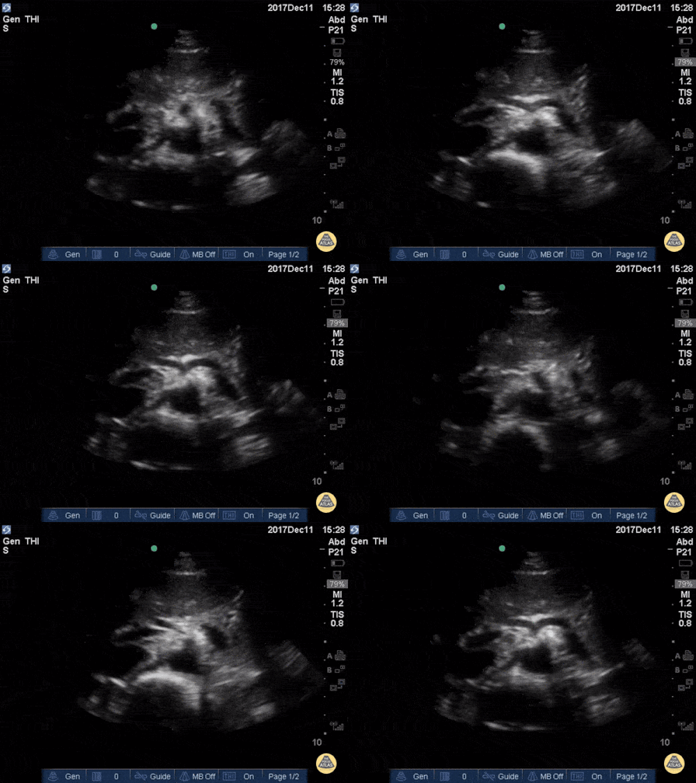
This clip represents a normal suprasternal view. The brachiocephalic artery (BCA), left carotid artery (LCA) and left subclavian artery (LSA) emerge from the aortic arch (AA). The left subclavian artery (LSA) marks the division between the proximal thoracic aorta and the distal descending aorta (DA).
Dr. Felipe Urriola P., Emergency Unit, Puerto Aysen Hospital, Chilean Patagonia.

Normal Celiac Trunk & SMA - Longitudinal
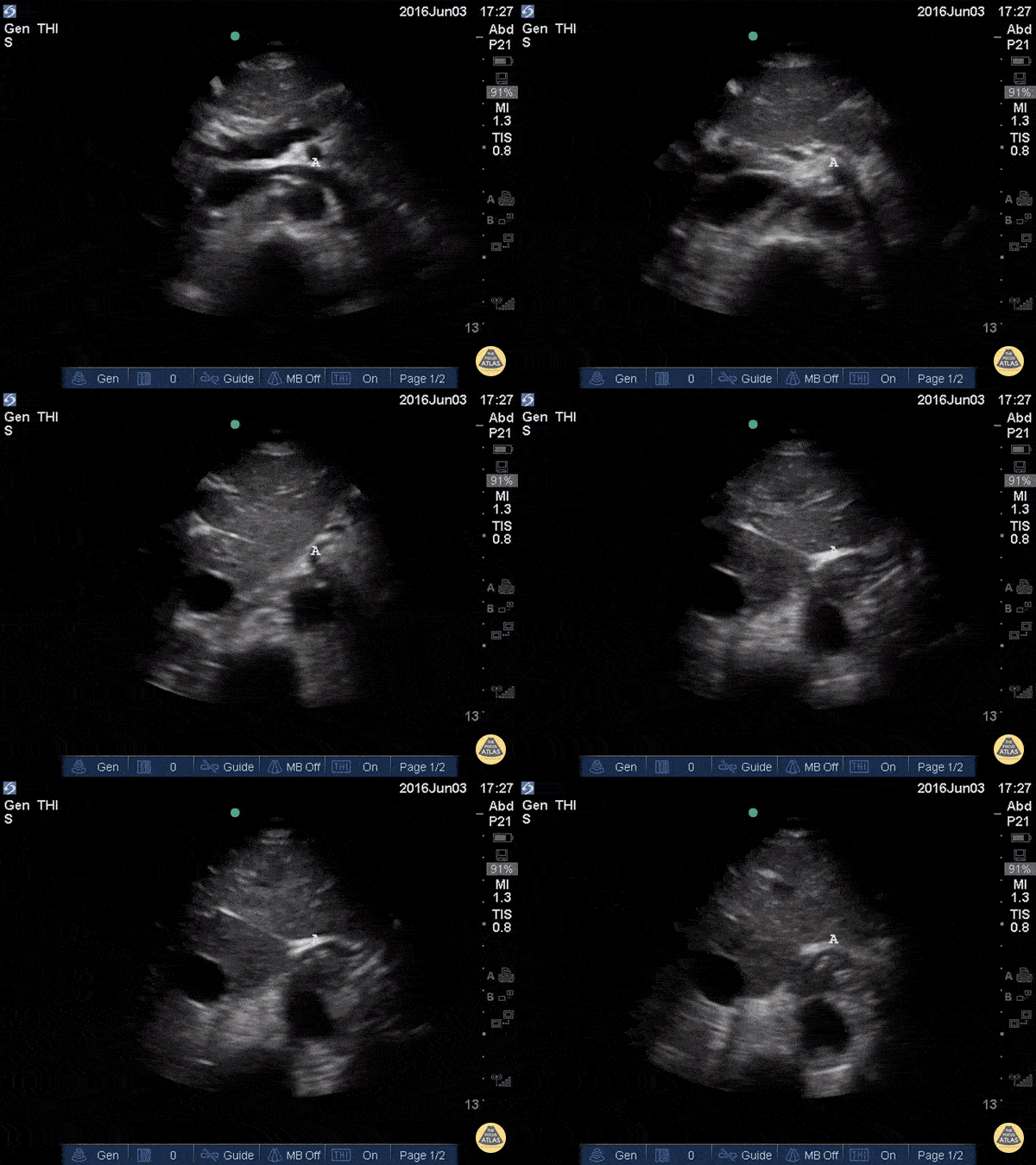
At the center of the screen, the proximal aorta can be identified by the emergence of the coeliac trunk and the superior mesenteric artery. This clip is taken at the subxiphoid level, longitudinal to the body’s axis, and with the probe marker oriented towards the head.
Dr. Felipe Urriola P., Emergency Unit, Puerto Aysen Hospital, Chilean Patagonia.

Normal Aorta & Iliac Arteries - Transverse
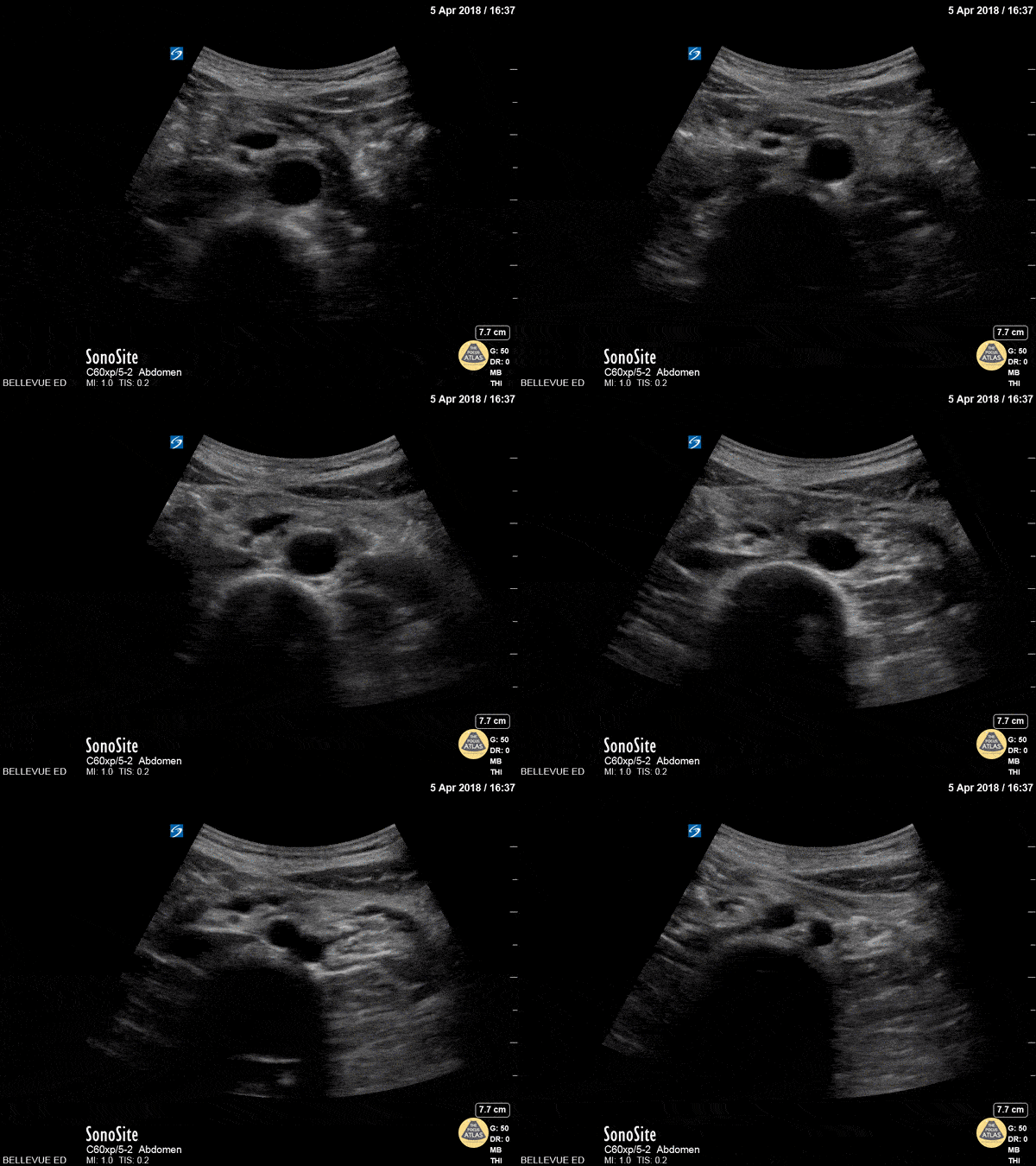
At the center of the screen, the distal aorta lies anterior to a vertebral body. Sliding the probe caudally reveals the emergence of the iliac arteries. This clip is taken at the umbilicus level, transverse to the body’s axis, and with the probe marker oriented to the patient’s right.
Dr. Felipe Urriola P., Emergency Unit, Puerto Aysen Hospital, Chilean Patagonia.

Normal IVC and Aorta - Transverse view
The two round, anechoic structures laying on top of a hyperechoic vertebra are the IVC to the left of the screen (patient’s right), and the thicker, contractile aorta to the right (patient’s left). Deep inspiration or a sniff test demonstrates a collapsible IVC in normal subjects. Consider, however, that visualization of both these vessels may certainly be obscured by very obese patients or those with subcutaneous emphysema, significant bowel gas, ascites, or a large ventral hernia.
This clip is taken over the subxiphoid region, transverse to the body’s axis, and with the probe marker oriented to the patient’s right.
Dr. Felipe Urriola P., Emergency Unit, Puerto Aysen Hospital, Chilean Patagonia.

Normal IVC to AA lateral drag - Longitudinal View
While maintaining its orientation, the probe is dragged to the left of the patient, revealing the abdominal aorta (AA) and the emergence of the coeliac trunk. Notice the contractile, thicker aortic walls and how both the common hepatic artery and splenic artery form the “seagull sign”. Also notice that a normal IVC frequently demonstrates transmitted pulsations from the RA; hence, pulsation is not the proper method for differentiating it from the aorta.
This clip is taken over the subxiphoid region, longitudinally to the body’s axis, and with the probe marker oriented to the patient’s head.
Dr. Felipe Urriola P., Emergency Unit, Puerto Aysen Hospital, Chilean Patagonia.

Proximal Aorta Anatomy
In the beginning of this clip we see several major structures in one view. From superficial to deep: liver, pancreas, splenic vein draining into the portal confluence, superior mesenteric artery in transverse (surrounded by a hyperechoic layer of fat creating the “mantle clock sign”), IVC with the left renal vein branching off, and the abdominal aorta. A vertebral body is visible at the bottom of the screen. As the probe is moved caudally, most of these vessels move out of plane and we are left following the course of the IVC and aorta inferiorly.

Seagull Sign
The vertebrae, seen here as the deepest structure as a hyperechoic arch with posterior shadowing (horseshoe sign), is a key anatomic reference when scanning the aorta. The aorta is located immediately anterior to the vertebrae and to the right side on the screen (patient’s left). Contrast this with the inferior vena cava, which is seen to the left of the aorta, often in an oval or teardrop shape. In the center of the screen we see the celiac trunk branching off the proximal abdominal aorta. The Y-shaped “seagull sign” is created by the celiac trunk as it branches into the hepatic artery (left) and splenic artery (right). The portal venous confluence is visible between the IVC and the hepatic artery.

Distal Aorta
In the center of the screen we see the distal aorta in transverse view. As the probe is moved caudally, the aorta bifurcates into the two common iliac arteries. Deep to the aorta we see the round hyperechoic edge of the vertebral body and to the left of the aorta we see the more compressible inferior vena cava.


