1
2
3
4
5
6
7
8

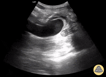
Aortic Aneurysm with Endoleak - Measurement
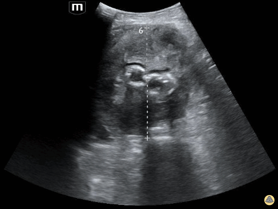
A patient in their 80s with PMH of abdominal aortic aneurysm s/p grafting with type 2 endoleak presented to the ED for dizziness. Bedside ultrasound demonstrated an 8.55 cm aortic aneurism with large thrombus. Endoleak is the hypoechoic area seen above the hyperechoic endograft.
Mehtab Galeh, MD; Bayley Espinoza, MD
Aortic Aneurysm with endoleak
A patient in their 80s with PMH of abdominal aortic aneurysm s/p grafting with type 2 endoleak presented to the ED for dizziness. Bedside ultrasound demonstrated an 8.55 cm aortic aneurism with large thrombus. Endoleak is the hypoechoic area seen above the hyperechoic endograft.
Mehtab Galeh, MD; Bayley Espinoza, MD
Thoracic Aortic Aneurysm Rupture Seen on PLAPS View
An elderly woman presented to the ED with dyspnea and left mid back pain for 1 week. Left PLAPS (Posterolateral Alveolar and/or Pleural Syndrome) view revealed a round-shaped pulsatile mass surrounded by pleural effusion & atelectatic lung. CTA later confirmed a partially ruptured, thrombosed thoracic aorta aneurysm. This case illustrates the possibility of quickly diagnosing life-threatening conditions with POCUS.
Contributed by: Caio Sangirardi, Emergency Medicine Resident Quinta D'Or Hospital, Rio de Janeiro - Brazil, @SangirardiMD
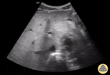
Bilateral Iliac Artery Aneurysms
This patient originally complained of right back pain as well as numbness along the thigh and knee. Starting from the abdominal artery, gliding inferiorly will eventually reveal the bifurcation of the abdominal aorta into the left and right common iliac arteries. In this scan, you can see both iliac arteries have enlarged diameters, indicating aneurysms in both.
Image courtesy of Robert Jones DO, FACEP @RJonesSonoEM
Director, Emergency Ultrasound; MetroHealth Medical Center; Professor, Case Western Reserve Medical School, Cleveland, OH
View his original post here
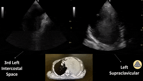
Subacute Aortic Trauma
Patient with a subacute history of chest trauma (occurred approximately 30 days prior) presented reporting intermittent chest pain. He was noted in the ED to have persistent pain and to be sweating. Bedside evaluation included lung ultrasound that identified the cystic structure seen here with a hypoechoic interior with visible swirling. Chest CT shown confirmed this to be sequelae of aortic trauma.
Renato Melo, Emergency Physician Brazil. PocusJedi affiliated.
@Renato_Melo_
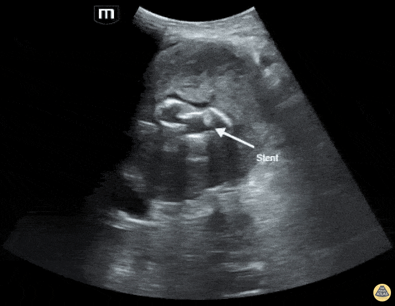
Aortic Endograft Leak - Transverse
Aortic Endograft Leak - Dr. Lindsay Howe Dr. Tim Scheel Dr. Paul Pelletier - Denver Health
Aortic Endograft Leak Long Axis
Aortic Endograft Leak - Dr. Lindsay Howe Dr. Tim Scheel Dr. Paul Pelletier
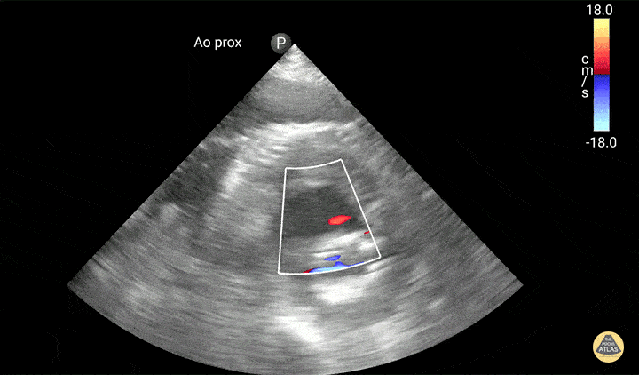
Aortic Graft Endoleak
Active extravasation is seen through endograft into false lumen. Known chronic endoleak, but CT 30 minutes prior to this study showed no active extravasation or impending rupture. Keep an open mind when reassessing your patients, and try not to anchor too much on prior results or others' opinions!
Dr. Jaffa