
Orbital Jr
Orbital Jr
Note: These search bar results are only for the Orbital Jr image archive. To search the entire site, click here.
Recently Added

Retinoblastoma in a 3-year-old is noticed on the right side of the clip. Associated retinal detachment on the left side.
Contributor: Peter Gutierrez, MD, FAAP Emory University School of Medicine/Children's Healthcare of Atlanta, @pocuspete
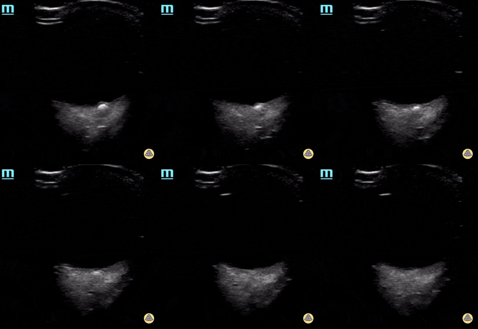
Note the hyperechogenic area that represent the drusen in the optic disc.
Contributor: Maher M. Abulfaraj, MD, @mahermabulfaraj
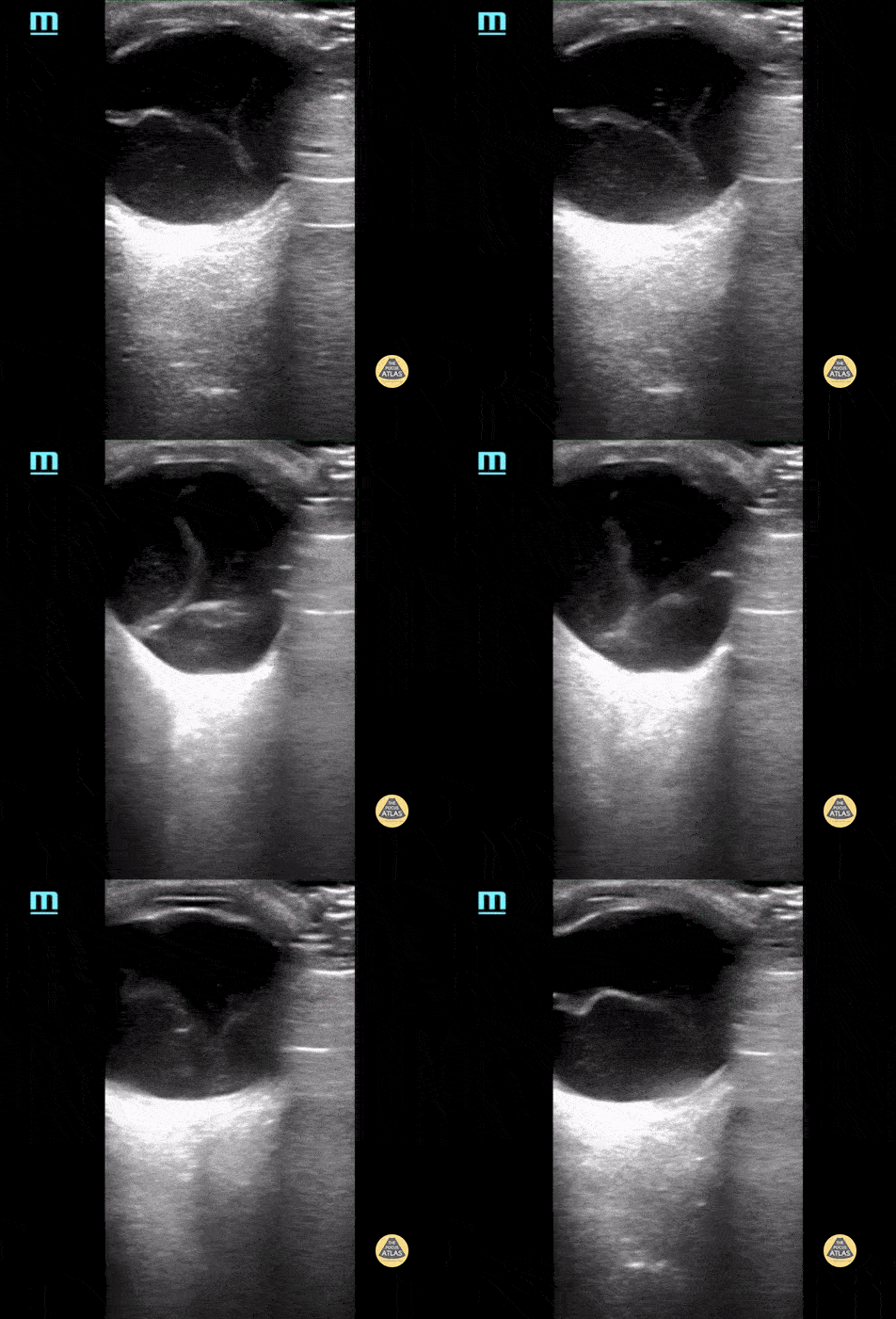
Retinal detachment. Please note how the retina is floating the posterior chamber and is anchored to the optic disc posteriorly.
Contributor: Maher M. Abulfaraj, MD, @mahermabulfaraj
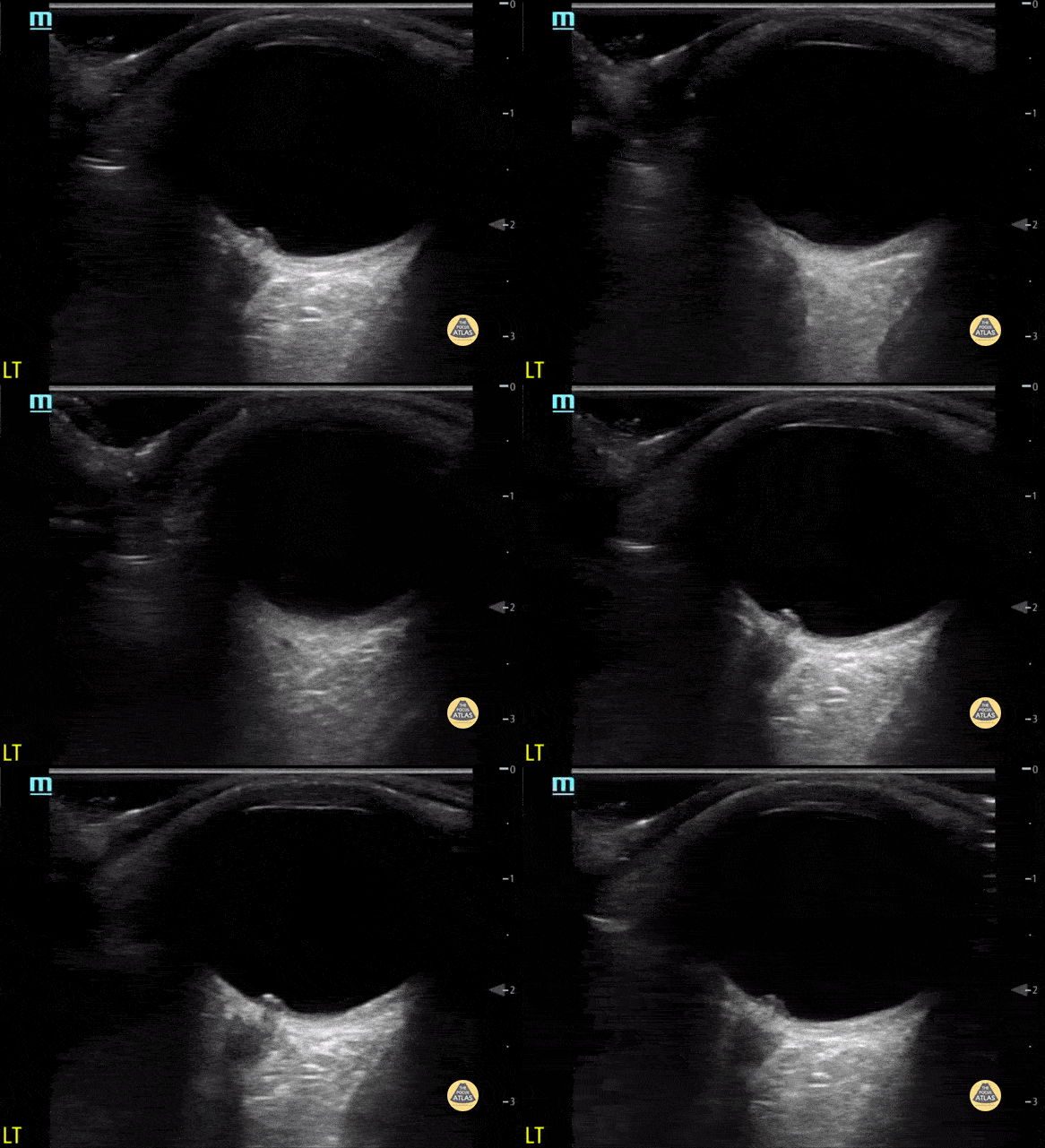
Optic disc elevation representing papilledema
Contributor: Maher M. Abulfaraj, MD, @mahermabulfaraj
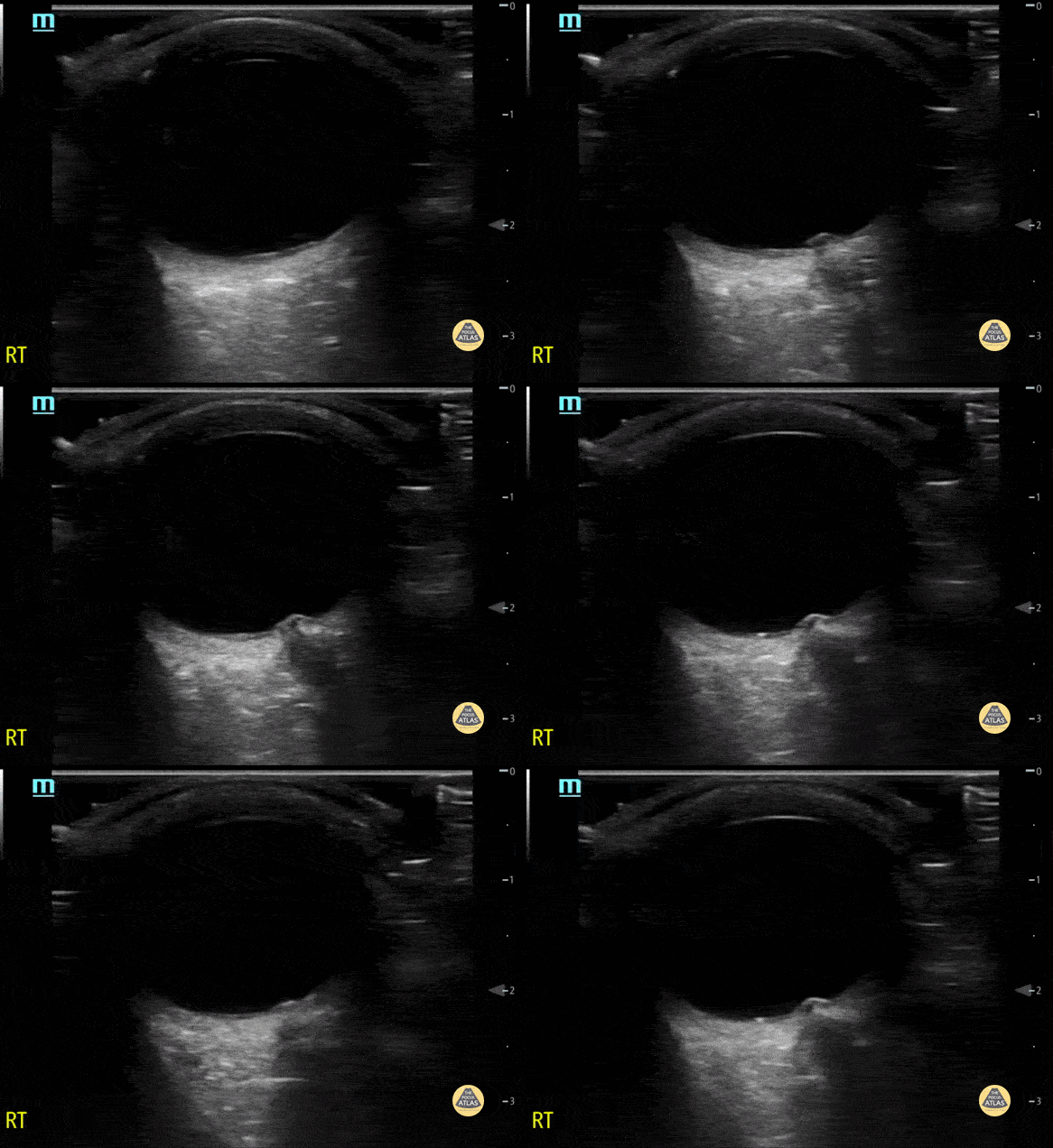
Optic disc elevation representing papilledema
Contributor: Maher M. Abulfaraj, MD, @mahermabulfaraj

Normal ocular anatomy, note the cornea, iris and lens anteriorly
Contributor: Maher M. Abulfaraj, MD, @mahermabulfaraj
Normal Anatomy
Normal ocular anatomy, note the cornea, iris and lens anteriorly
Contributor: Maher M. Abulfaraj, MD, @mahermabulfaraj
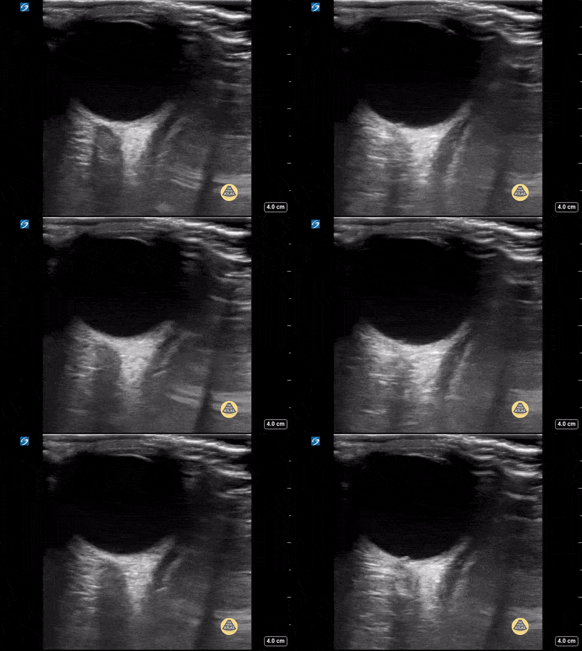
8 year old female presented with headache for 3 days, ocular ultrasound revealed no increased optic nerve sheath diameter. Measured 3 mm from the posterior border of the eye, the diameter was 3.7 mm, and there was no visualized crescent sign.
Contributor: Zach Boivin, MD, @ZachBoivinMD
Retinal Detachment
Retinoblastoma in a 3-year-old is noticed on the right side of the clip. Associated retinal detachment on the left side.
Contributor: Peter Gutierrez, MD, FAAP Emory University School of Medicine/Children's Healthcare of Atlanta, @pocuspete
Retinal detachment. Please note how the retina is floating the posterior chamber and is anchored to the optic disc posteriorly.
Contributor: Maher M. Abulfaraj, MD, @mahermabulfaraj
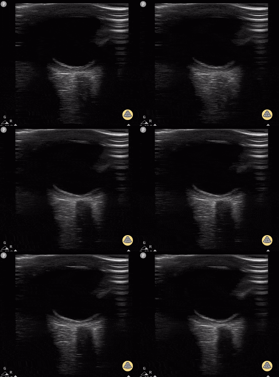
12 year old with subtle retinal detachment (vision 20/400 in affected eye). Dilated eye exam with Inferior retinal detachment from 3 o'clock to 9 o'clock.
Contributor: Antonio Riera, MD
Vitreous Detachment
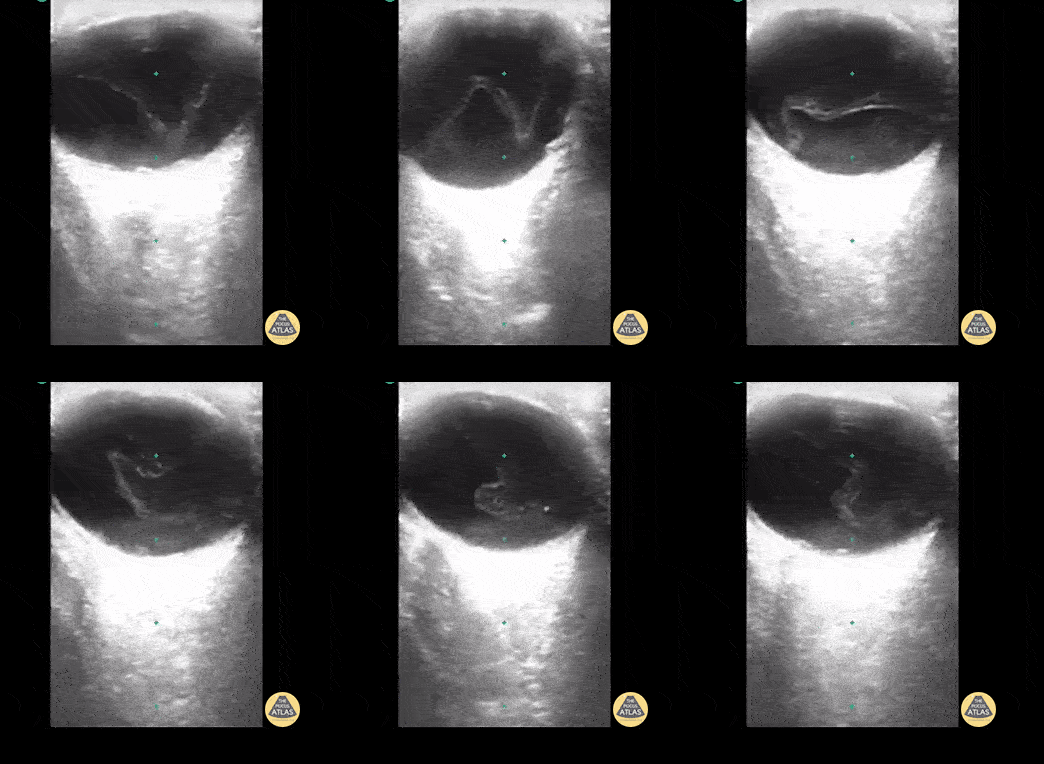
12 y/o with blurry vision for 1 month. POCUS shows thickening of vitreous in middle of eye / vitreous detachment. Note there is no point of fixation at the base/optic nerve when patient is asked to move eye side to side. This finding differentiates vitreous detachment from retinal detachment.
Contributor: Rahul Shah, MD
Vitreous Hemorrhage
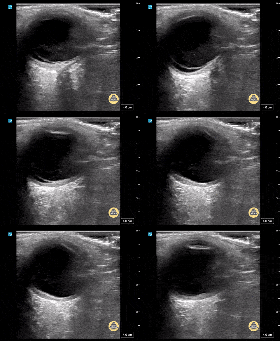
8 yo male was on a scooter and struck his head on handle. He presented to the ed with blurry vision. POCUS shows vitreous hemorrhage with a retinal detachment.
Contributor: Richard Ramirez, MD Nicklaus Children's Hospital
Dilated Optic Nerve
Optic disc elevation representing papilledema
Contributor: Maher M. Abulfaraj, MD, @mahermabulfaraj
Optic disc elevation representing papilledema
Contributor: Maher M. Abulfaraj, MD, @mahermabulfaraj
Other Pathology
Retinoblastoma in a 3-year-old is noticed on the right side of the clip. Associated retinal detachment on the left side.
Contributor: Peter Gutierrez, MD, FAAP Emory University School of Medicine/Children's Healthcare of Atlanta, @pocuspete
Note the hyperechogenic area that represent the drusen in the optic disc.
Contributor: Maher M. Abulfaraj, MD, @mahermabulfaraj
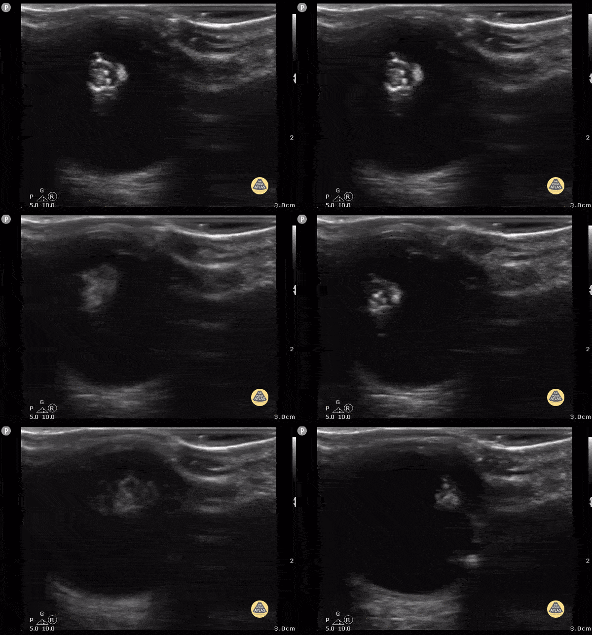
19 year old female with glaucoma presents with head trauma and abnormality of the lens on CT (calcification) that was subsequently visualized by POCUS.
Contributor: Julie Leviter, MD