Lumbar Erector Spinae Plane (ESP) Block
The lumbar erector spinae plane block deposits local anesthetic at the corner of a lumbar transverse process, providing analgesia for low back pain and radicular symptoms such as sciatica.
Indications: Low back pain, sciatica
Considerations & Technical Details
Considerations:
Document a neurovascular exam prior to performing the block.
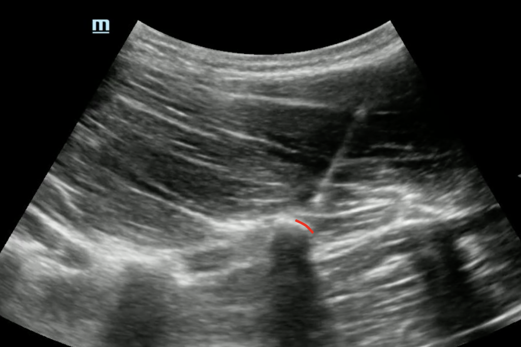
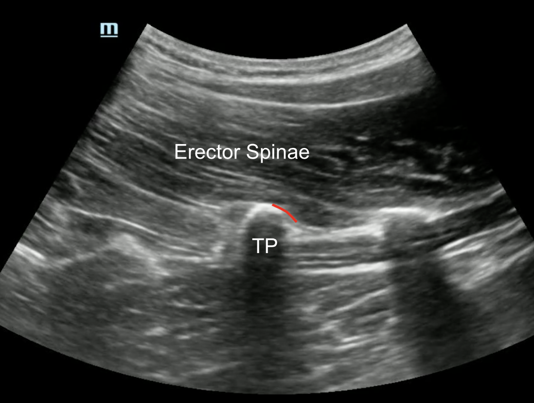
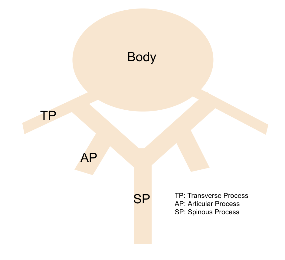
Lumbar vertebrae have a transverse process laterally, a spinous process medially, and an articular process in between. These articular processes are often more superficial and larger than the transverse processes and can be easily mistaken for your target. Aim for the most lateral bony structure that you encounter.
Use half-dose anesthetic for thoracolumbar blocks due to higher risk of LAST given the increased likelihood of vascular absorption in these regions.
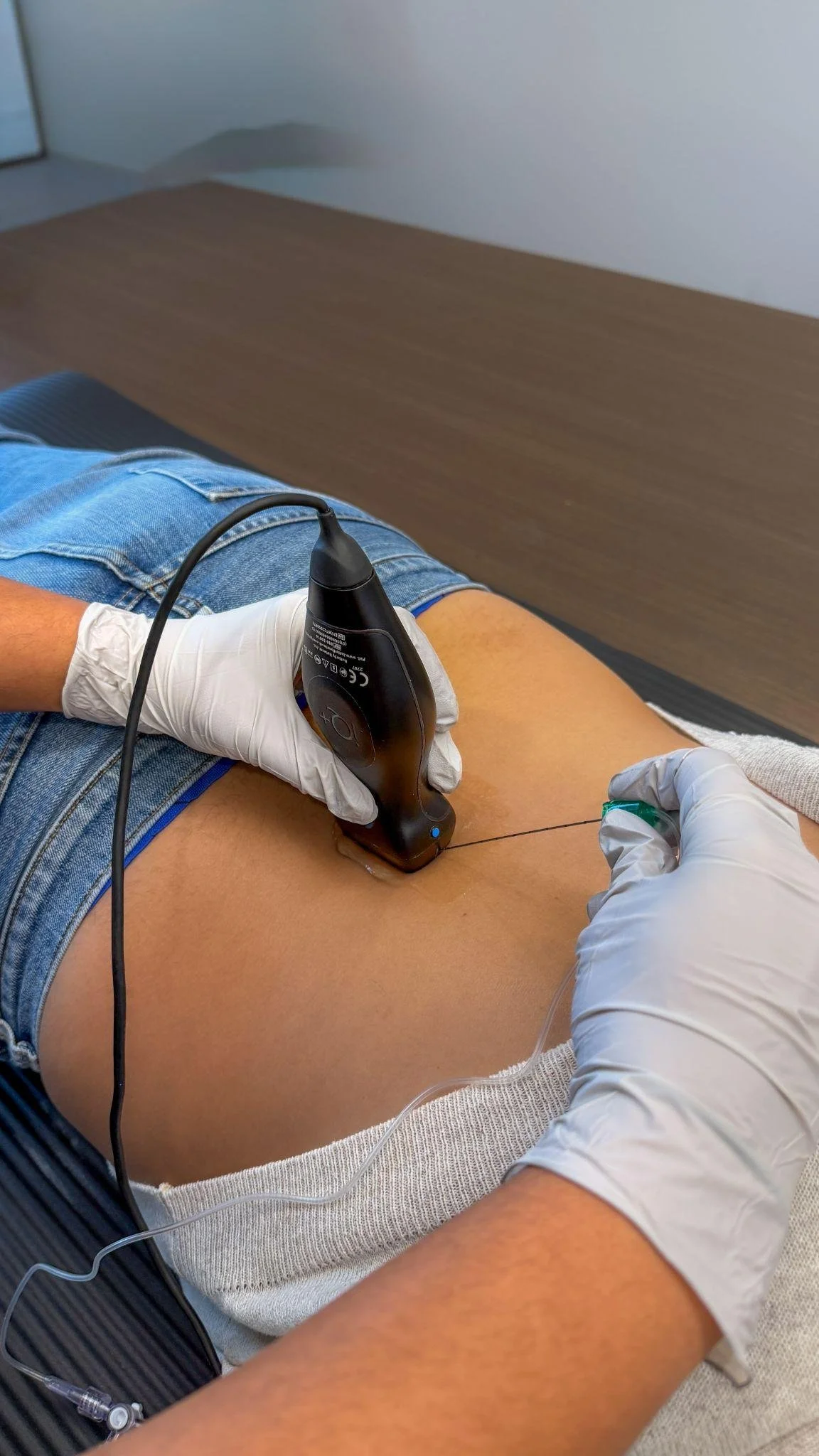
Transducer: Low-frequency curvilinear probe
Needle: 20 g, 10 cm nerve block (or spinal) needle
Anesthetic Volume: 20–30 mL
Target of Anesthetic: Anesthetic spread along the corner of the transverse process of the lumbar vertebrae
Area of anesthesia
Positioning
Position the patient prone (preferred) or sitting. Place the curvilinear probe in a cranial-to-caudal (parasagittal) orientation.
Target Identification
Begin medially over the superficial spinous process, then slowly move the probe laterally past the articular process (which can look very similar to the transverse process) until you identify the last and deepest bony structure — this is the transverse process and your target.
Needle Approach
Using a steep needle angle, advance the needle in-plane targeting the corner of the transverse process. Deposit anesthetic against the periosteum to lift the erector spinae fascial plane off the bone.