
Biliary Pathology
Biliary Pathology
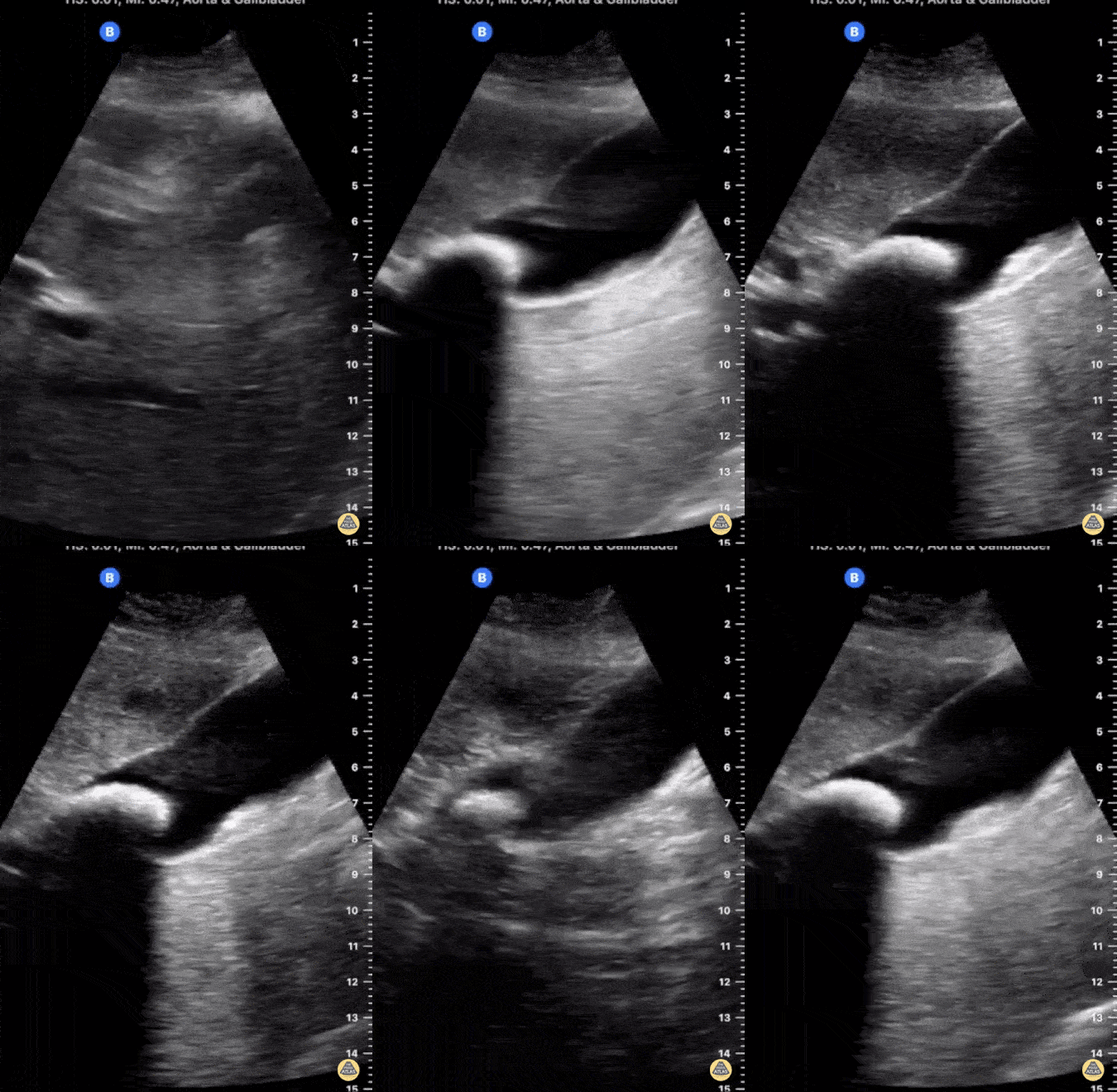
This image demonstrates a large gallstone lodged in the neck of a distended gallbladder with posterior shadowing. There is no evidence of gallbladder wall thickening seen here.
Jonny Wilkinson, Consultant in ICU and Anesthesia
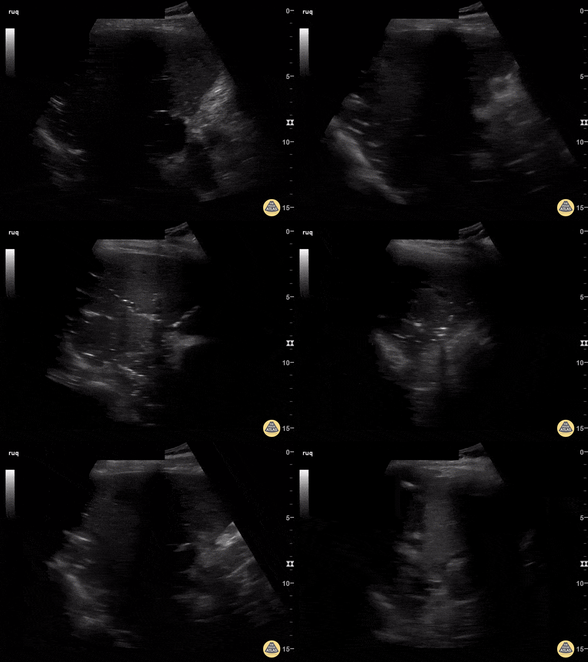
82-year-old male with history of recurrent cholangitis presenting to the ED with abdominal pain. US imaging of the RUQ reveals presence of pneumobilia. Click here to read the full caption.
Andrew Morris, M.D. Gaurav Patel, M.D. Vu Huy Tran, M.D. - Aventura Hospital & Medical Center, Emergency Medicine Program
This non-inflamed gallbladder contains many stones. The stones are hyperechoic with posterior shadowing. The gallbladder wall is not thickened, there is no pericholecystic fluid, and no sonographic Murphy's sign, thus cholecystitis is unlikely.
Justin Bowra MBBS, FACEM, CCPU Emergency Physician, RNSH et al. (Dr. Ken Lee)
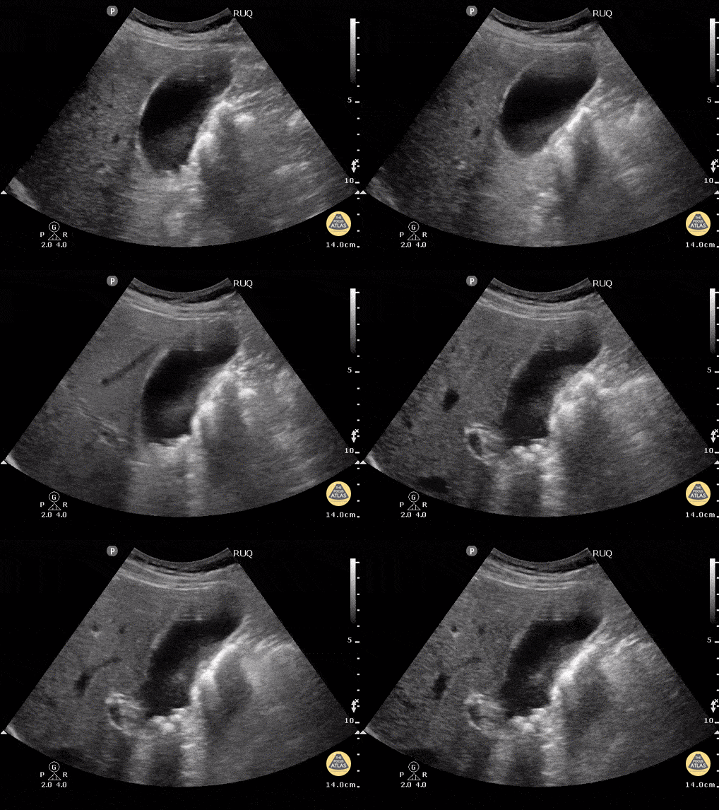
This patient w/o cholecystitis clearly demonstrates cholelithiasis. A 2002 study showed that ER doctors can diagnose gallstones with a sensitivity of 88% and specificity 96% with POCUS.
Gallstones can be identified by hypoechoic "shadowing" behind hyperechoic stones. If there isn't shadowing, hyperechoic structures could represent polyps or sludge. In this image the stones are resting mainly in the neck of the gallbladder.
Kendall JL, Shimp RJ. Performance and interpretation of focused right upper quadrant ultrasound by emergency physicians. J Emerg. Med. 2001; 21(1):7-13
Submitted by Justin Bowra MBBS, FACEM, CCPU Emergency Physician, RNSH et al.
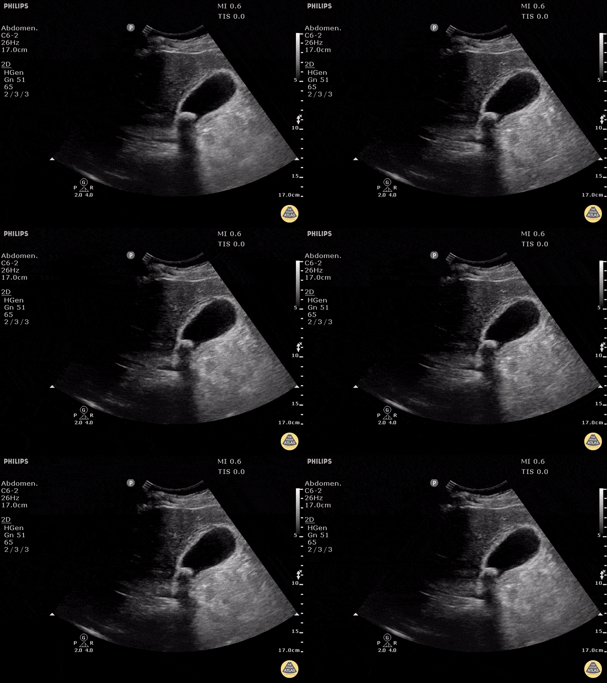
This clearly demonstrates a stone in the gallbladder neck as a hyperechoic structure with posterior shadowing. The neck is not always clearly visualized on first glance so it is important to scan through the gallbladder in two planes to exclude stones in the neck. Other signs of acute cholecystitis include pericholecystic fluid, gallbladder wall thickening (>3mm) common bile duct dilatation, and positive sonographic Murphy's sign.
Justin Bowra MBBS, FACEM, CCPU Emergency Physician, RNSH et al. (Mo)
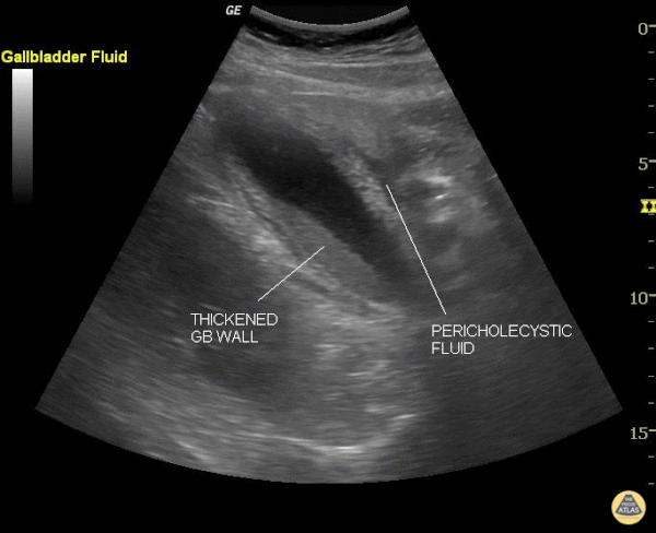
This still image shows two clear signs of acute cholecystitis. The gallbladder wall is thickened and there is pericholecystic fluid. Of note, when measuring the gallbladder wall it's better to measure the anterior wall as the posterior wall can appear enlarged due to artifact. The cut off for wall thickening is 3mm. The anterior wall is thickened in this case but is not labeled. Other signs to look for are stones, sludge, common bile duct dilatation, and overall gallbladder enlargement.
Of note, a positive sonographic Murphy's carries a sensitivity of 63%, specificity of 93.6% and positive predictive value of 72.5%.
Justin Bowra MBBS, FACEM, CCPU Emergency Physician, RNSH et al. (Sharon)
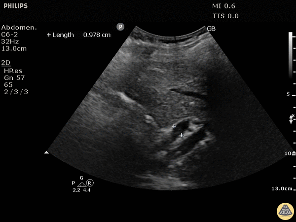
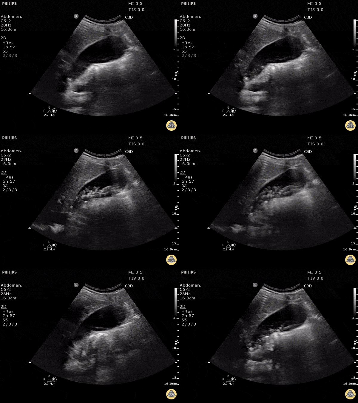
This patient has a dilated common bile duct and obstructive pattern LFTs, highly suggestive of choledocholithiasis or other biliary obstruction. No stone is visualized on this study.
A normal common bile duct should be <4mm plus 1mm per decade after 40 years of age. Here it is 9.78 mm.
POCUS is highly sensitive for acute cholecystitis but as many people already know, it can be hard to find the CBD, which is reflected in the lower sensitivity and specificity for this indication in many studies.
Justin Bowra MBBS, FACEM, CCPU Emergency Physician, RNSH et al. (Dr. Ken Lee)
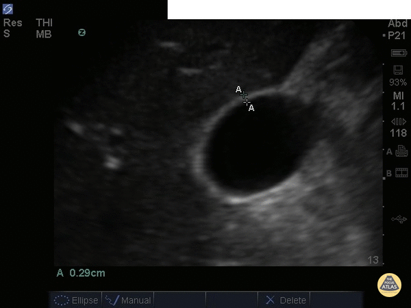
This normal gallbladder image shows how to measure the anterior wall of the gallbladder. A normal gallbladder wall should be less than 3mm. The posterior wall should not be measured as it can appear enlarged due to artifact.
Justin Bowra MBBS, FACEM, CCPU Emergency Physician, RNSH et al.
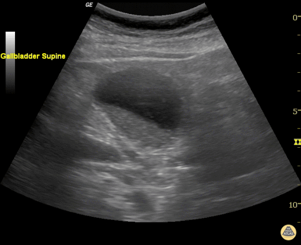
Echogenic material can be seen within this gallbladder but there are no stones. It is not bright white (it is not highly echogenic) and furthermore there isn't any shadowing. When there is no shadowing consider polyps or in this case biliary sludge.
Justin Bowra MBBS, FACEM, CCPU Emergency Physician, RNSH et al. (Sharon)
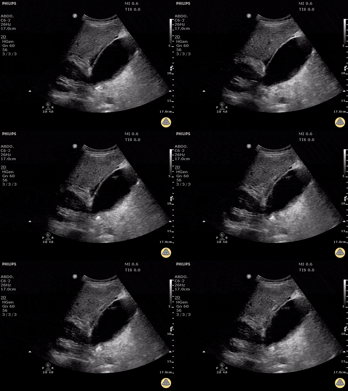
Free fluid can be appreciated around this gallbladder. This is called pericholecystic fluid. However there are no stones, nor gallbladder wall thickening, and the sonographic Murphy's is negative. This free fluid is unlikely to be caused by cholecystitis. In this case, the free fluid came from a different source.
Justin Bowra MBBS, FACEM, CCPU Emergency Physician, RNSH et al.
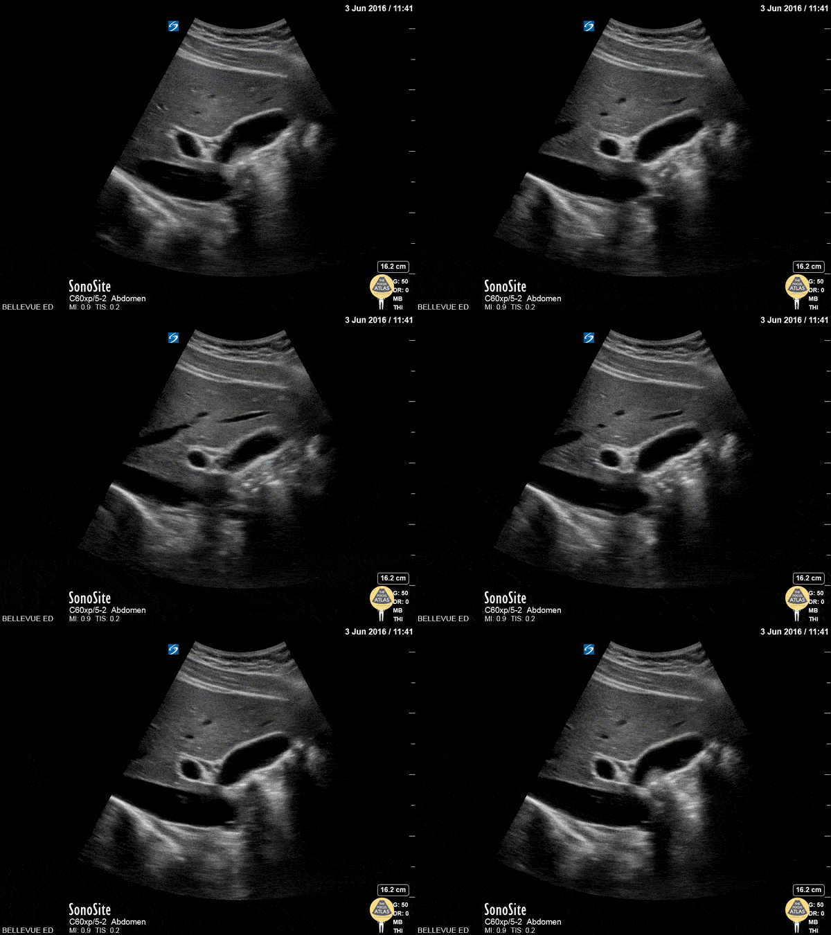
In this clip we see the liver on the left, and the IVC inferior to the liver with a hepatic vein draining into it. The ovoid anechoic gallbladder is in long axis in the center of the screen. To the left of the gallbladder we see the portal vein in cross-section. Running just superior to the portal vein, in long axis, is the narrow common bile duct.
Hannah Kopinksi and Dr. Lindsay Davis - NYU Emergency Medicine