
Lung Pathology
Lung Pathology
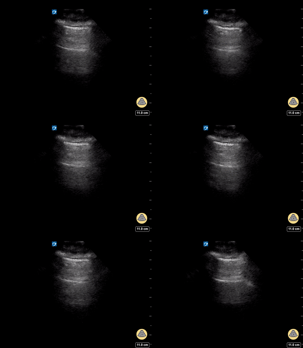
A lines appear as horizontal lines that represent normal aerated lung (dry interlobular septa). They are a reverberation artifact caused by the sound waves bouncing off the highly echogenic pleura and back to the probe, and repeating.
Hannah Kopinski (MS4) and Dr. Lindsay Davis - NYU Emergency Medicine, Matthew Riscinti - Kings County Emergency Medicine
26 yo male presents to ED stating he was kicked in the chest. He went home to “try to relax and smoke some weed” now short of breath and with pleuritic chest pain after smoking. POCUS demonstrating decreased lung slide on the left.
What are the signs of pneumothorax on ultrasound?
Decreased lung sliding - In normal lungs, lung sliding refers to the parietal pleura moving against the visceral pleura - described as “ants marching.”
Lack of B-lines or comet tails – These artifacts will not be present if there is a pneumothorax and the presence of B-lines or comet tails can rule out a pneumothorax.
No Lung pulse – the visceral pleura moving along a stationary parietal pleura due to cardiac motion when lung sliding is not present. These are so called “T lines” on M-mode. These signify that the parietal and visceral pleura are opposing one another and therefore that there is no pneumothorax
Lung point – 100% specific for pneumothorax, this is the cutoff point above which you can appreciate the lung sliding and below which there is no lung sliding. The lung point is the pneumothorax border.
Dr. Stacey Frisch, Dr. John F Kilpatrick - Kings County Emergency Medicine
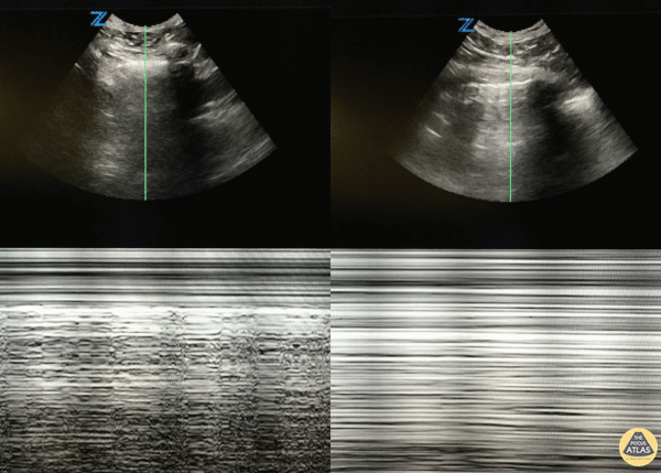
46 y/o M with 20 pack year smoking history with sudden onset right sided chest pain that woke him from sleep. Decreased breath sounds on right side. POCUS with decreased lung slide (right of image) with normal lung slide in left lung (left side of image).
Lung slide can often be appreciated by watching the pleural surfaces move along each other but if you're uncertain, putting the US in m-mode and looking for the classic "seashore sign" (left image) versus the "barcode sign" (right image) can help you figure it out.
Dr. Eric Roseman - Resident Physician, Kings County Emergency/Internal Medicine
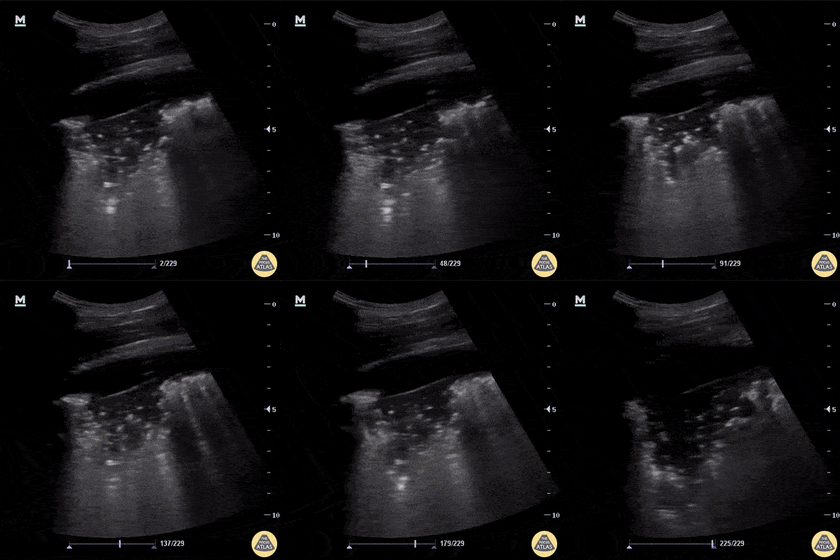
Pictured here are classic ultrasound findings for pneumonia including a shred sign, lung consolidation with dynamic air bronchograms and a small associated parapneumonic effusion. Note also adjacent B-lines.
A shred sign represents the distinction between the consolidated lung and the aerated lung and is seen in this clip as the irregular “shredded” border just posterior to the consolidation.
Aaron Inouye, PA-C, North Canyon Medical Center
@PAintheED
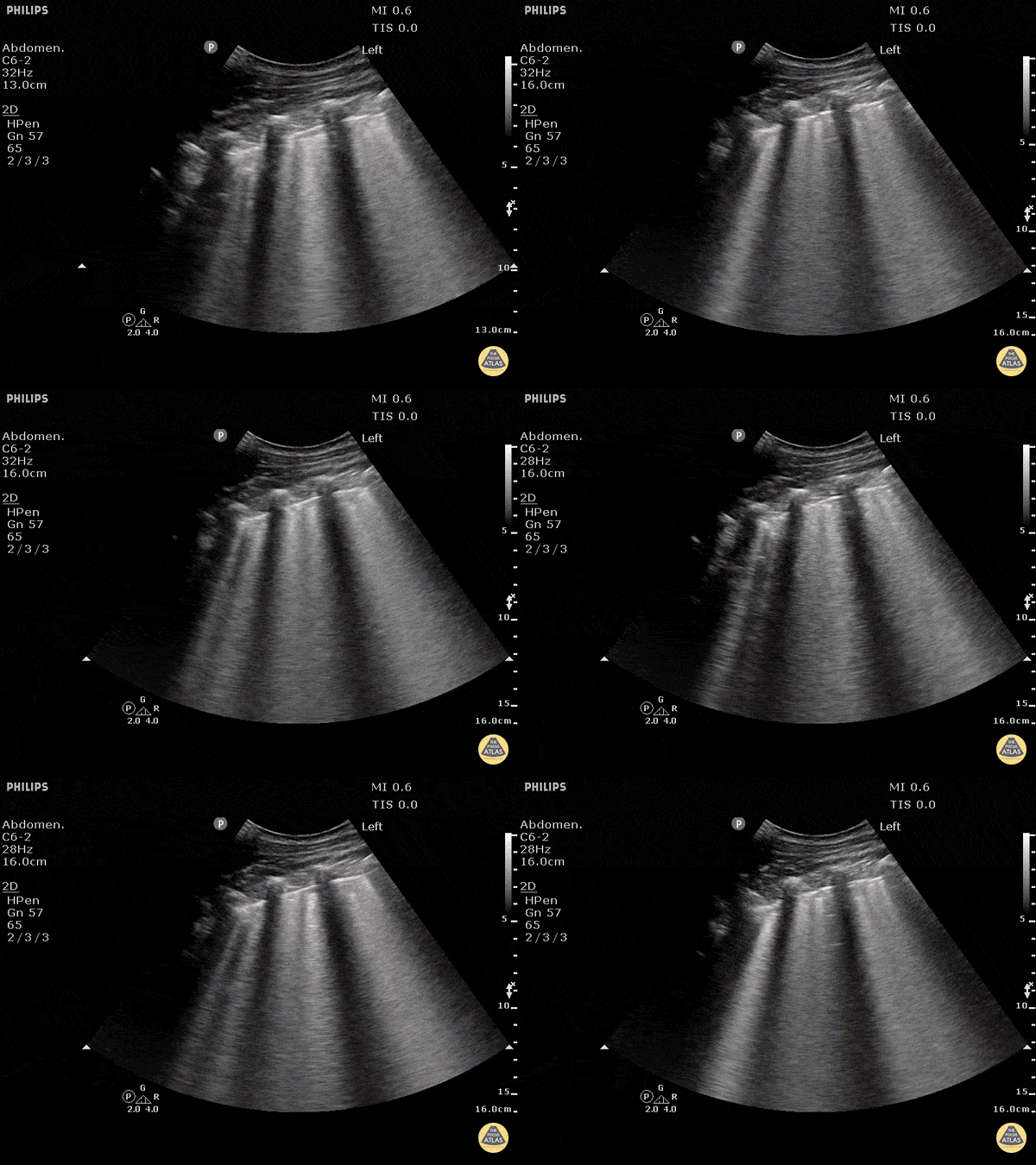
B-lines obtained with curved probe.
B-lines are vertical artifacts that move with respiration from the pleural surface. They represent increased water in an area of the lung. In the right clinical context this could represent pulmonary edema. An increase in B-lines correlates with the degree of pulmonary edema.
3 B-lines in an intercostal space represent a "positive" region of the lung, and if there are two regions of the lung that are positive, you can diagnose pulmonary edema.
Dr. Justin Bowra et al. (Dr. D Browne and Dr. J Knights)
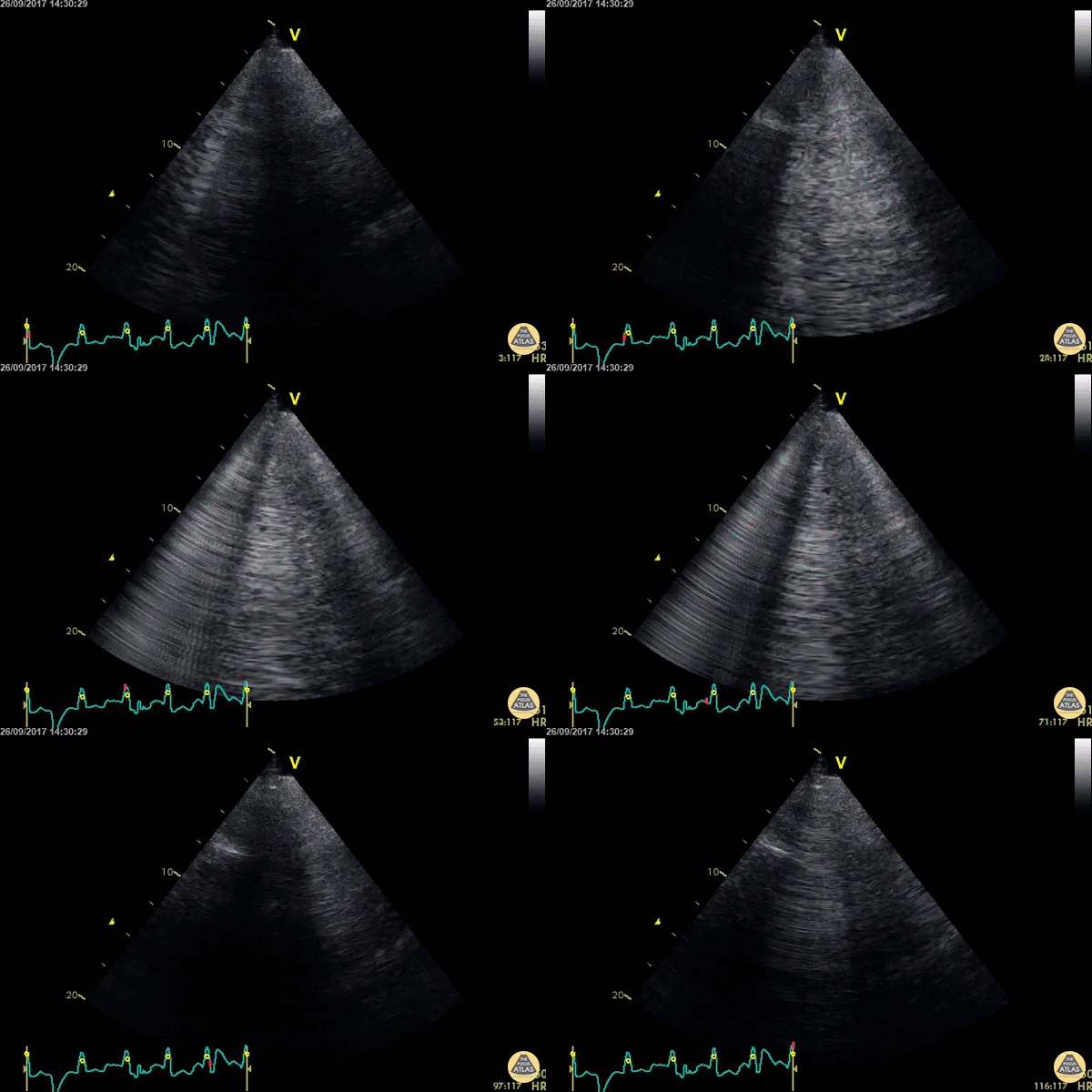
WCUME 2017 Submission for "Best POCUS"
An acutely dyspnoeic patient presents with ventricular tachycardia and has no response to initial chemical cardioversion. Lung POCUS shows widespread bilateral confluent B lines indicating acute pulmonary edema. Unstable tachycardia terminated using synchronized electrical cardioversion.
Dr. Cian McDermott - Dublin, Ireland
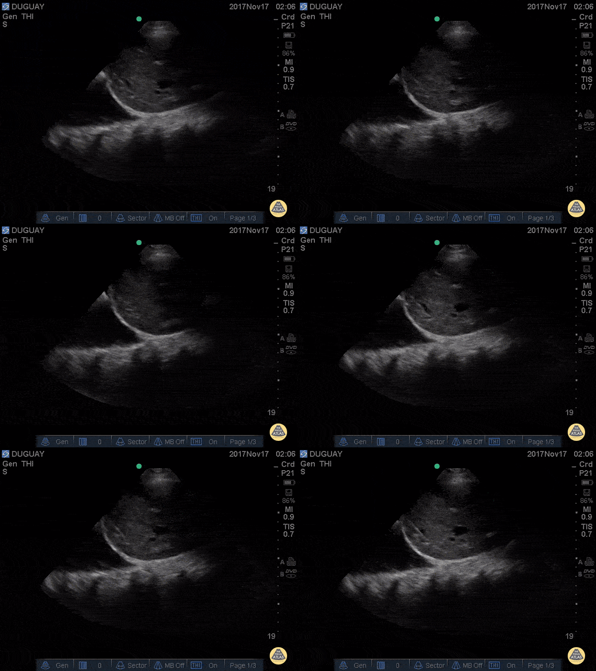
RUQ scan with large R pleural effusion. Spine sign+ (clear view of several thoracic vertebrae through the effusion)
Gary Duguay
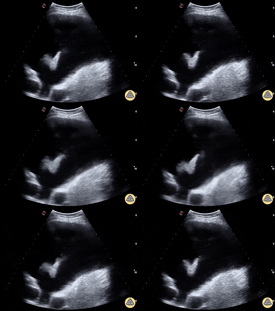
WCUME 2017 Submission for "Creative Caption"
"In these days of violent extremist and warmongers, can it be a a good omen to find a dove flying in the pleural fluid?"
Marco Garrone, MD - Torino, Italy
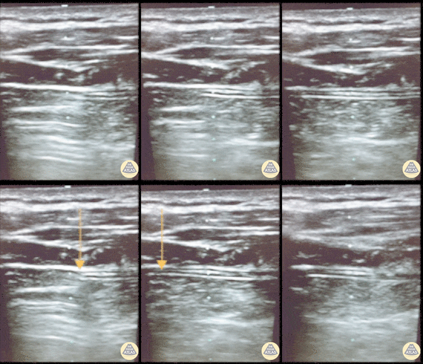
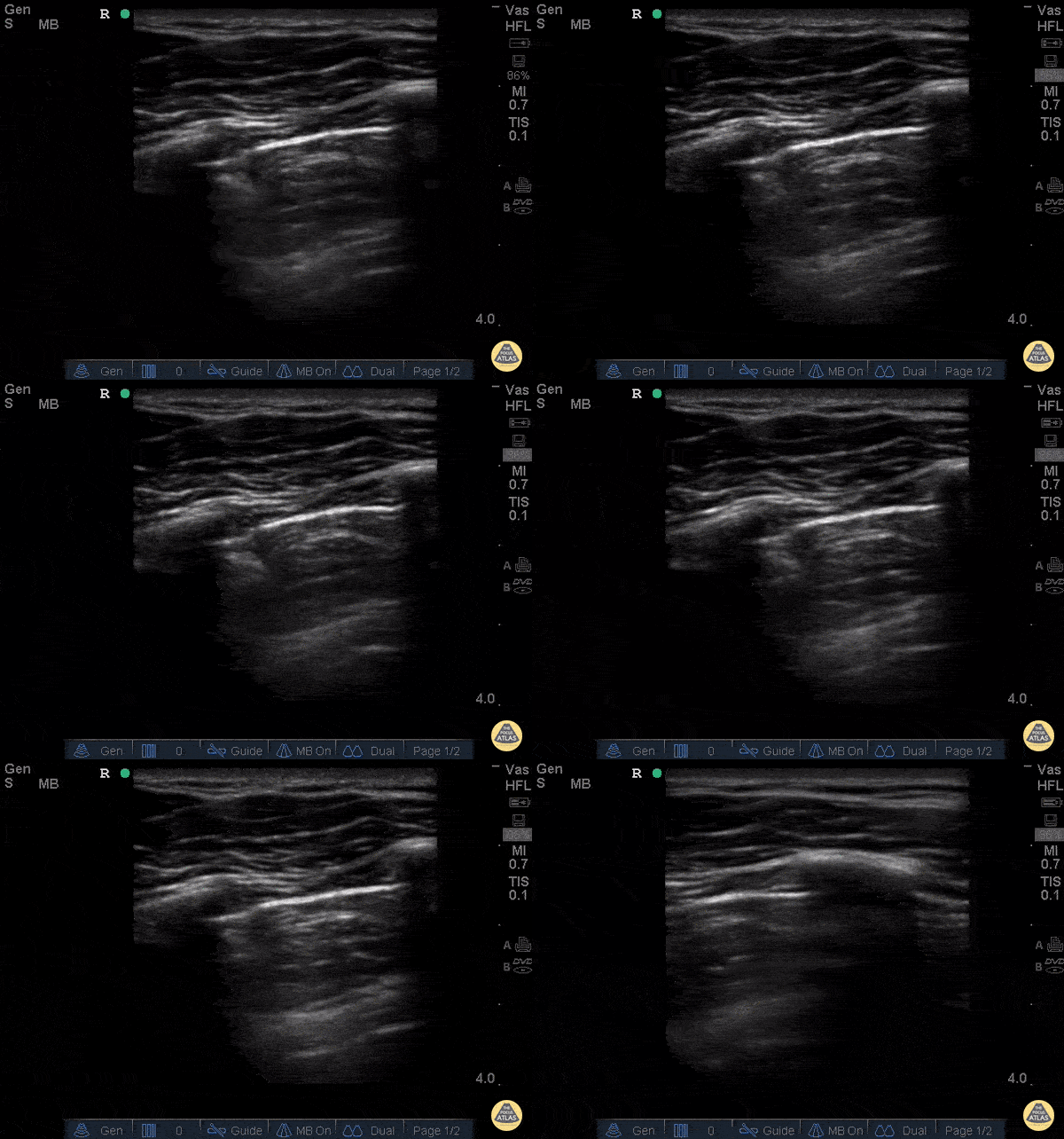
This is an image of a patient's chest wall using a high frequency transducer, with the transducer oriented in a transverse plane between rib spaces. The patient had a pneumothorax and a chest tube was placed. This clip illustrates what happens when the suction is turned 'on'. You will see the pleura slide from right to left as the pneumothorax resolves.
- Jason Tanguay, DO; Ultrasound Leadership Academy Graduate
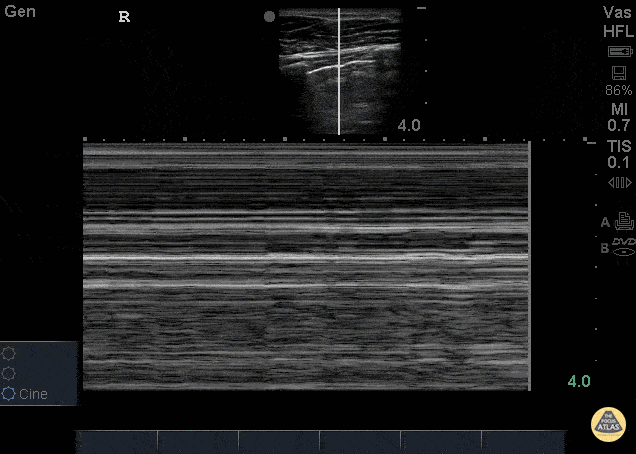
26 yo male presents to ED stating he was kicked in the chest. He went home to “try to relax and smoke some weed” now short of breath and with pleuritic chest pain after smoking. POCUS demonstrating decreased lung slide on the left.
This can be seen as decreased lung sliding - In normal lungs, lung sliding refers to the parietal pleura moving against the visceral pleura - described as “ants marching.” M-mode can be used to evaluate lung sliding. Remember, normal lung slide will look like a seashore on M-mode whereas a pneumothorax will appear as horizontal lines termed Bar Code sign (pictured here). Make sure to check in the most anterior fields as well at lateral lung fields.
Dr. Stacey Frisch, Dr. John F Kilpatrick - Kings County Emergency Medicine
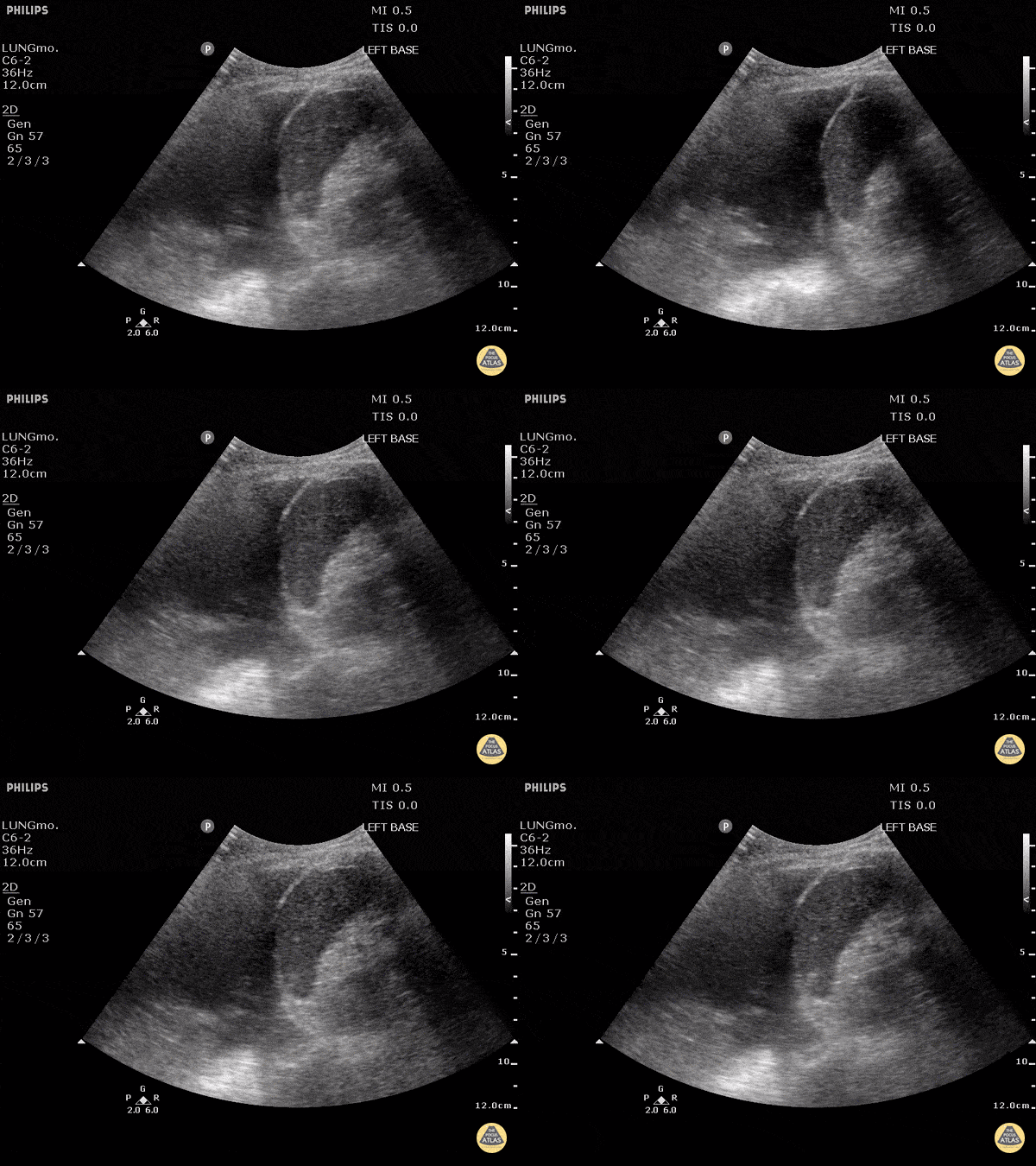
The left lung can be seen freely floating in anechoic fluid on the left side of the screen. Also pictured, the diaphragm, spleen, and edge of the beating heart.
Justin Bowra MBBS, FACEM, CCPU Emergency Physician, RNSH
et al.
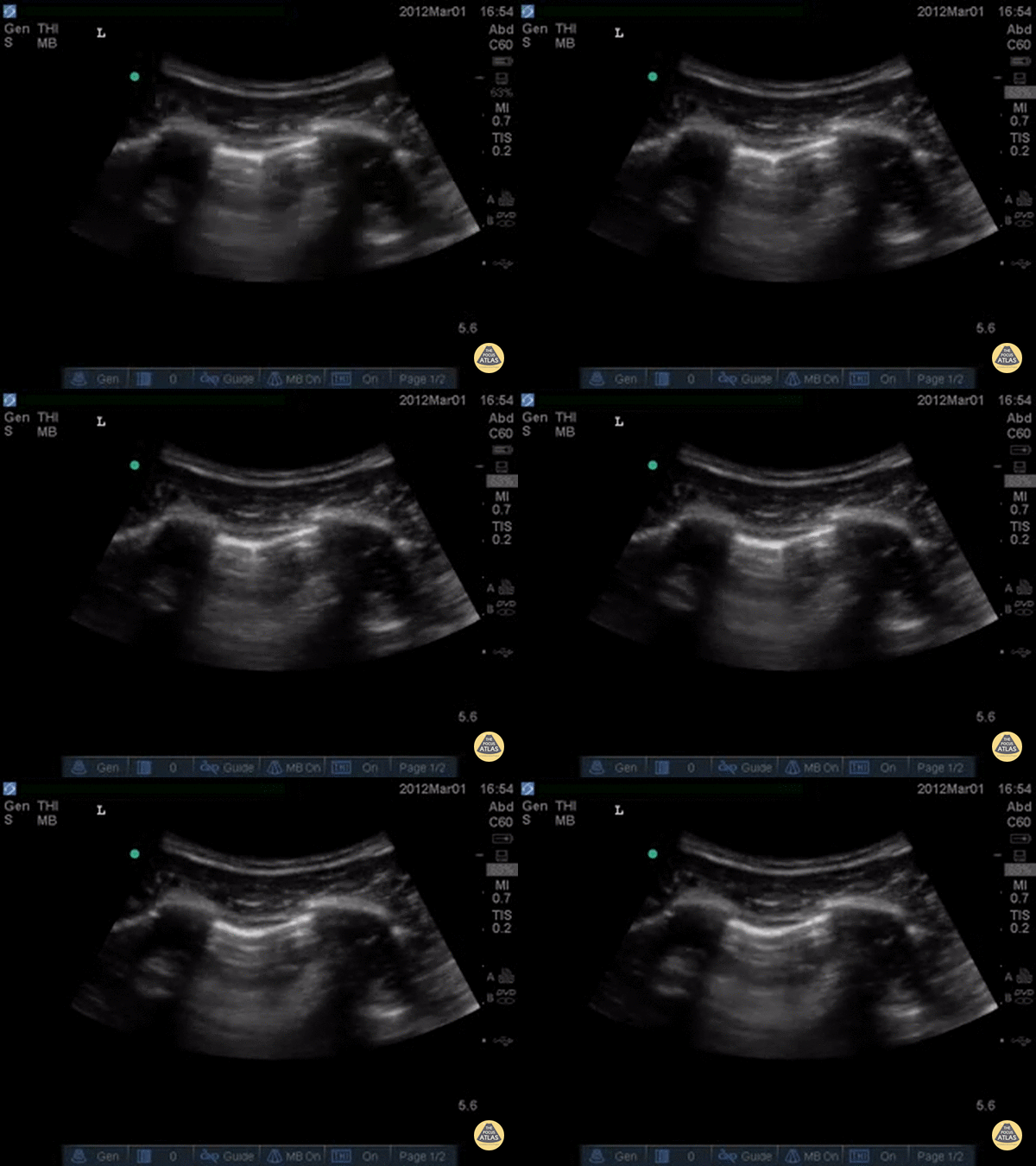
Decreased lung slide is highly sensitive, it lacks specificity. Lung point however, is a highly specific finding indicating a pneumothorax.
Lung point indicates the transition point between normal pleura with normal lung sliding and where there is air disrupting the pleural space with decreased lung sliding.
In this intercostal space, one can see lung with normal lung slide on the left, and decreased lung slide on the right, and a point where the lung slide changes, which is moving with inspiration. This is the lung point.
Dr. Justin Bowra et al.
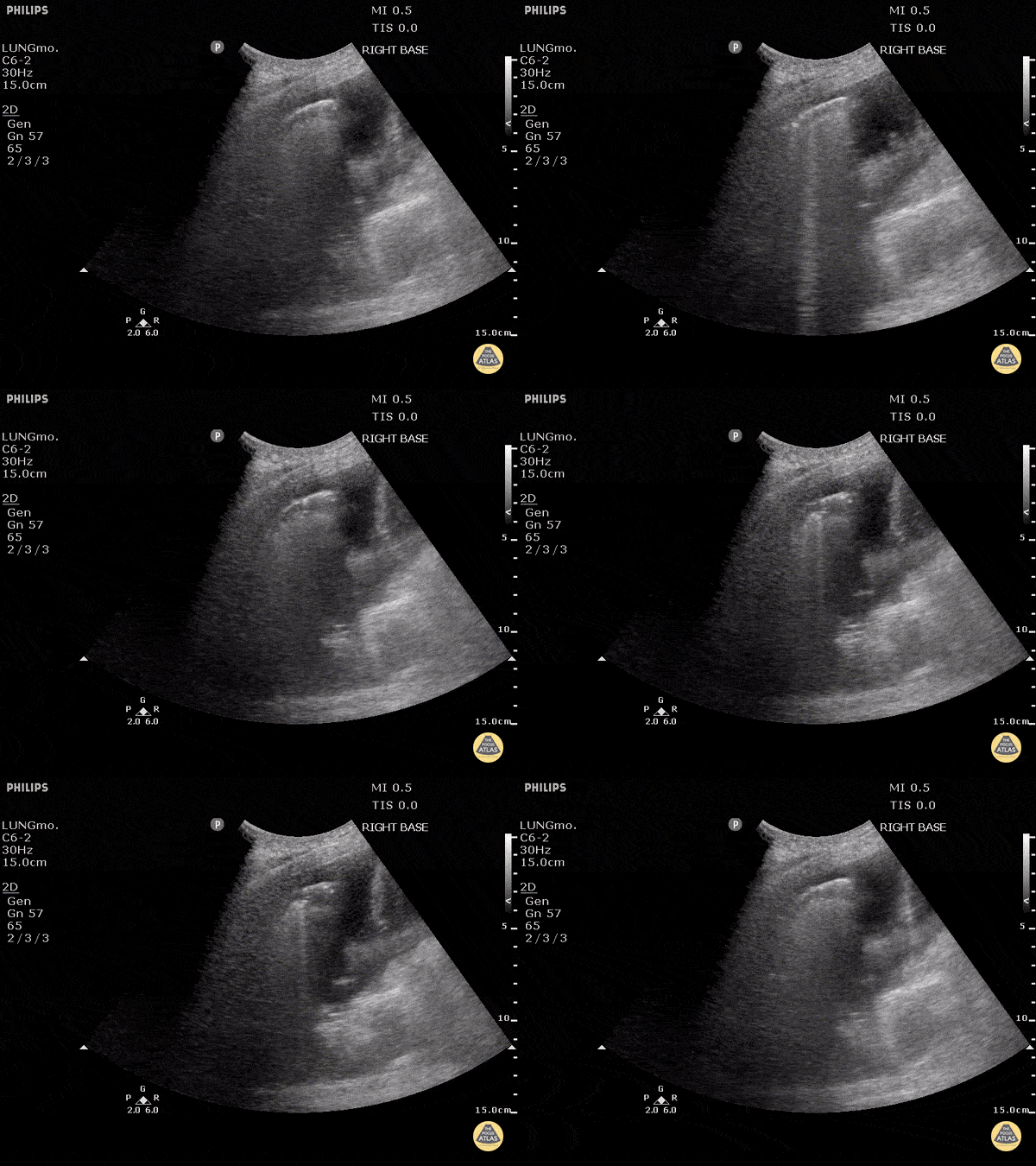
To the left of the image, the lung can be seen clearly floating in anechoic fluid representing a pleural effusion.
B-Lines can be seen radiating from the surface of the lung to the far left especially as this patient inspired.
B lines (also known as comet tails) are white lines that emanate from the pleural surface of the lung. They have been shown to be highly sensitive for pulmonary edema.
Justin Bowra MBBS, FACEM, CCPU Emergency Physician, RNSH
et al.
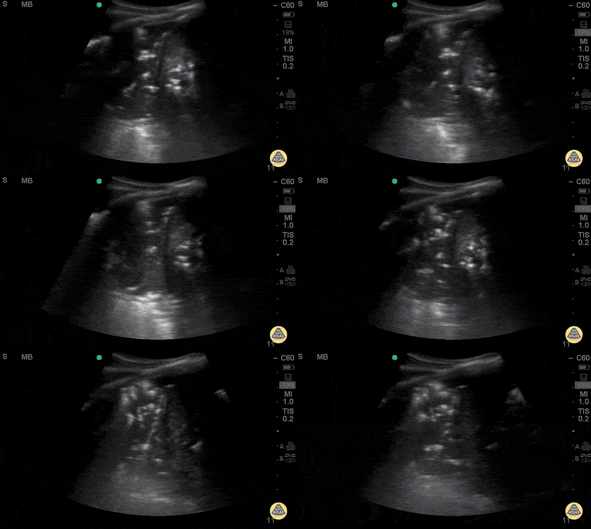
Middle aged female with history of HIV, asthma, and polysubstance use who presents with progressively worsening dyspnea over 3-4 days, found to have diffuse rales, worse in left mid to lower lung fields. AP CXR with bilateral lower lobe patchy infiltrate, left greater than right.
POCUS with curvilinear probe revealed B lines in left mid lung fields and consolidation with air
bronchograms in left lower lung. Air bronchograms is one of the most specific signs for the diagnosis of pneumonia with a specificity (93%) and a positive LR (12.14). Ultimately, CT chest is the gold standard for diagnosis of pneumonia, which was consistent with the CT in this patient.
Priscilla Chao, MD, Matthew Riscinti, MD - Kings County Emergency Medicine