
MSK Pathology
Basic MSK Pathology
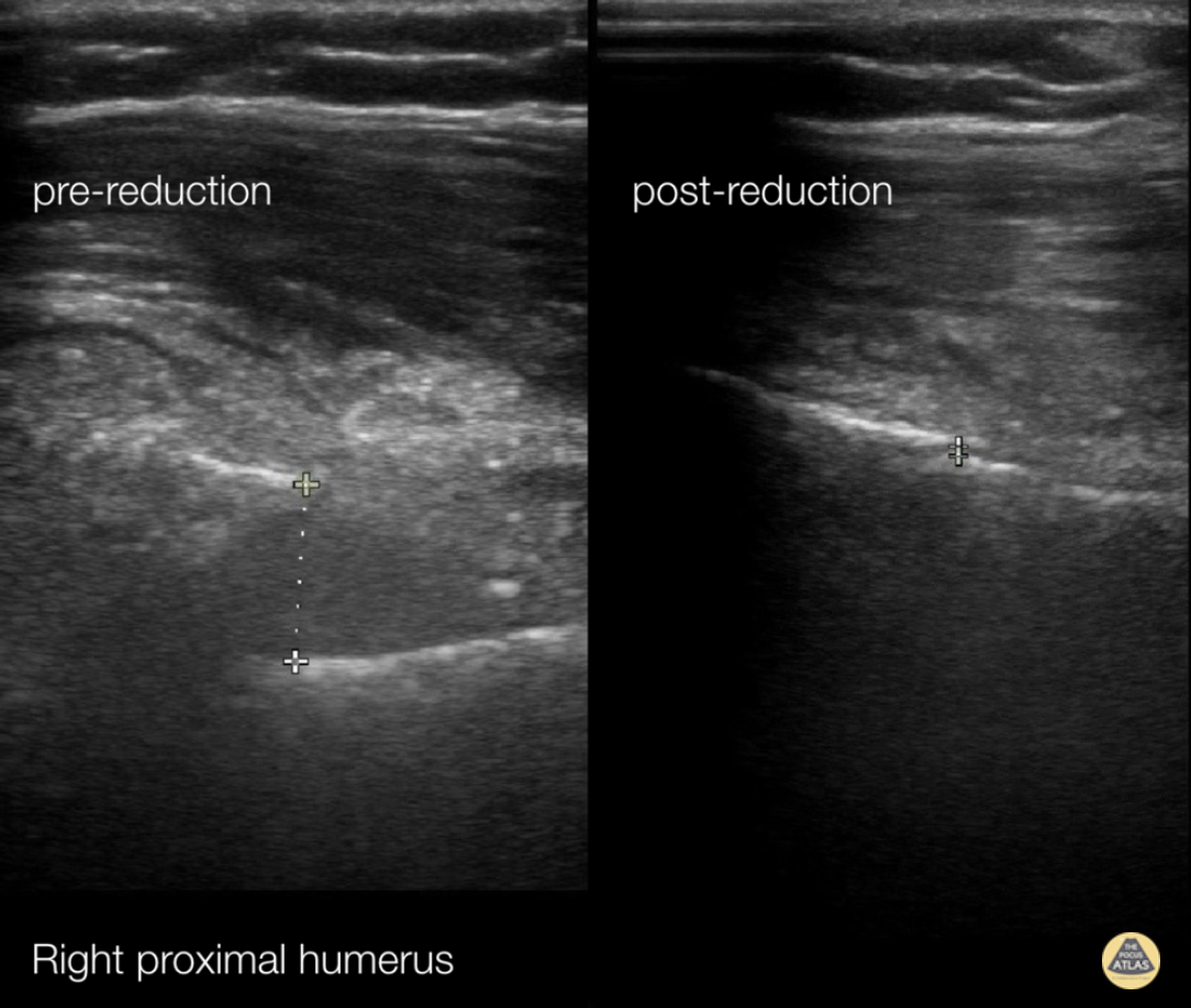
72 y/o female presents with right upper arm pain after a mechanical fall from standing.
A longitudinal coronal image was obtained at the right proximal humerus. Imaging showed displacement of bone fragments with hyperechoic lines 1.17cm apart. Pictures were obtained intermittently throughout reduction until displacement was reduced to 0.06cm.
This case demonstrates the utility of ultrasound in fracture reduction. Traditionally, care teams perform repeat X-rays until the fracture is reduced. In comparison, ultrasound can be quicker and reduce exposure to ionizing radiation.
Crozer Chester EM
Arthur Strzepka (MS4), Damarcus Ingram (MS4), Dr. Max Cooper
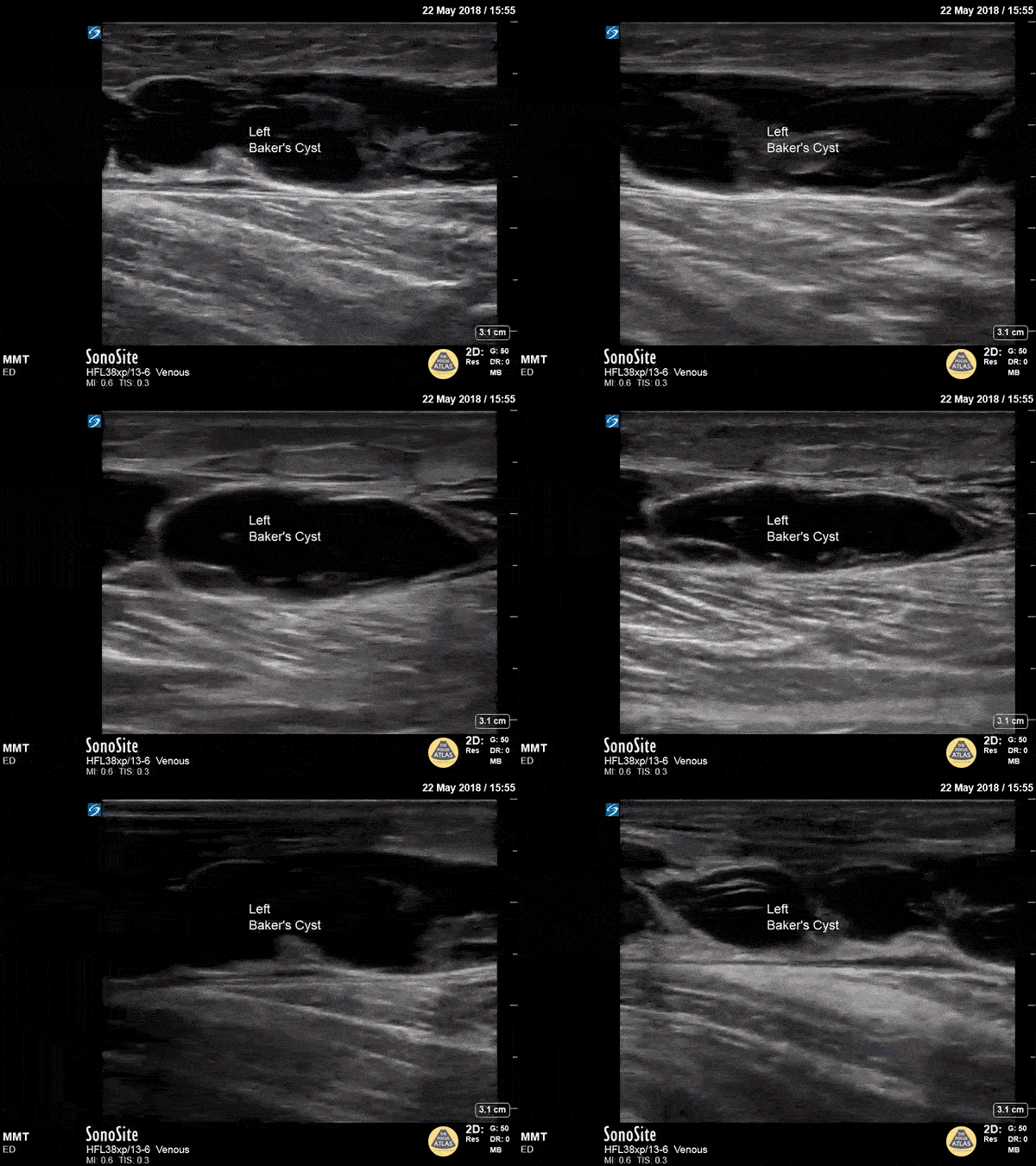
A longitudinal view of a ruptured Baker's cyst. When performing a DVT scan, always look out for incidental findings that may explain the patient's presentation!
Dr. Michael Trauer
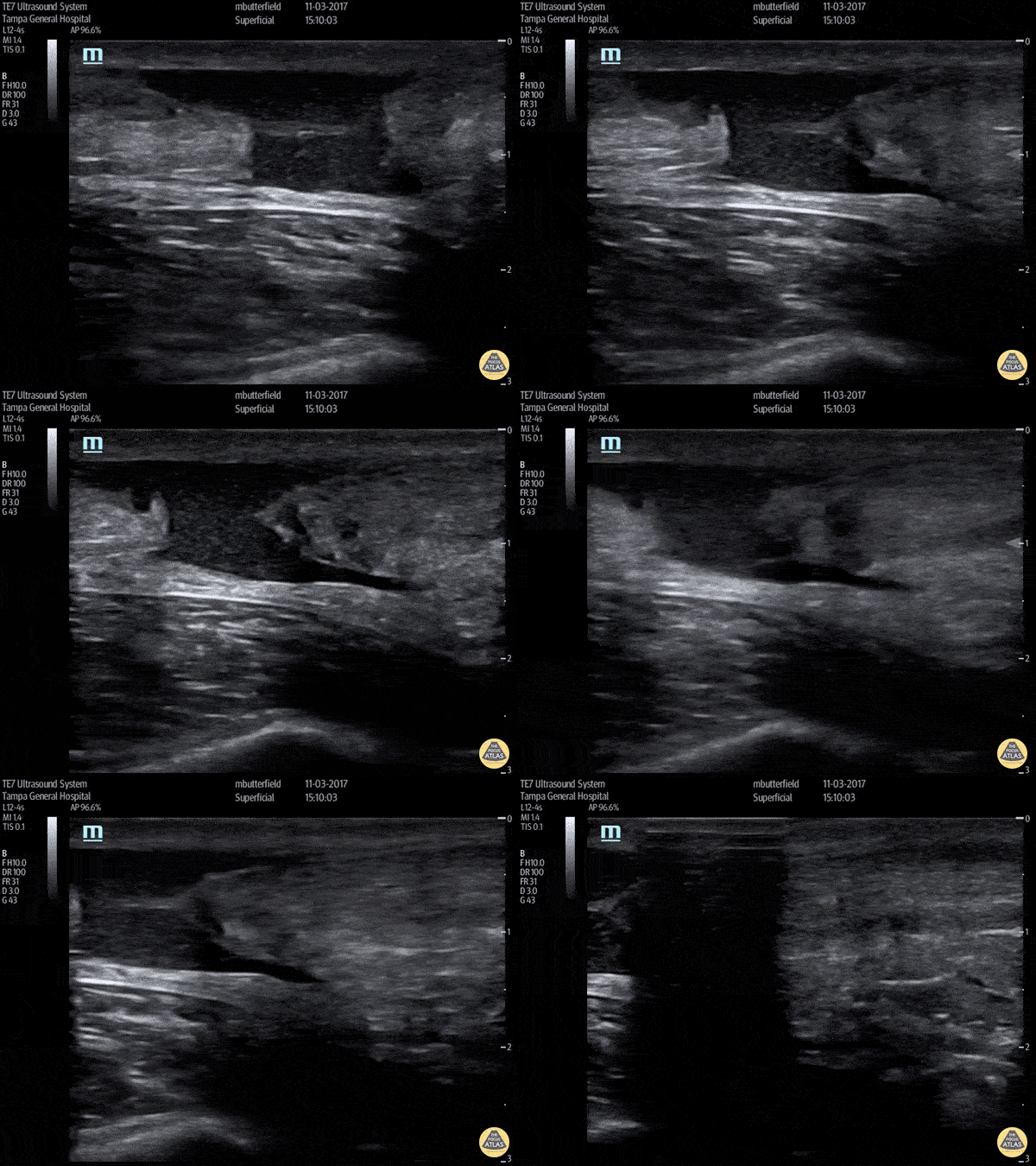
Full thickness tear of right achilles tendon after a skateboarding accident. (Long Axis)
Dr. Mike Butterfield
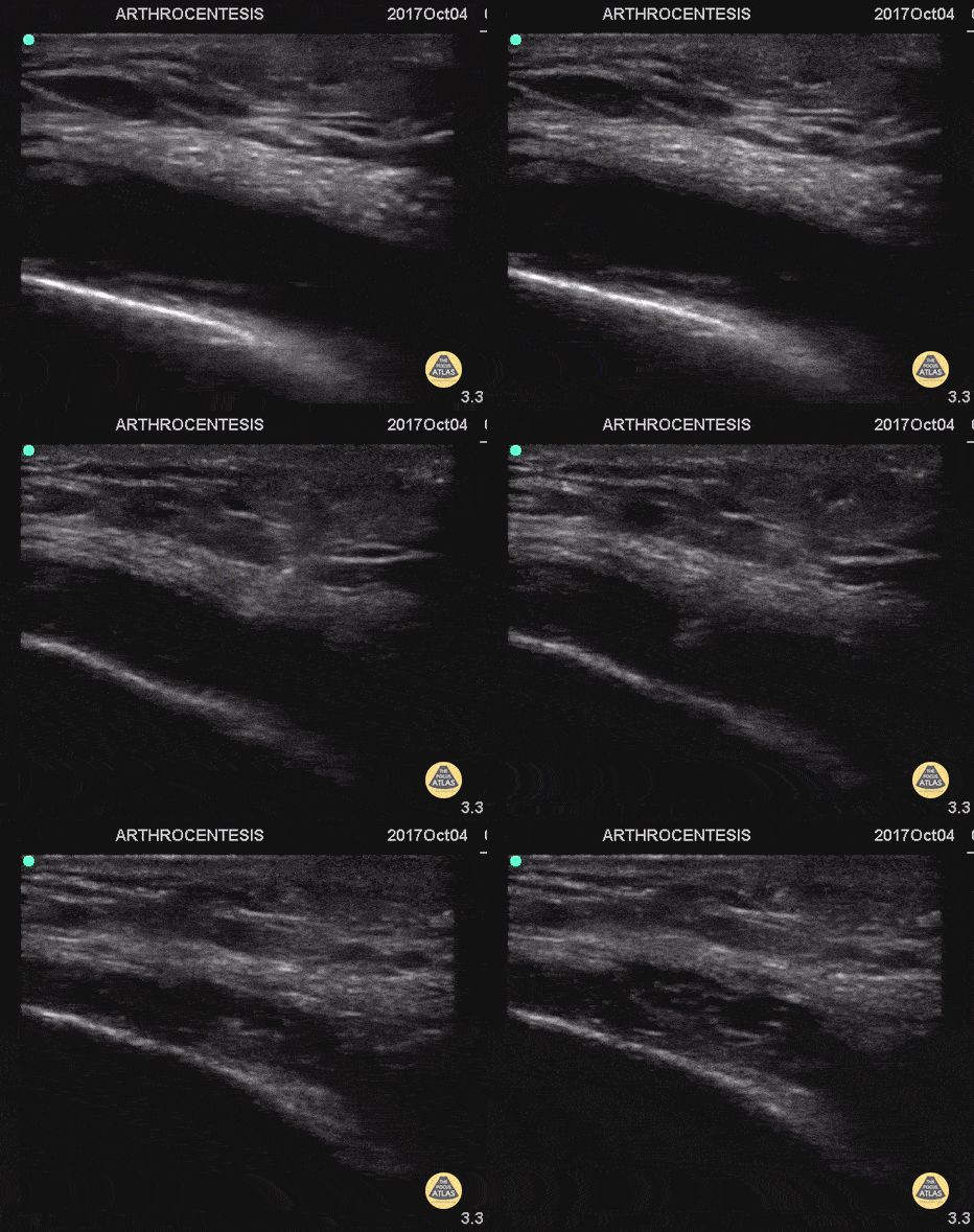
55 y/o with history of gout and osteoarthritis with an effusion. Join tapped and triamcinalone injected at the end.
Going to tap a joint and unsure of the best spot? Grab POCUS to find the biggest fluid pocket. Many prefer the in-place US guidance technique for big targets such as a joint and watching the needle enter the whole way.
Matthew Riscinti, MD - Kings County Emergency Medicine
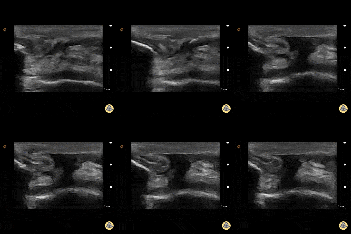
50 y/o M presents with acute left anterior knee pain after fall. On exam, patient noted to have high riding patella.
Longitudinal sonogram of the infrapatellar region showed marked discontinuity of the normally hyperechoic linear patterned tendon. The discontinuity is replaced with an anechoic collection indicative of hemorrhage. Comparison to normal knee can be seen on the next post.
Of note, anisotropy may be encountered when utilizing ultrasound, leading to artifact. Depending on the angle of the insonating beam, a normally hyperechoic structure may be falsely viewed as hypoechoic due to poor return of echo. Ensure US probe remains perpendicular to the tendon to minimize this artifact.
Dr. Hannah Moreira, Dr. Tareq Azad, Dr. Kyle Kelson - Kings County/SUNY Downstate Emergency Medicine
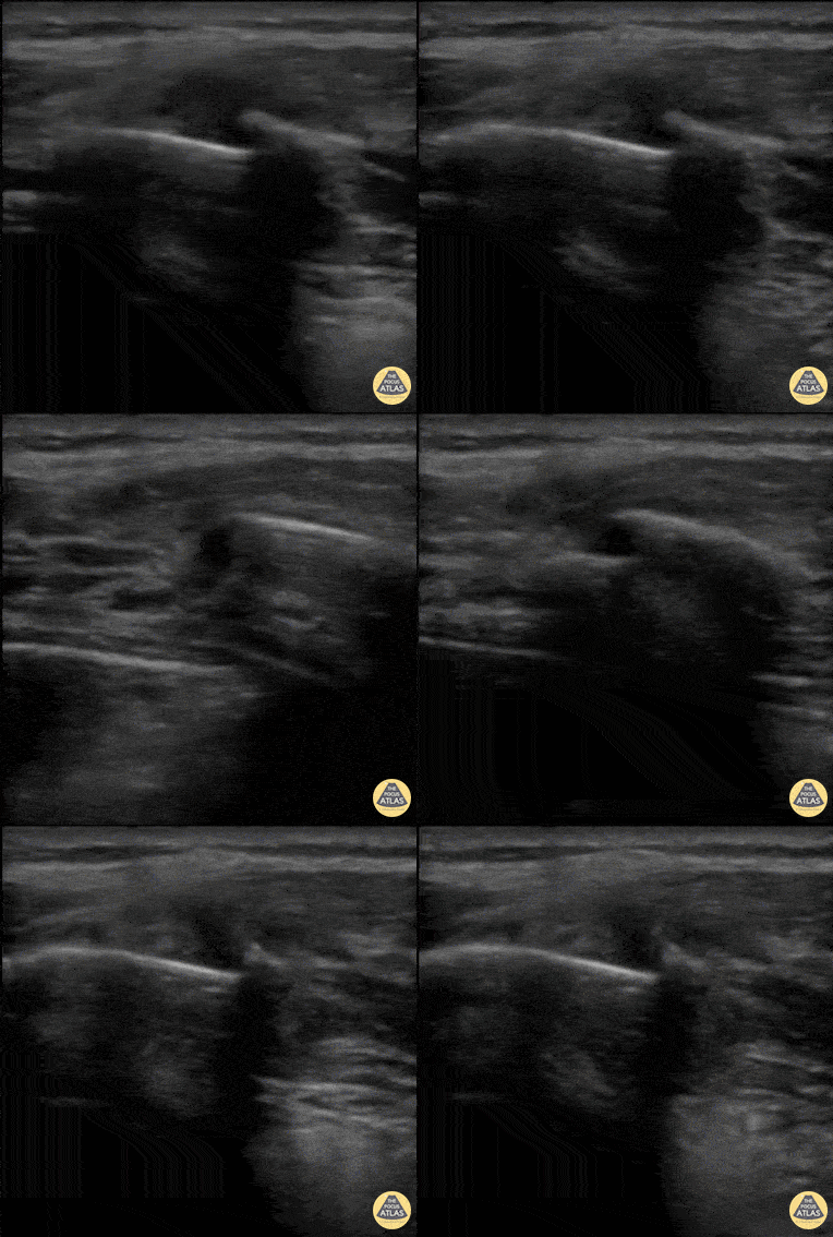
40 y/o M with polysubstance abuse, left-sided rib pain after a traumatic blow. Chest xray was equivocal. The patient was asked to "point to where it hurt", and the linear transducer revealed a displaced rib fracture. He complained of significant pain even after the resident gave two Percocet and was unwilling to leave the ED.
An intercostal nerve block, and that relieved the patient's pain and he went home.
Dr. Stephen Alerhand, Mt Sinai Hospital NYC
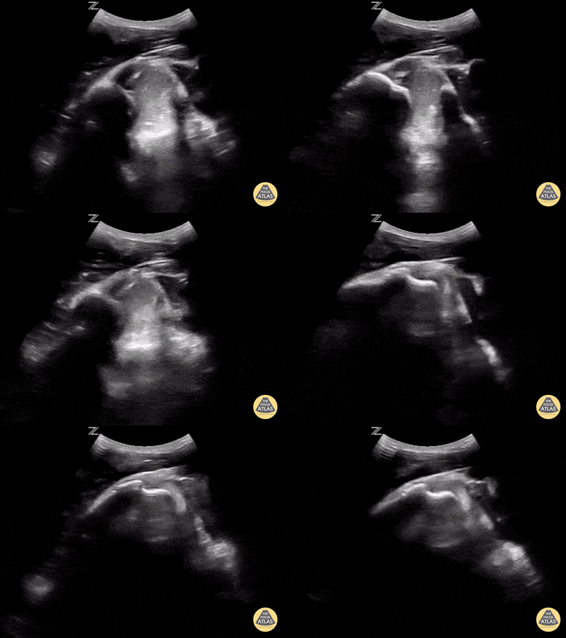
This is a posterior approach looking at the left shoulder showing an anterior dislocation. The dislocation was found before X-ray was shot. While the resident was performing the Cunningham technique to reduce the shoulder, the attending was able to, in real time, watch it go back in place. In between the humeral head and the glenoid rim is a large hematoma where I would aim my needle if I were to do an intra-articular block. It is a helpful technique in those recurrent dislocators. Saves the initial X-ray, but probably should get the post-reduction film at this point.
Matt Rutz, MD - Indiana University Department of Emergency Medicine, Ultrasound Division
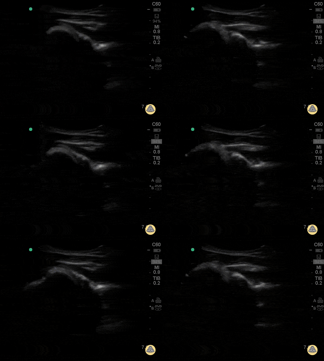
This clip of the shoulder is obtained by placing the probe in transverse plane on the posterior shoulder, and demonstrates the humeral head (left of screen) rotating in the glenoid. The scapular spine is to the right of the glenoid. The infraspinatus muscle is seen overlying the humeral head superficial to the infraspinatus is the deltoid muscle.
Hannah Kopinksi and Dr. Lindsay Davis - NYU Emergency Medicine
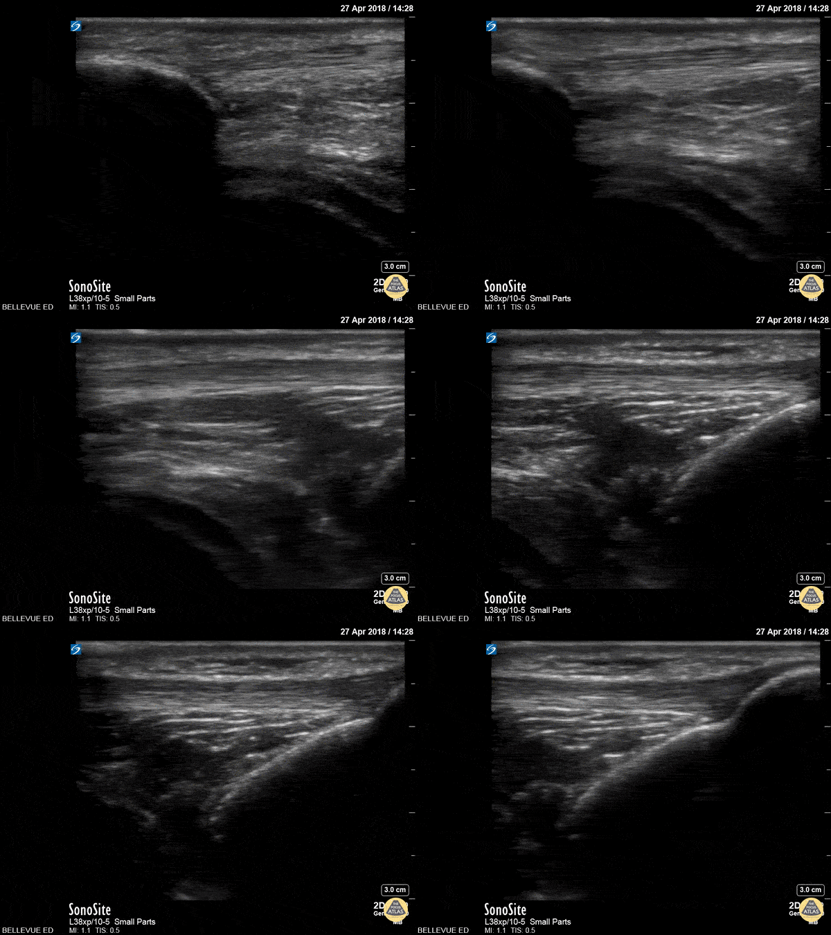
In this clip we see the linear, fibrillar, echogenic patellar tendon in long axis tracking along the top of the screen. It starts with the insertion point at the distal patella (left side of the screen). As the probe moves distally, we see where it attaches to the proximal tibia on the right of the screen.
Hannah Kopinksi and Dr. Lindsay Davis - NYU Emergency Medicine