
OB Pathology + Pregnancy
OB Pathology + Pregnancy
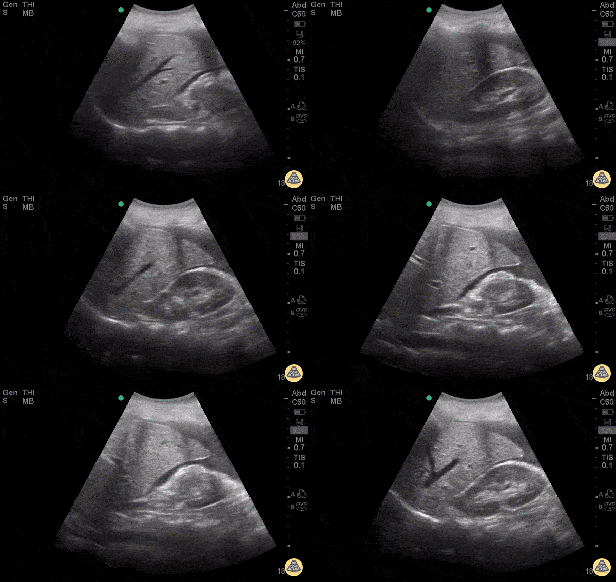
30 y/o F presented to ED for abdominal pain stating she had a recent miscarriage. Now she is having vaginal bleeding for the last 2 weeks. Borderline hypotensive and in severe distress due to pain. Abdomen diffusely tender with guarding. POCUS demonstrated +FAST with blood in the hepatorenal space. Remember to fan all the way to liver tip in FAST scan to fully evaluate for free fluid, it can be subtle and not simply in morrison’s pouch. The patient was rushed to OR by OBGYN for ex-lap based on this scan and was found to have ruptured tubal ectopic pregnancy.
Dr. Stacey Frisch - Kings County Emergency Medicine
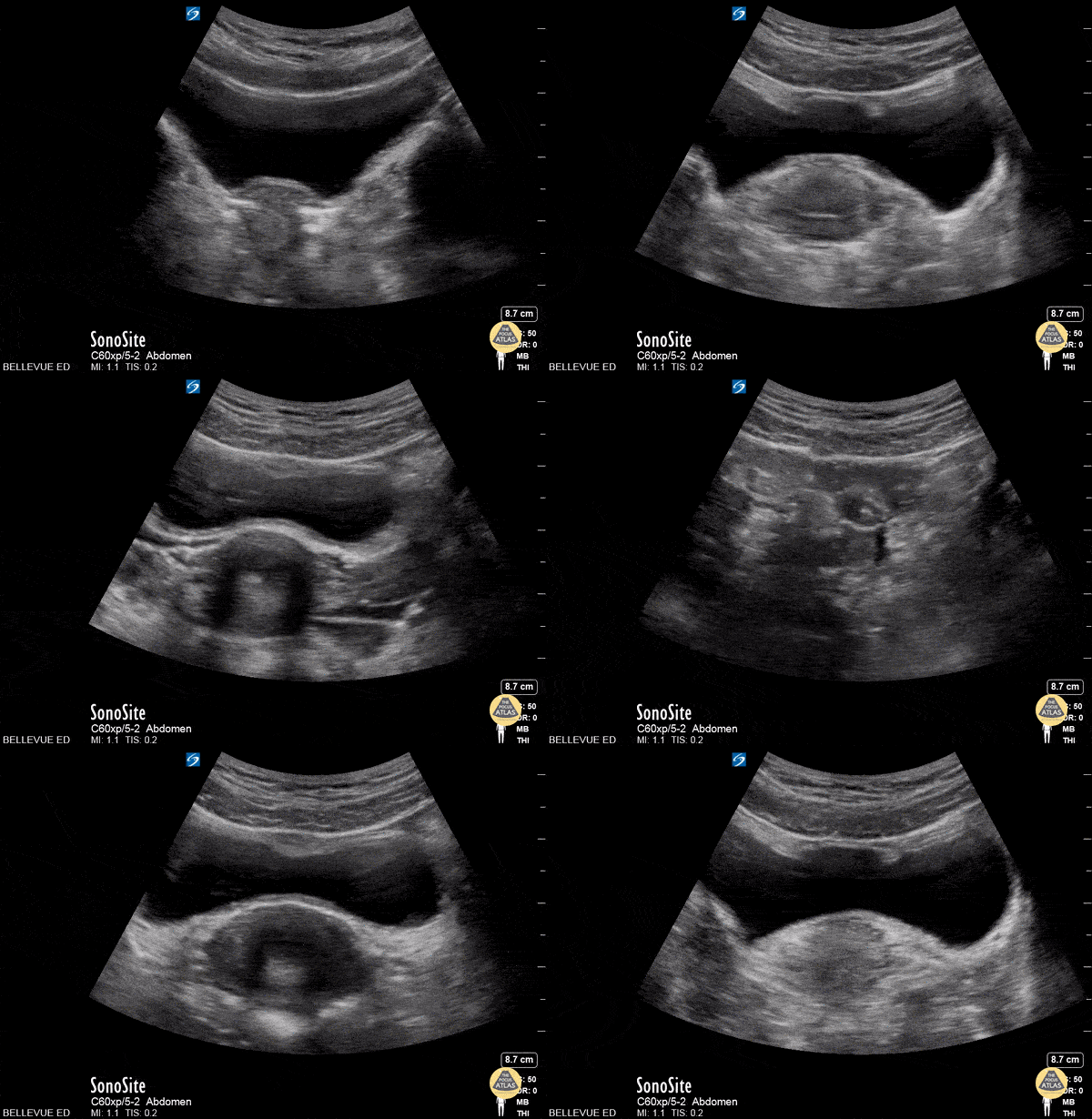
This is a clip of the uterus in transverse view using the transabdominal approach. The large anechoic structure in the center is the bladder in transverse view, and immediately deep to the bladder is the round uterine fundus. As the probe fans through the uterus we see some dark shadowing within it, likely due to artifact created by an IUD. The fallopian tubes are also faintly visible bilaterally branching off the uterus.
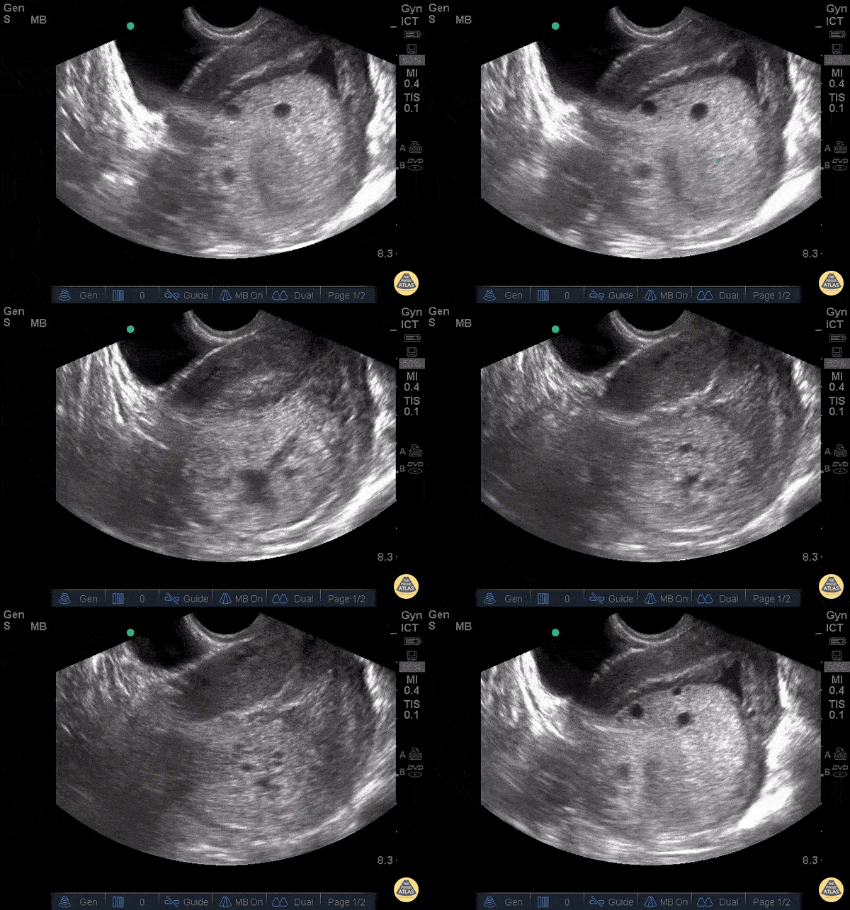
23 y/o F, gestational age 10 weeks by LMP referred to ER for suspicion of molar pregnancy. TV US shows hydropic vesicles within uterus, represented by anechoic regions scattered throughout a hyperechoic mass in uterine cavity. This is classically described as a “bunch of grapes” or “snowstorm pattern”.
Patients diagnosed with molar pregnancy can present with vaginal bleeding or symptoms similar to hyperemesis gravidarum, making ER POCUS a useful evaluation tool that can expedite disposition.
Dr. Bryan Flores, Dr. Teresa Smith - Kings County Emergency Medicine
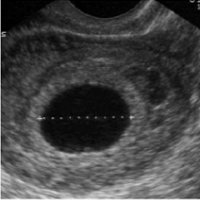
Seen at 4-6 weeks:
- Not diagnostic for IUP
- Ectopic may have pseudogestational sac, though this is rare
- It might be early pregnancy but they will need close follow up

Seen at 5-7 weeks:
- First evidence of IUP
- Appears as ring like structure within the gestational sac

Seen at 6-8 weeks:
- Tissue with similar echogenicity as uterus seen within the gestational sac
- If you see a fetal pole, you should see fetal cardiac activity
- Early on, the yolk sac and fetal pole may be present simultaneously
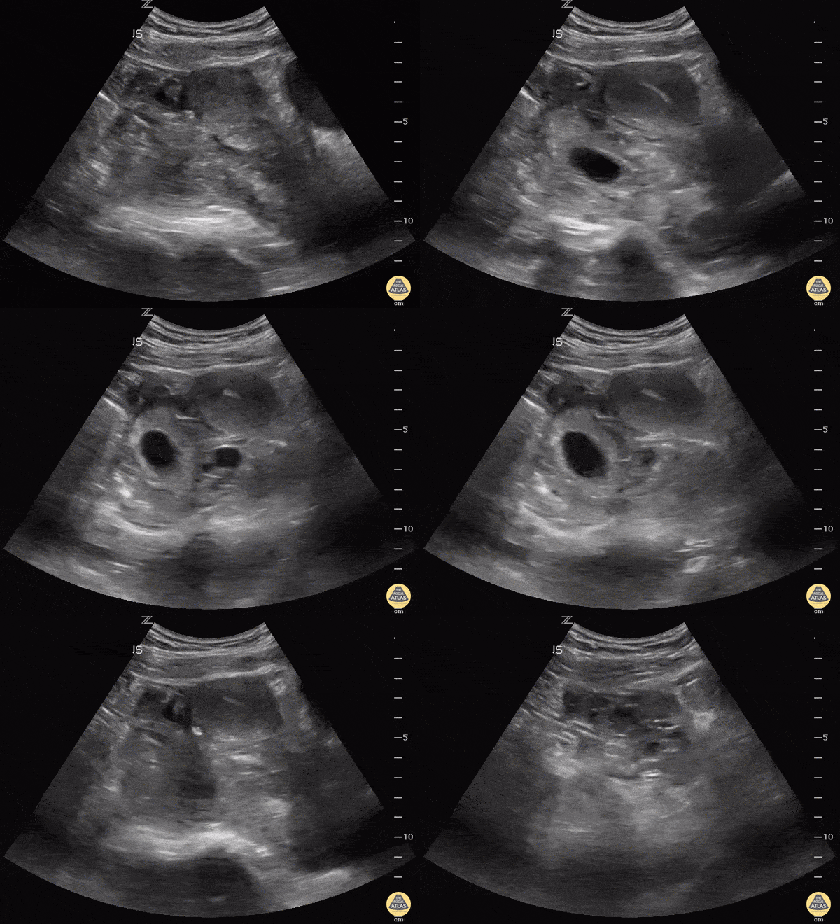
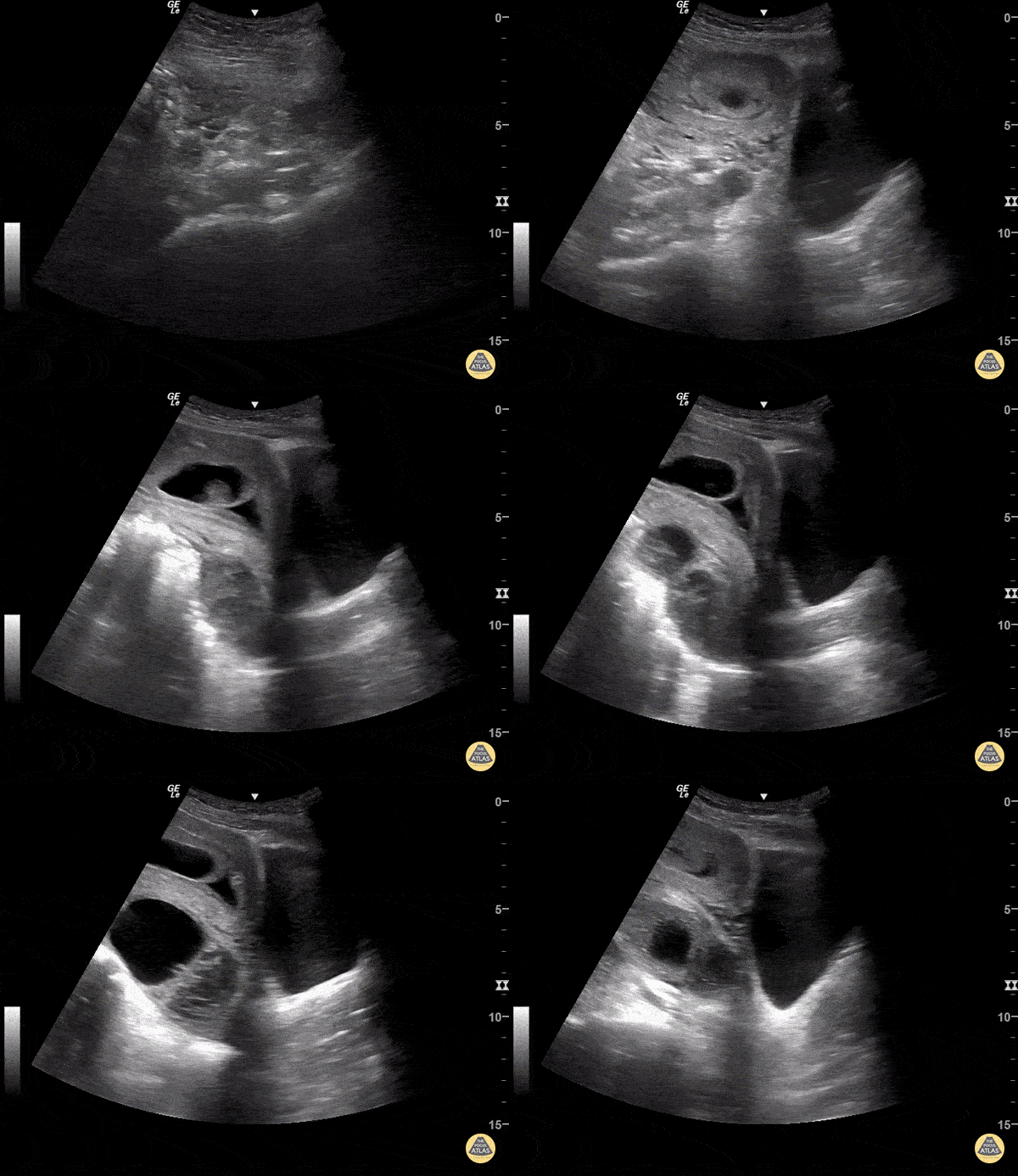
Always make sure the "IUP" is actually in the uterus by observing the uterus superior to the bladder in the sagittal view. This young lady could have been diagnosed easily with IUP and her abdomen pain dismissed as pain with pregnancy instead of a 10 week ectopic.
Matt Rutz, MD
@IUEM_Ultrasound
Indiana University School of Medicine
Department of Emergency Medicine
Division of Ultrasound