
Ocular Pathology
Ocular Pathology
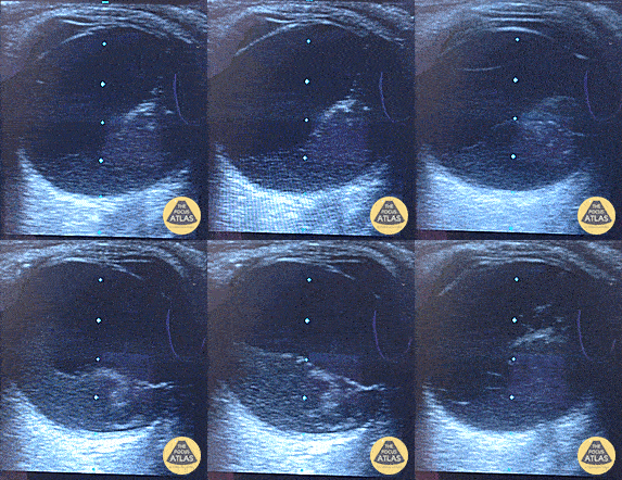
Posterior Vitreous Hemorrhage
Painless visual field cut with position change. Posterior vitreous hemorrhage with organization and dynamic movement gong over the top of the optic nerve, but not involving it to suggest retinal detachment.
Dr. Dustin Morrow
Here a hyperechoic linear density is seen arising from the location of the fundus, indicating retinal detachment. Posterior vitreous detachment and vitreous hemorrhage can look similar to retinal detachment. Given the imminent risk of blindness in these conditions it is essential to identify them as soon as possible. POCUS is an excellent way to do so given the time and skills required to perform a proper fundoscopic exam. Literature has shown sensitivity for this test to be 97-100% and specificity 83-100%. So next time ask yourself... is your fundoscopic exam that good?
Sukh Singh, MD
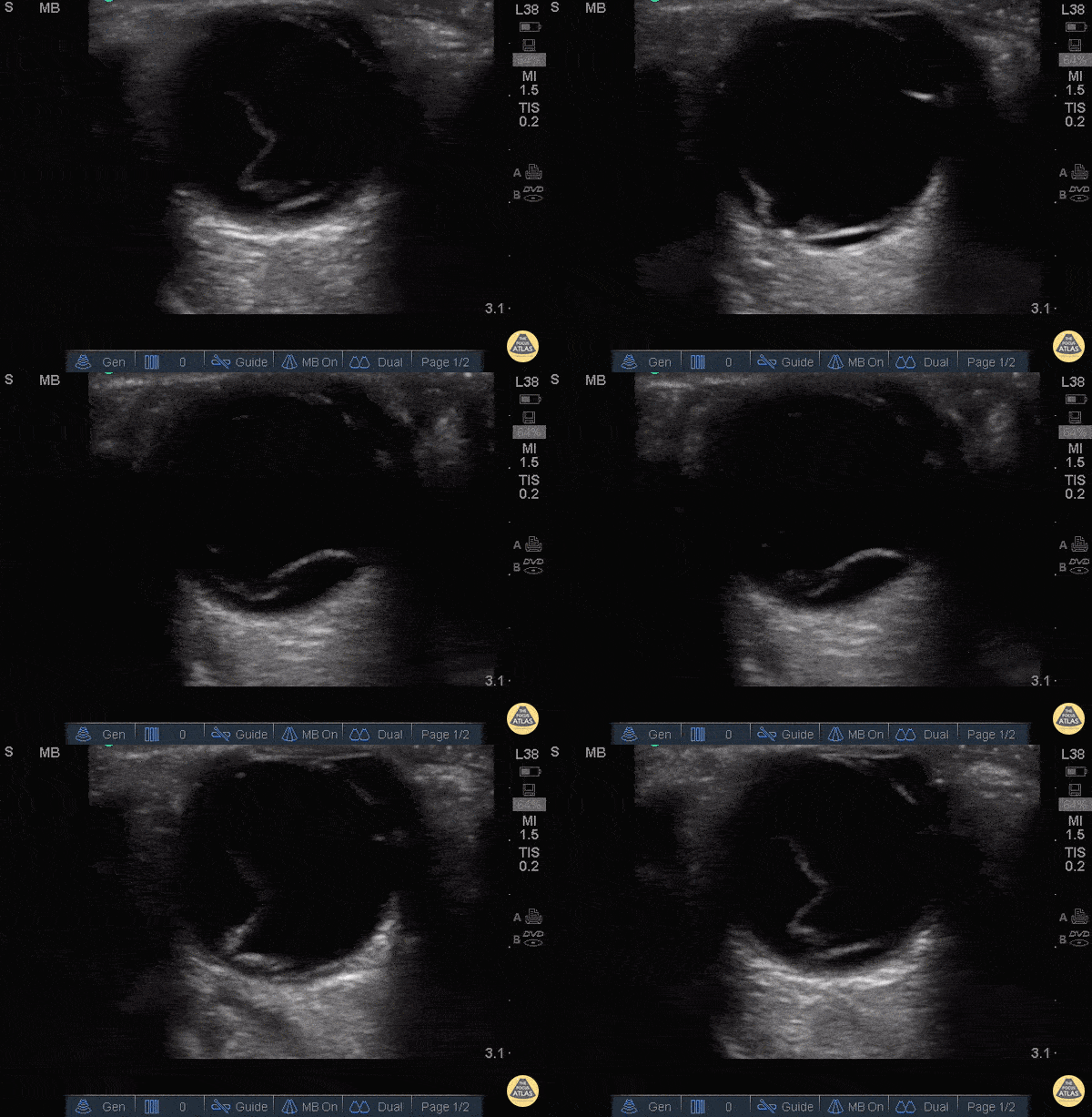
65 y/o F PMH DM with sudden onset flashes of light and floaters followed by progressively worse blurred vision in her right eye. She has history of cataracts surgery as well as DM retinopathy. POCUS of affected eye shows a prominent hyper-echoic linear density floating freely above the posterior fundus as shown.
Retinal Detachment is considered one of few emergencies whereby the retina becomes detached from the choroid. Once separated from the choroid’s blood supply, retinal ischemia ensues which can result in complete blindness in the affected eye. Salvage of such a potentially devastating injury relies on quick and efficient diagnosis with subsequent referral to definitive care.
Bedside ultrasound has been shown to be 100% sensitive as a screening tool for RD [AEM 2010 Sep;17(9):913-7]. Have patient close eye and place liberal amount of gel either directly over eye or atop a tegaderm dressing. Use a linear probe and scan eye in axial plane to reveal findings demonstrated above.
Dr. Eric Roseman - Kings County Emergency Medicine
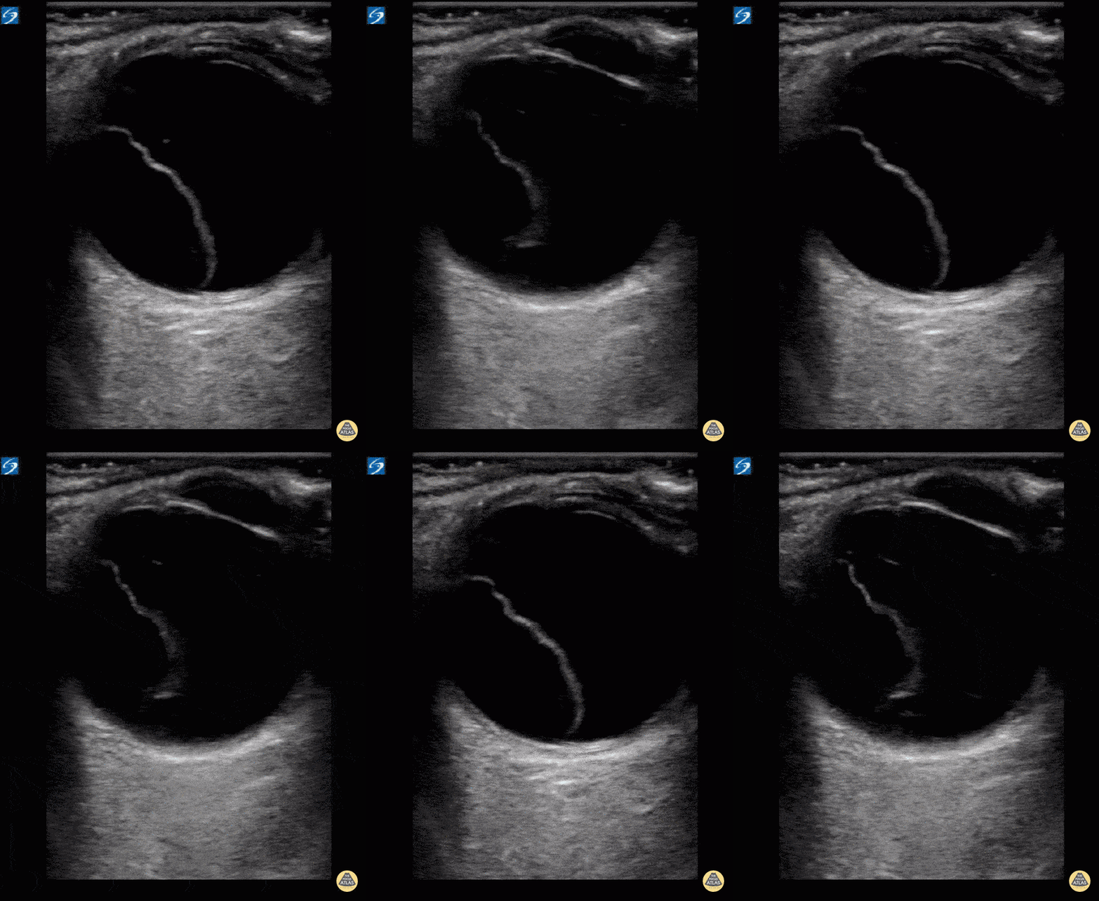
Retinal detachment in a patient with 1 day of a right sided visual field cut.Literature has shown sensitivity for this test to be 97-100% and specificity 83-100%. So next time ask yourself... is your fundoscopic exam that good?
Dr. William Scheels
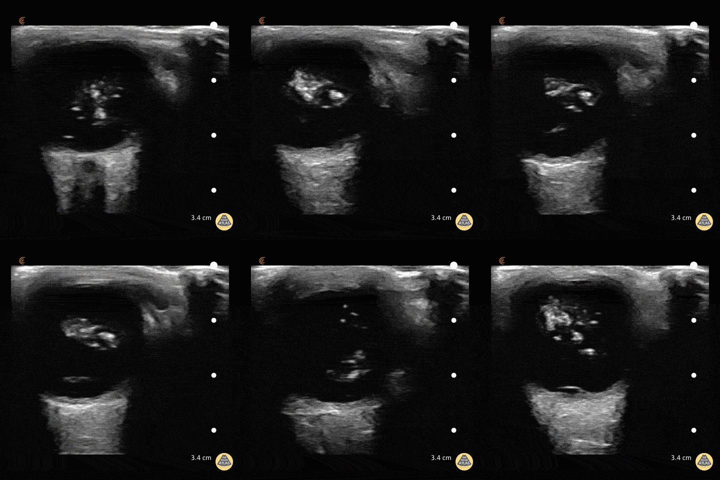
47 y/o M with coronary artery disease on aspirin/clopidogrel with 4 days of decreased vision in his right eye. POCUS demonstrates swirling, amorphous echogenicities known as "the washing machine sign." If the eye remains still the blood will settle with gravity.
Fundoscopic evaluation by ophthalmology confirmed the diagnosis of vitreous hemorrhage.
Dr. Matthew Riscinti and Dr. Taylor Conrad - Kings County/SUNY Downstate Emergency Medicine
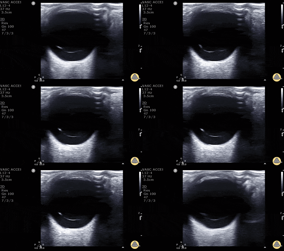
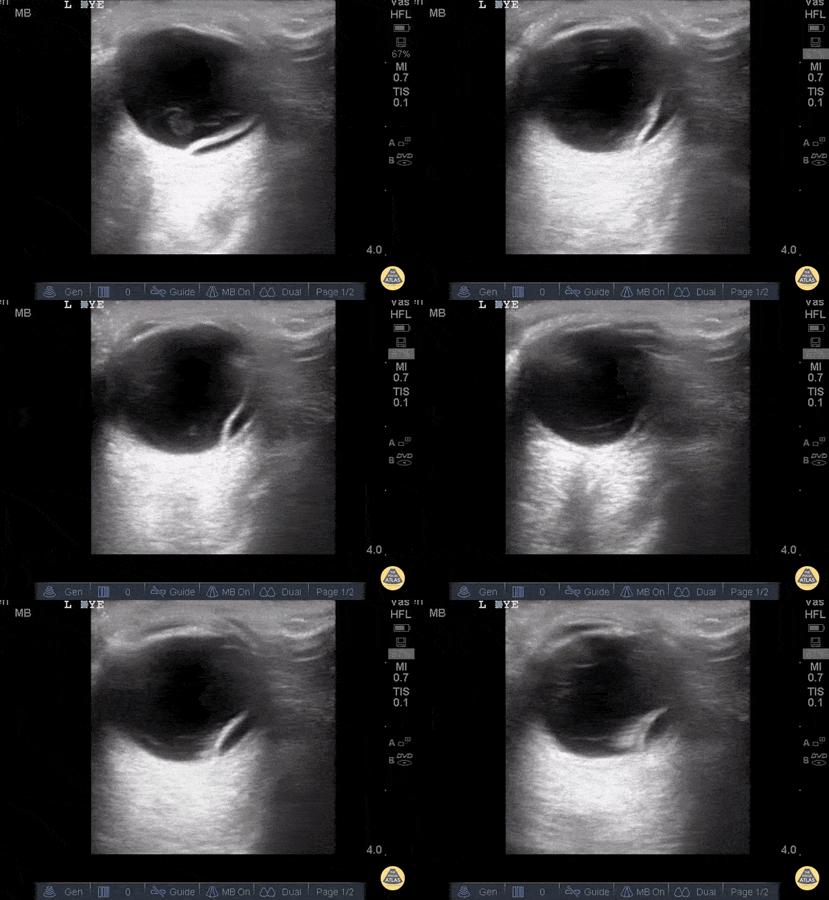
40 year old male with 6 months of decreased vision from his left eye. No pmh or history of trauma. VA was obviously decreased relative to the contralateral eye. POCUS rapidly revealed a freely floating hyperechoic membrane consistent with vitreal detachment. To differentiate between a vitreous and retinal detachment you must look to see if the membrane is attached to the hypoechoic optic nerve. The retinal is continuous with the optic nerve therefore stays attached to the optic nerve in a retinal detachment. A vitreous detachment would be completely untethered as seen in this image. You can also see the “washing machine sign” better pictured in other images in this gallery.
Matt Riscinti, MD Bryan Flores, MD - Kings County Emergency Medicine