
Soft Tissue Pathology
Soft Tissue Pathology
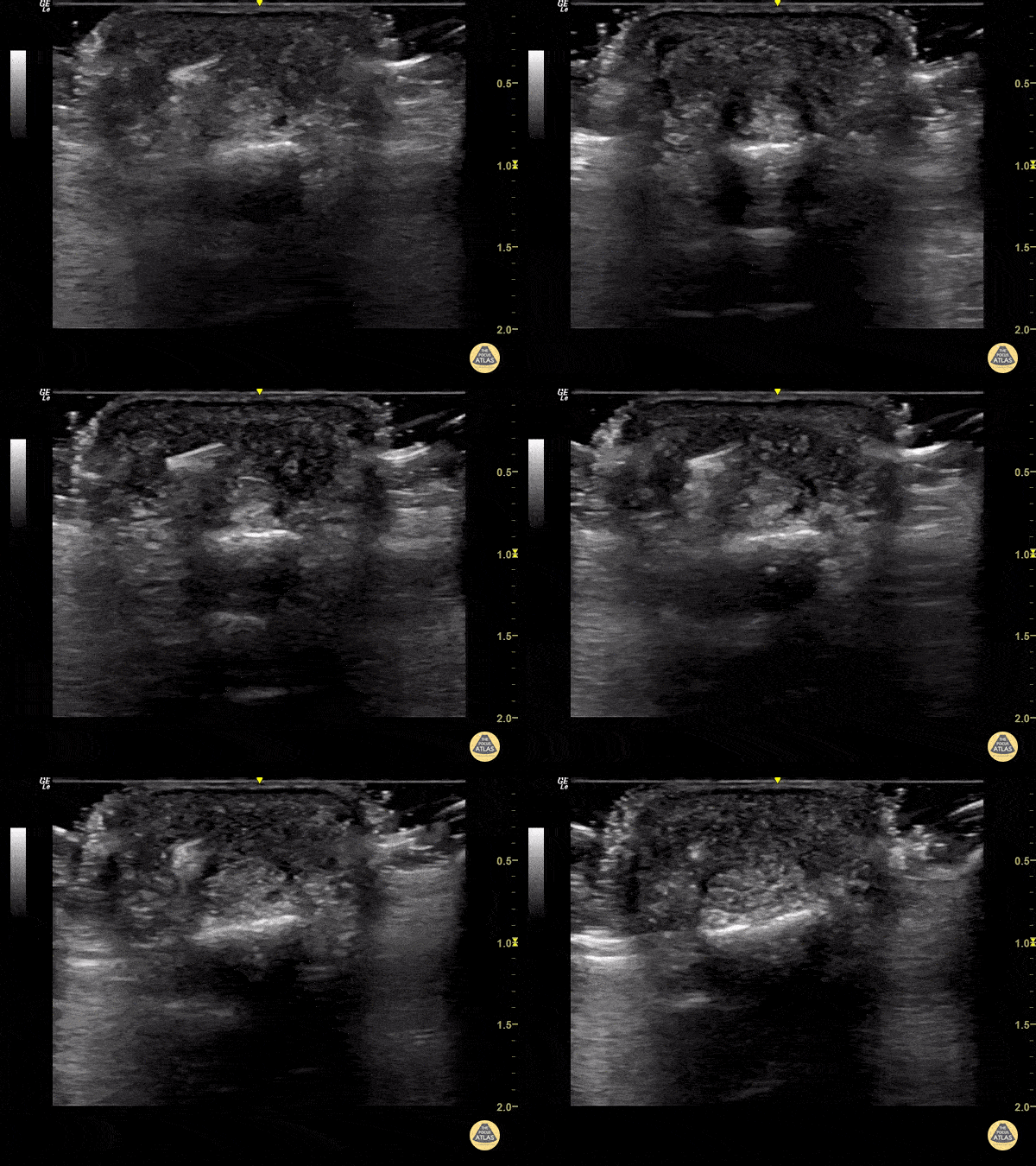
65 year old female with pain and swelling over 4th phalanx, no history of trauma. Hyperechoic density located above flexor tendon sheath. Patient sent to Ortho for surgical exploration.
Dr. Magner

Hyperechoic gas (screen left) obscure the typical architecture of soft tissues (screen right). There is a small amount of edema in the superficial soft tissues.
Dr. Bell
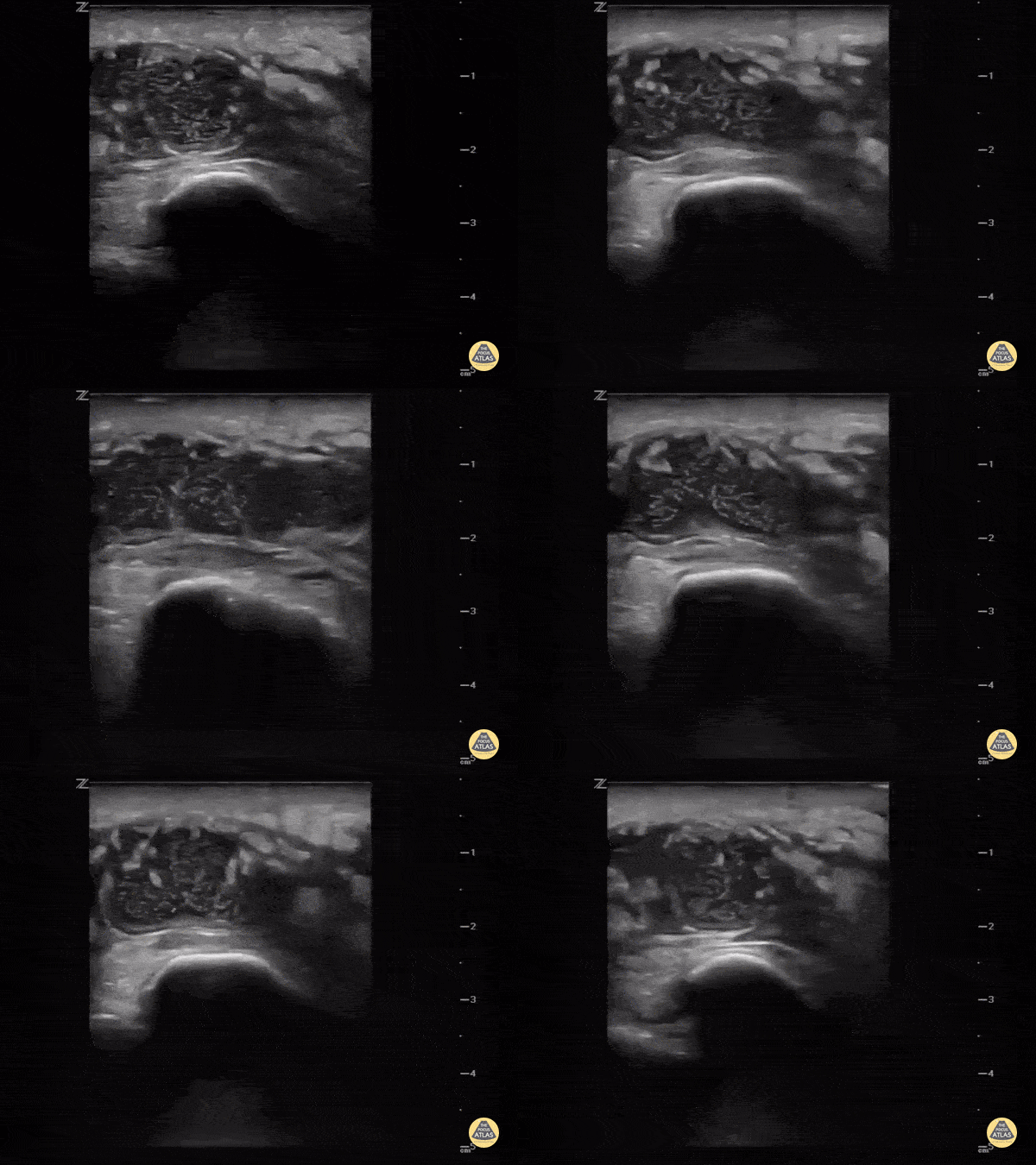
Sometimes it's hard to tell if the knee is septic or just the bursa. Ultrasound can help. This image demonstrates a hypoechoic fluid collection ABOVE the patella consistent with pre-patellar bursitis.
Image courtesy of IUEM Ultrasound
Original Twitter Post can be found here.
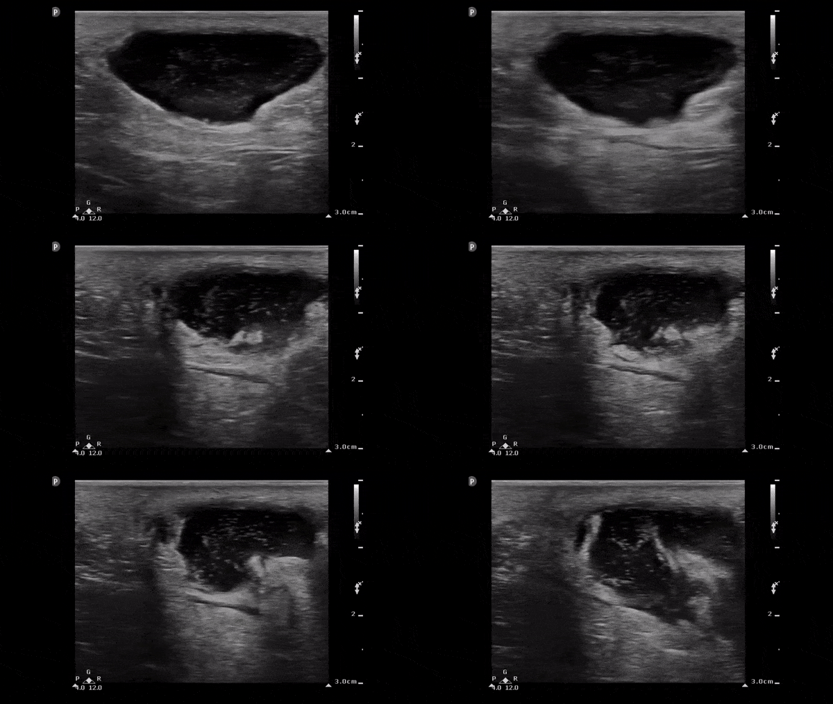
The image below demonstrates a well circumscribed fluid collection in the soft tissue consistent with an abscess. Note the scattered punctate echogenic densities moving within the abscess fluid which suggest high cell/protein content of the fluid.
Image obtained from 5 Minute Sono.
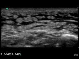
This is the classic appearance of subcutaneous edema which in the appropriate clinical setting (overlying redness and warmth) would be consistent with a cellulitis

While typically an abscess will appear as a hypoechoic fluid filled structure, sometimes it can be missed as its echotexture is similar to other surrounding soft tissue structures. To improve sensitivity, gentle compression should be applied to identify a subtle abscess as seen above.
Image obtained from Ultrasound of the Week.
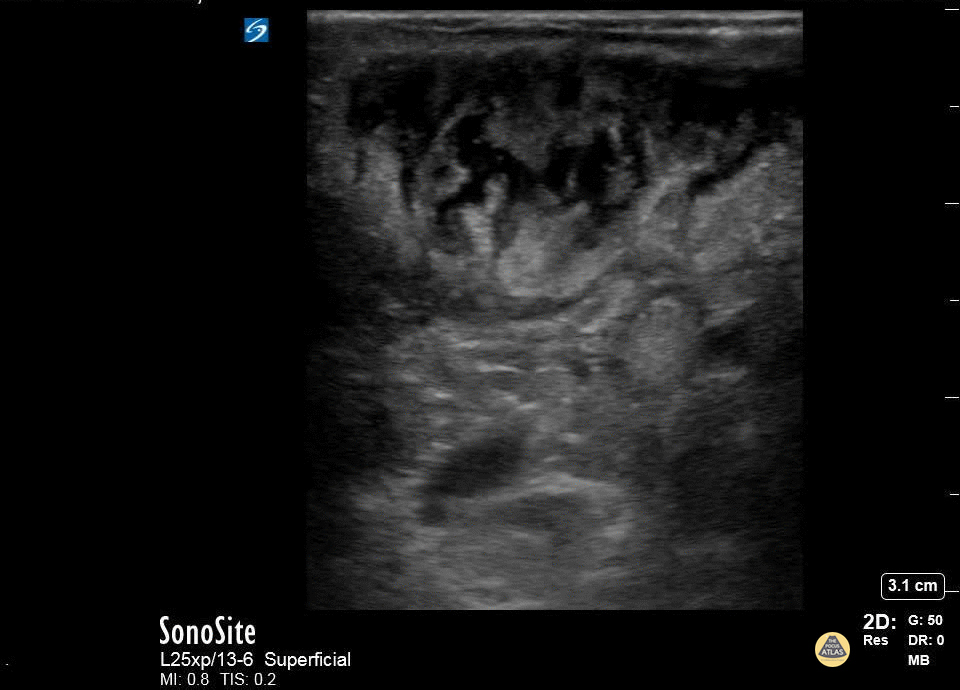
A patient with cerebral palsy and quadriplegia presented with her mother for evaluation of a fluctuant mass in her right axilla. Ultrasound imaging of the mass showed an abscess with multiple septations. The abscess was incised and drained, and the patient was discharged on antibiotics.
Katy Van Donselaar, Emergency Medicine Resident
Christopher Heberer, Emergency Medicine Resident
Simhadri Botta, 4th year Medical Student
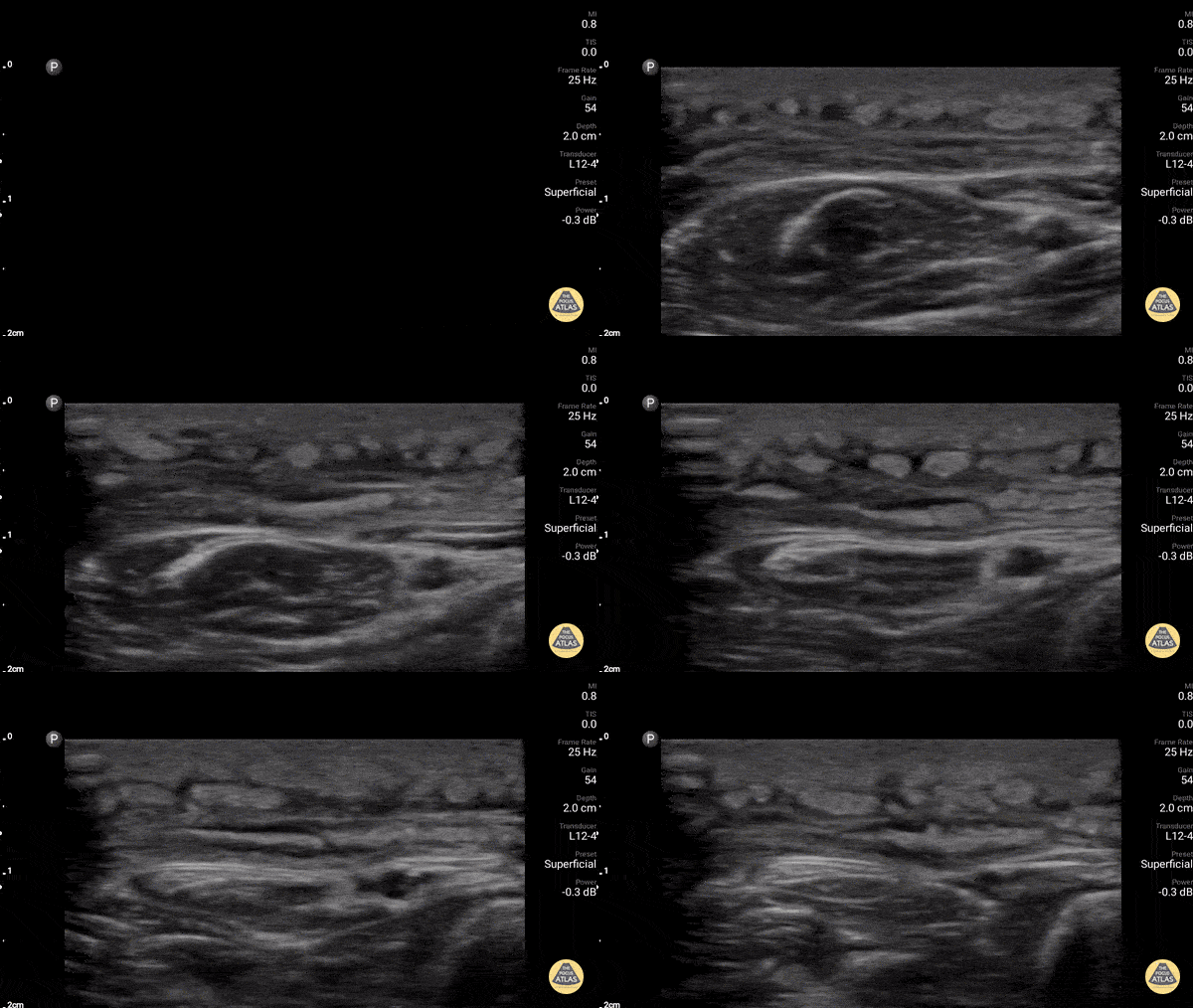
Diffuse cobblestoning typically seen with cellulitis.
In this case it is biopsy proven panniculitis.
Dr. Gordon Johnson
@pdxfutebol

The image above demonstrates a well circumscribed fluid collection within the soft tissue, without evidence of surrounding cellulitis. The above abscess was incised with immediate release of a large volume of purulent material. The patient did well.
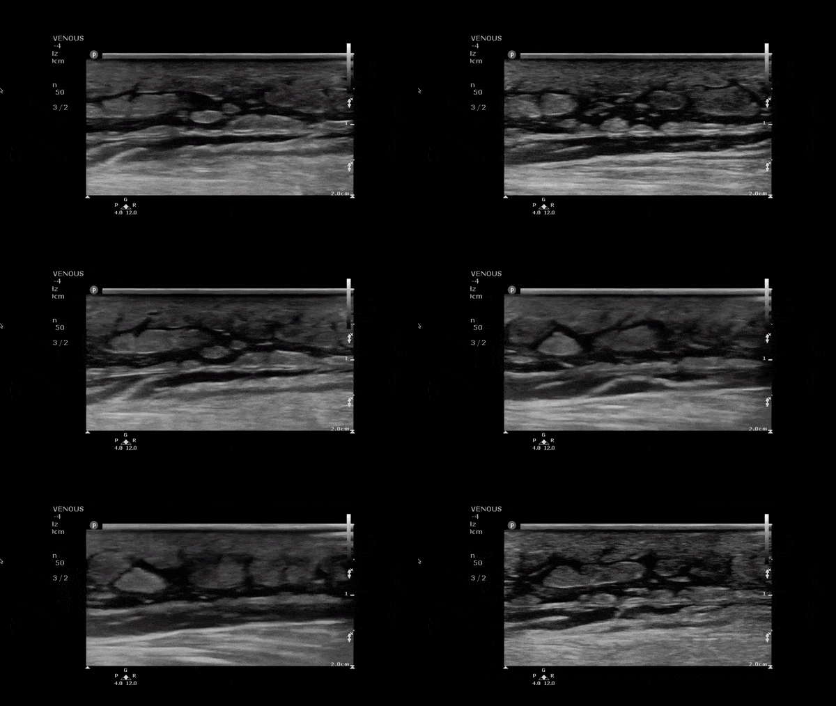
This image demonstrates anechoic fluid surrounding islands of soft tissue. In the setting of infectious signs and symptoms this is consistent with a cellulitis.
Image obtained from 5 Minute Sono.