Echocardiography
The best Echocardiography POCUS clips. This is free and open access medical education content. Right click to download.
Echocardiography Home
Normal Anatomy
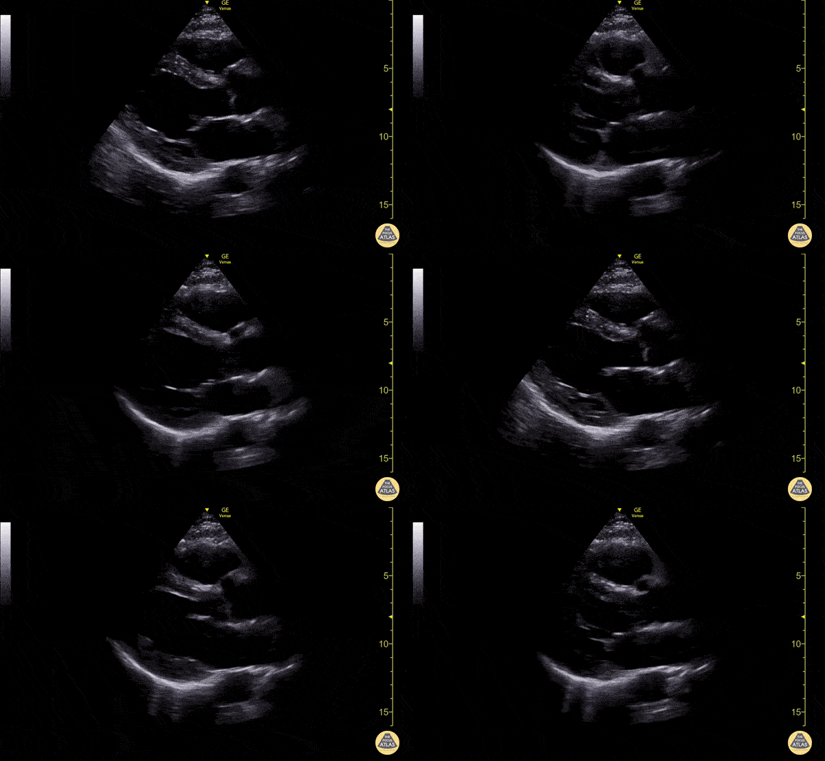
Parasternal long axis view with normal ejection fraction
Nigist Taddese MBChB. Division of Hospital Medicine, John H Stroger Hospital of Cook County
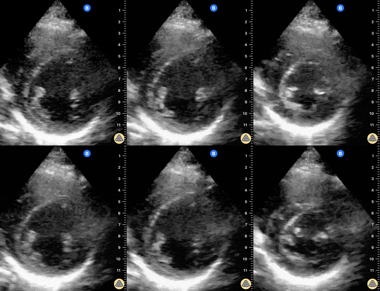
Normal PSAX view at a level of the papillary muscles. In the center of the screen is the muscular walled LV, which forms a perfect circle. The smaller, thin-walled RV is seen superficially and wrapped around the LV.
Dr. Felipe Urriola, Puerto Aysen Hospital, Emergency Department, Chilean Patagonia.
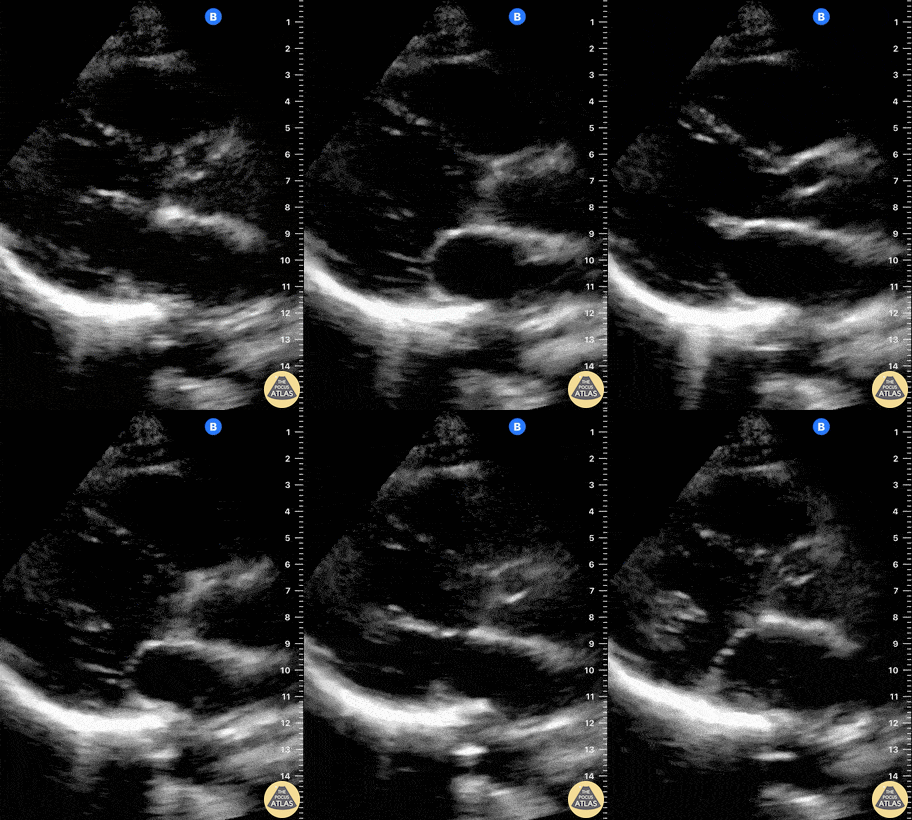
This is a normal parasternal long axis (PLAX) view. The right ventricle (RV) is at the top of the screen. Further down and from left to right: left ventricle (LV), outflow tract, aortic valve, ascending aorta. The actively moving mitral valve separates the LV from the left atrium (LA). At the bottom of the screen, the circular, anechoic image is the descending aorta.
Dr. Felipe Urriola. Puerto Aysen Hospital Emergency Department, Chilean Patagonia.
Colorized Normal Anatomy
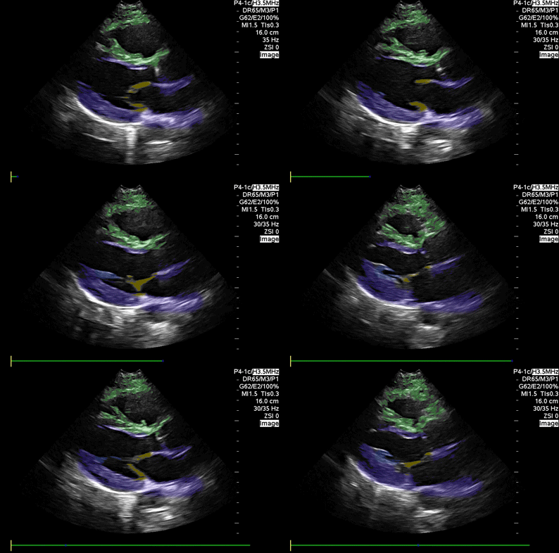
Parasternal Long Axis
Blue : Left atrium and ventricle, Yellow: Mitral valve, Green: Right ventricle
Images: Dr. Lindsay Davis, Dr. Hannah Kopinski. Image Editing: Michael Amador and Dr. Matthew Riscinti
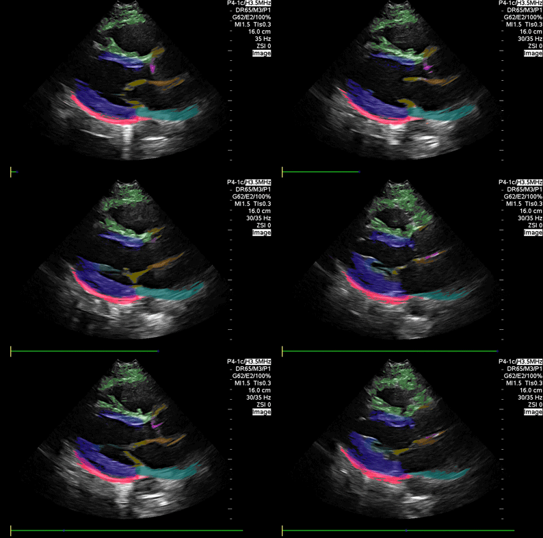
Parasternal Long Axis (detailed)
Blue: Left ventricle, Green: Right ventricle, Orange: Aortic outflow Pink: Aortic valve; Yellow: Mitral valve; Red: Pericardium; Light orange: Left atrium
Images: Dr. Lindsay Davis, Dr. Hannah Kopinski. Image Editing: Michael Amador and Dr. Matthew Riscinti
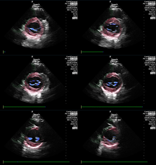
Parasternal short axis
Red: Left ventricle, Blue: Mitral valve, Green: Right ventricle
Images: Dr. Lindsay Davis, Dr. Hannah Kopinski. Image Editing: Michael Amador and Dr. Matthew Riscinti
Left Ventricular Dysfunction
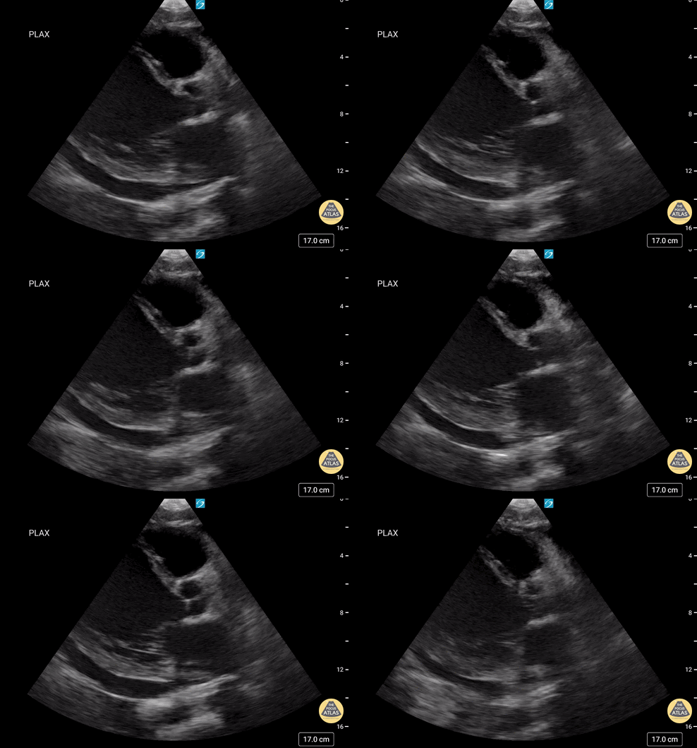
47 year-old male with no cardiac history presenting with shortness of breath and leg swelling for two weeks found to have new onset cardiomyopathy. POCUS echocardiogram showed significant dilation of the LV on parasternal long axis view with reduced EF; EPSS of 23.7 mm. Formal echocardiogram confirmed findings with additional findings including EF <20%, mild dilation of the LA, RA, and RV. While LV is grossly dilated, the significantly increased EPSS correlates with the dramatically reduced EF.
Brent Oldham, MD, MPH, PGY-3, Central Michigan University College of Medicine, Emergency Medicine
Dan Dunaske, DO, PGY-1, Central Michigan University College of Medicine, Emergency Medicine
Brad Buska, MS4, Central Michigan University College of Medicine
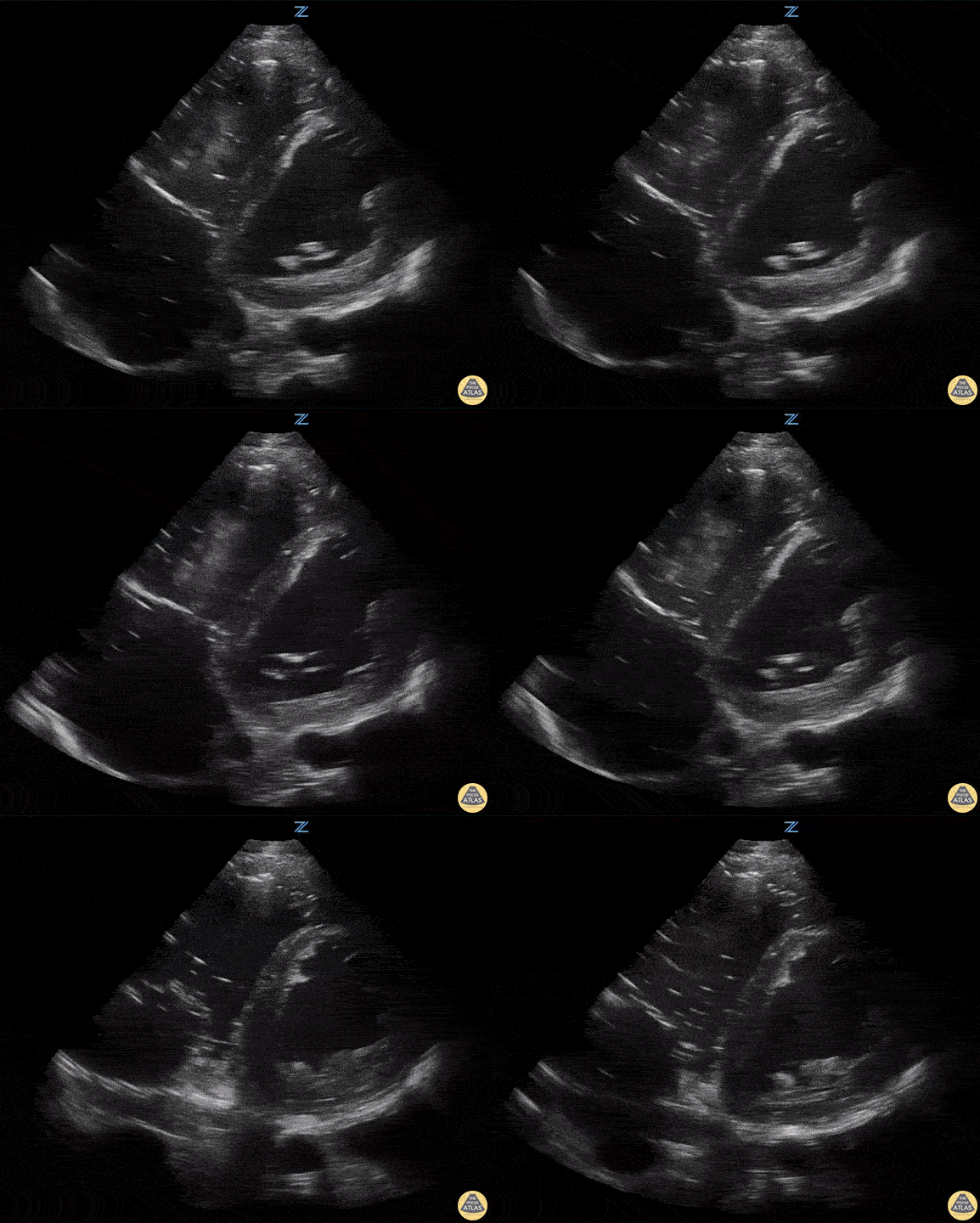
90 y/o F presents with hypotension. Patient was signed out to me by the overnight team with "sepsis" and received 30 cc/kg bolus. Patient began to have SBPs in the 70s again. She was immediately placed on vasopressors and managed for a CHF exacerbation. Image is a slightly obliqued PSAX.
John Bowling, DO, Cleveland Clinic Akron General, @BModeBowling
The image is slightly oblique giving the LV an oval appearance
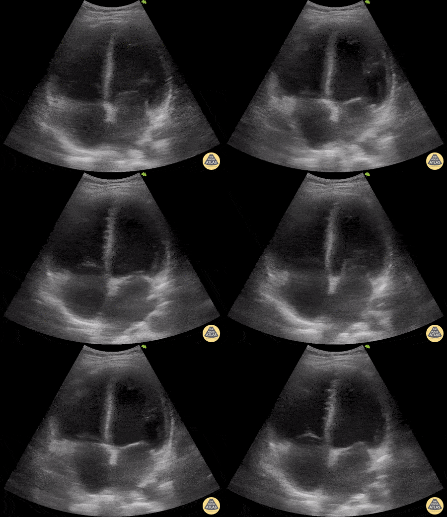
A 29-year-old patient, with a recent history of mild COVID 19 infection, consulted for a 5-day history of fever, associated with abdominal pain, arthralgia and myalgia. Hypotensive on admission (80/50 mmHg), with HR 100 bpm and echocardiography performed with a convex transducer: LVEF 12%, with global hypokinesia and dilated cavities. The diagnosis of post-COVID myocarditis is made.
Dr. Libardo Valencia Chicué
Right Ventricular Dysfunction
![Large RV Mobile Thrombus [1/2-Long Axis]](https://images.squarespace-cdn.com/content/v1/58118909e3df282037abfad7/1782483242211-L8244HHBG9ZDC7YCX7JJ/image-asset.gif)
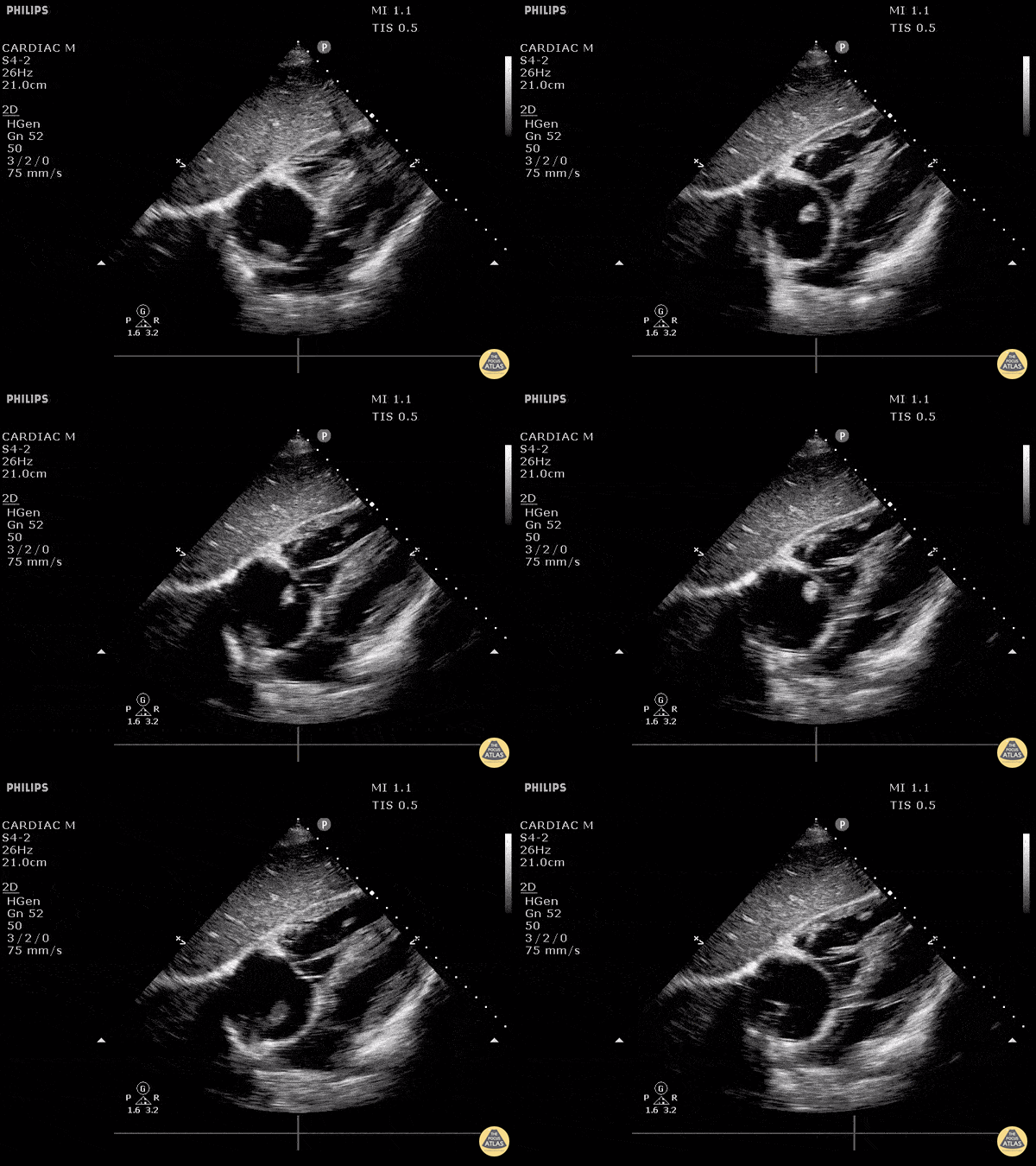
45-year-old male presented to the ED with sudden onset shortness of breath. His medical history was notable for a gastric sleeve procedure performed less than two weeks prior. On arrival, he was hypoxic with oxygen saturations in the 80s on room air, and his oxygen requirements progressively increased over a 10-minute period while IV access was obtained. Bedside cardiac ultrasound revealed a large, mobile, serpiginous clot in transit within the right atrium and right ventricle (RV). The thrombus was not adherent to the chamber walls and was visualized in both the RV inflow and long axis views. A parasternal short axis view demonstrated a positive D-sign, consistent with right ventricular overload and strain, further supporting the diagnosis of massive pulmonary embolism. Immediately following completion of the ultrasound, the patient experienced a PEA arrest. Tenecteplase was administered during resuscitation, resulting in return of spontaneous circulation.
Contributed by Dr. Thomas O'Mara, DO; Dr. Irving Chung, DO; Dr. Nicole Yuzuk, DO
St. Joseph’s University Medical Center, Paterson, NJ
![Large RV Mobile Thrombus [2/2-Short Axis]](https://images.squarespace-cdn.com/content/v1/58118909e3df282037abfad7/1782483537233-3VS1FHTMMV6IKYYOJYRU/image-asset.gif)
45-year-old male presented to the ED with sudden onset shortness of breath. His medical history was notable for a gastric sleeve procedure performed less than two weeks prior. On arrival, he was hypoxic with oxygen saturations in the 80s on room air, and his oxygen requirements progressively increased over a 10-minute period while IV access was obtained. Bedside cardiac ultrasound revealed a large, mobile, serpiginous clot in transit within the right atrium and right ventricle (RV). The thrombus was not adherent to the chamber walls and was visualized in both the RV inflow and long axis views. A parasternal short axis view demonstrated a positive D-sign, consistent with right ventricular overload and strain, further supporting the diagnosis of massive pulmonary embolism. Immediately following completion of the ultrasound, the patient experienced a PEA arrest. Tenecteplase was administered during resuscitation, resulting in return of spontaneous circulation.
Contributed by Dr. Thomas O'Mara, DO; Dr. Irving Chung, DO; Dr. Nicole Yuzuk, DO
St. Joseph’s University Medical Center, Paterson, NJ
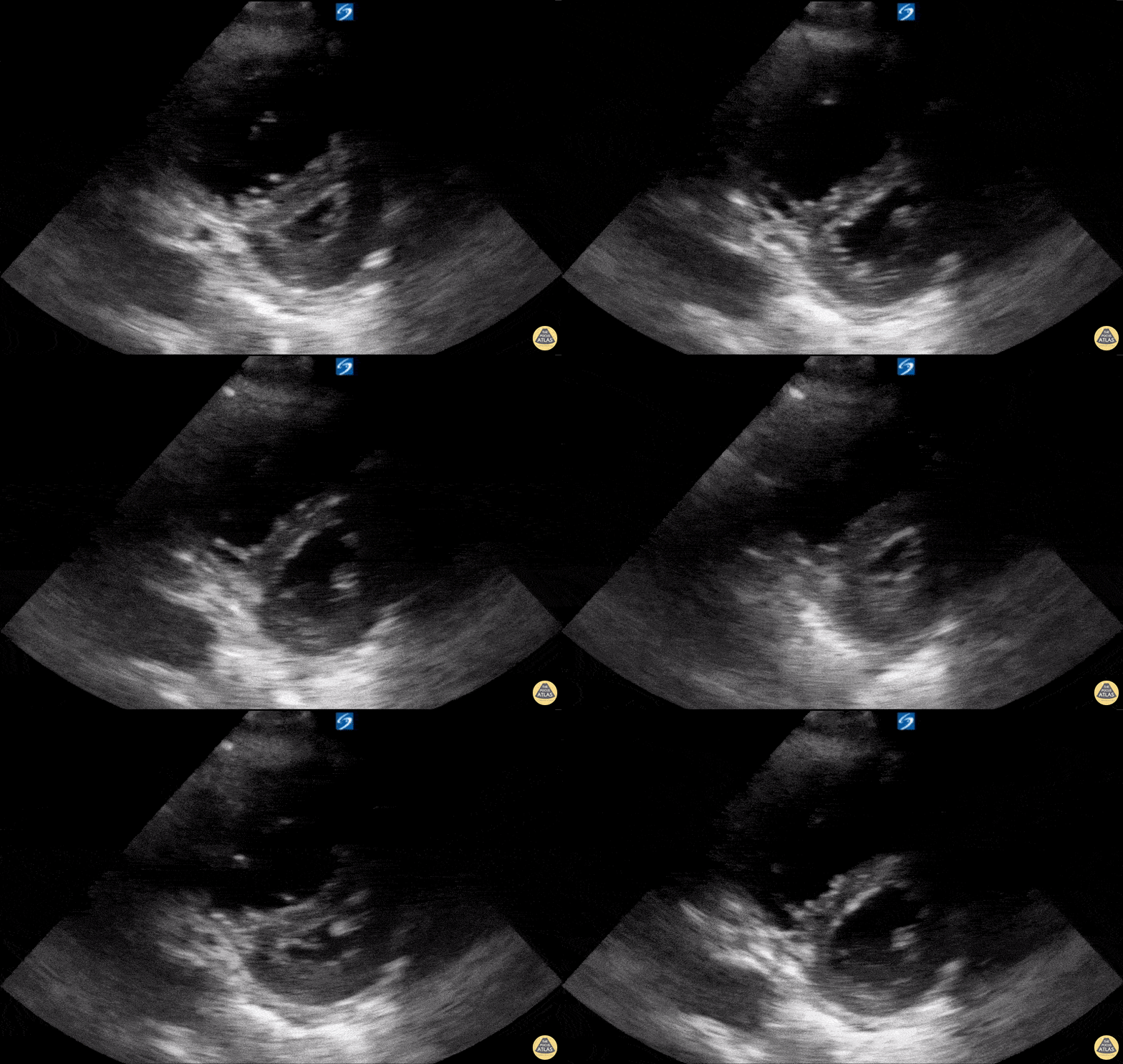
A 74-year-old male patient with Chronic Obstructive Pulmonary Disease (COPD) (not on home oxygen) presented to the ED with acute worsening dyspnea with persistent hypoxia despite supplemental oxygen, tachypnea, and increased work of breathing. There was no infectious or environmental exposure to explain the patient’s presentation. The patient’s differential diagnosis included acute pulmonary embolism (PE) and COPD exacerbation/progression. Parasternal long-axis (PSLA) and short-axis (PSSA) views showed RV dilatation with RV strain. The RV strain was demonstrated by flattening of the interventricular septum, creating a D-shaped LV during systole (D-sign). Also, it shows hyperdynamic LV from tachycardia with near obliteration of the LV cavity in systole. CT angiography scan showed no evidence of PE. The patient was admitted and diagnosed with a progression of severe COPD with pulmonary hypertension and right ventricular remodeling.
Contributed by: Hassan Alshaqaq, MBBS, Emergency Medicine Resident at King Saud University Medical City, @HassanAlshaqaq
Thoracic Aortic Dissection & Aneurysm
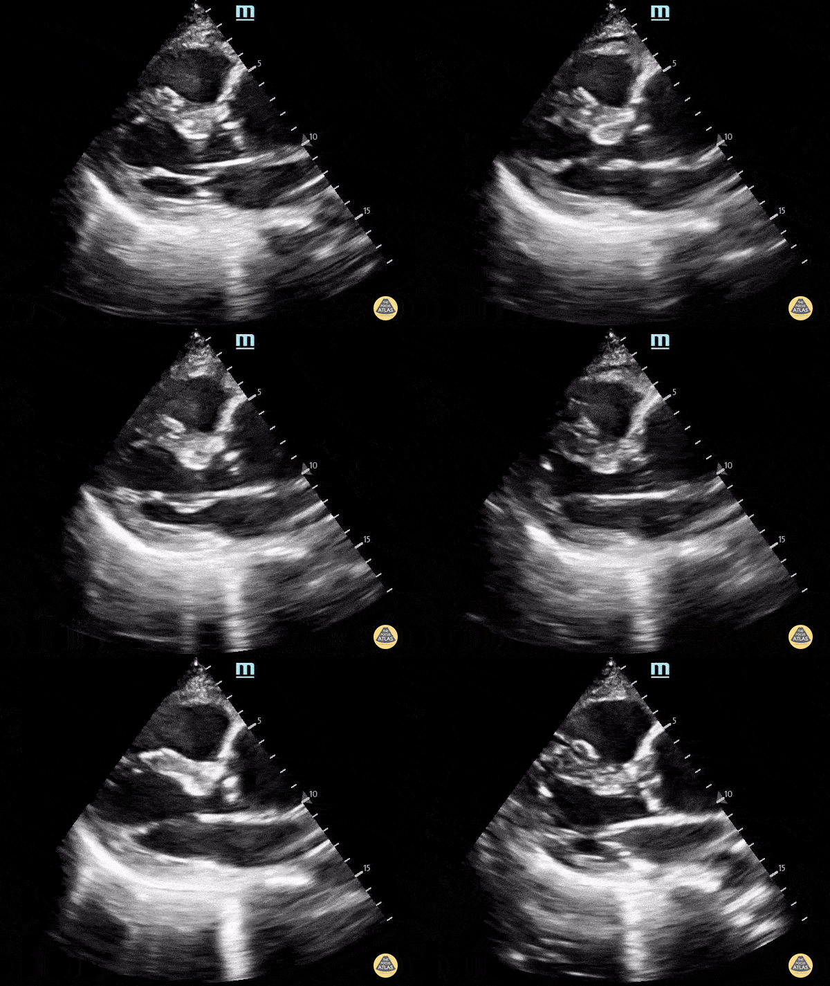
A 70 y.o. male presented with altered mental status.
POCUS echocardiogram performed in the ED revealed an ascending thoracic aortic aneurysm measuring approximately 5 cm in diameter (parasternal long-axis view shown here). A chart review revealed that the patient does indeed have a history of TAA, and a comparison of our findings to a prior CTA demonstrates no significant increase in diameter. Nevertheless, this study demonstrates the utility of POCUS in the rapid and early detection of ascending aortic abnormalities.
Alex Schlangen, D.O. PGY-1 EM Resident at Central Michigan University; Andrew Namespetra, MB BCh BAO. @AndrewNamespet1 PGY-3 EM Resident at Central Michigan University
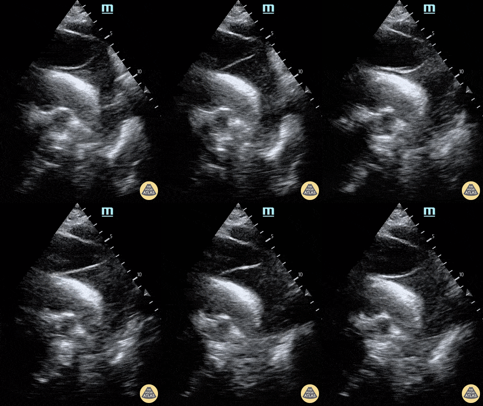
A 65-year-old male presents with shortness of breath (no chest pain) and was found to have a dilated aortic root on CT pulmonary angiogram. POCUS (supra sternal view) showed a dissection flap in the arch of aorta; a finding subsequently confirmed on CT aortagram. Patient was sent for emergency surgical intervention.
Dr.Rajasutharsan Kathirgamanathan, Emergency Physician
The Northern Hospital, Melbourne, Australia
@raj_kathir007
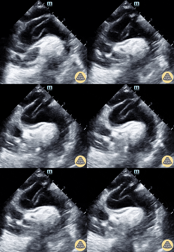
Suprasternal notch view shows a mobile intimal dissection flap in the aortic arch.
Michael Cover, MD
@michaelc0ver
Pericardial Disease
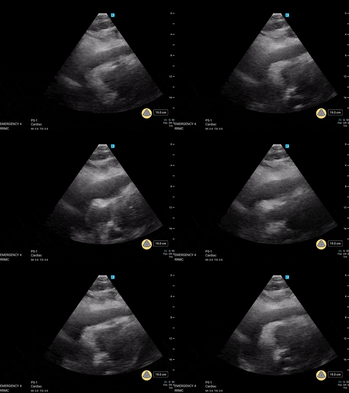
Here is an excellent of example of utilizing Rapid Ultrasound for Shock and Hypotension (RUSH). This was from a 77 year old patient who presented initially presented with progressive weakness and a fall while in the bathroom. His initial blood pressure was labile but not hypotensive. Workup revealed leukocytosis in the presence of anuria and was eventually admitted with broad spectrum antibiotics. Shortly after admission, he became increasingly hypotensive and required norepinephrine. RUSH performed initially with the intention of assessing IVC for fluid status however the image above was discovered. There is obvious right ventricular diastolic collapse in the presence of pericardial effusion, consistent with cardiac tamponade.
Dr. Austin Shanks, MD, PGY-2
Riverside Regional Medical Center Emergency Medicine Residency (Newport News, VA)
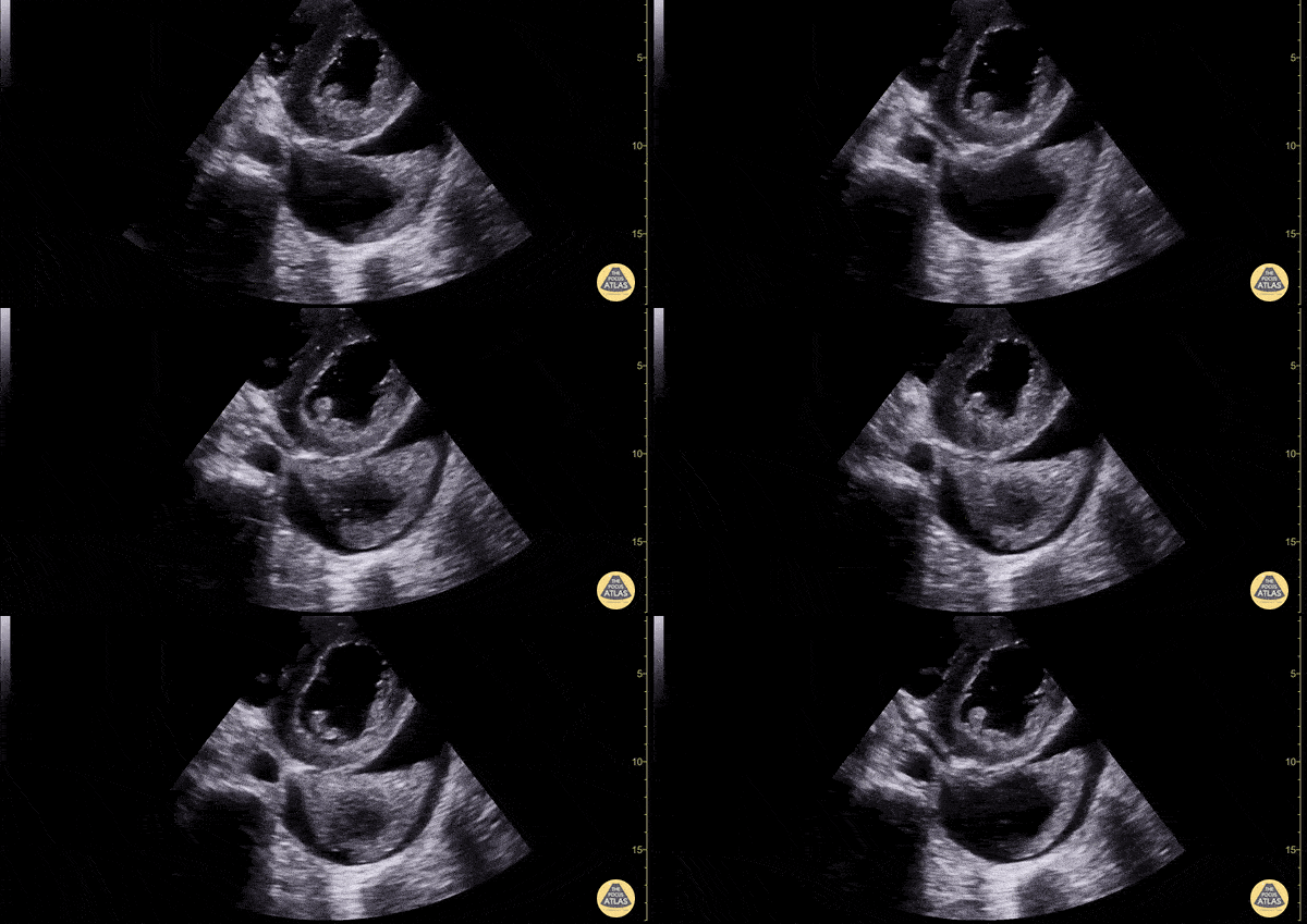
Patient with both a pericardial and pleural effusion in cardiac short axis view. You can see a large anechoic effusion surrounding the lung (pleural effusion) and a trace anechoic effusion spreading anterior to the descending aorta (pericardial effusion). Pleural effusions are never anterior to the aorta.
Dimitri Livshits DO, Ultrasound Fellow, Kings County/SUNY Downstate; Jane Belyavskaya MD, Ultrasound Fellow, Kings County/SUNY Downstate; Chris Hanuscin MD, Ultrasound Division Director, Kings County/SUNY Downstate;
![Pericardial Effusion Vs Fat Pad [1/2]](https://images.squarespace-cdn.com/content/v1/58118909e3df282037abfad7/1706570222652-Z6J3FZ0G4CX2KG8QEL57/image-asset.gif)
Pericardial fat pad is often mistaken for a pericardial effusion, this clip demonstrates both in the same clip. Pericardial fat pad moves in concert with the heart, while an effusion is circumferential, stationary and does not move in concert with the heart. Multiple cardiac views are helpful in making the diagnosis.
Dr. Dimitri Livshits Ultrasound Fellow;Dr. Jane Belyavskaya Ultrasound Fellow;Dr. Chris Hanuscin Ultrasound Fellowship Director
Valvulopathy
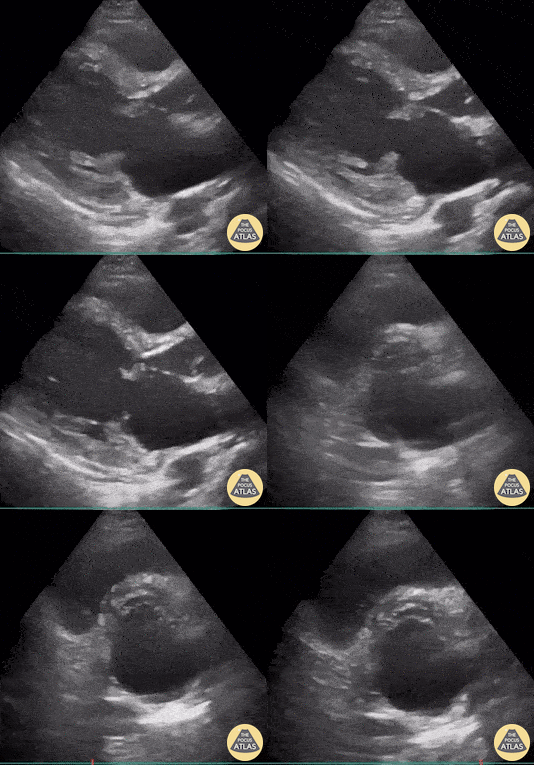
32-year-old woman presented to ED with clinical signs of subacute stroke - confirmed via Brain CT . In the investigation of etiology of cerebral injury, POCUS identified this large hyperechoic vegetation on the mitral valve (seen here both in PLAX and PSAX views). As a result of these images, a diagnosis of infectious endocarditis causing cerebral septic emboli injury was considered.
Renato Tambelli; @JediPocus
Emergency Physician (HCFAMEMA /Sao Paulo, Brazil)
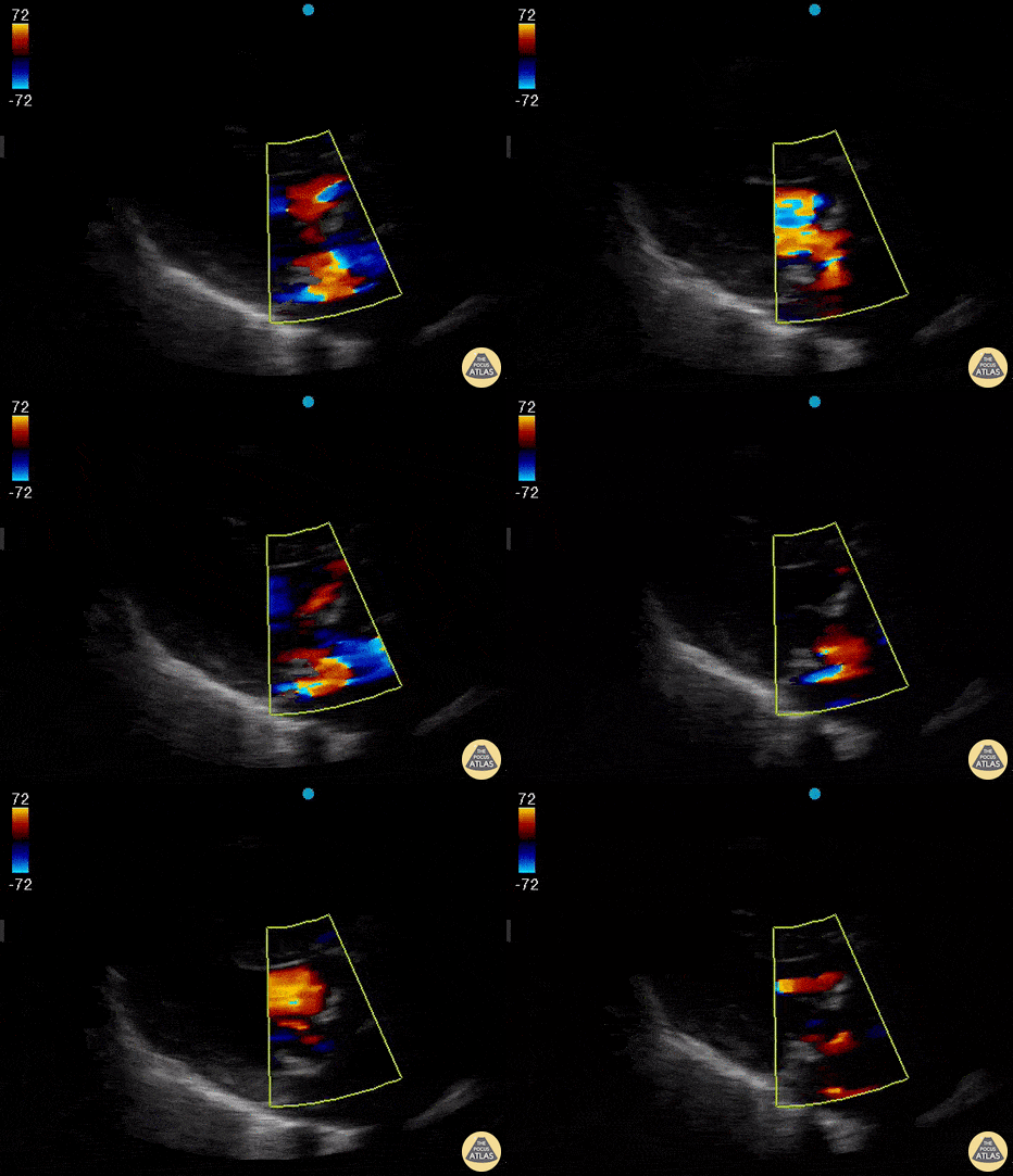
Parasternal long axis view in a patient with severe mitral regurgitation.
Rohan Rastogi, MD
@RohanRastogiMD
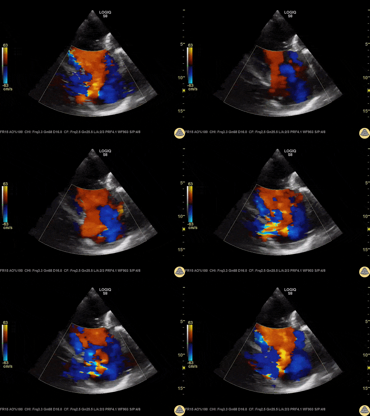
A patient with IVC and hepatic congestion on CT also had a holosystolic murmur at the left lower sternal border, worsened with inhalation. POCUS revealed severe tricuspid regurgitation on parasternal short axis view with color doppler.
Image courtesy of Robert Jones DO, FACEP @RJonesSonoEM
Director, Emergency Ultrasound; MetroHealth Medical Center; Professor, Case Western Reserve Medical School, Cleveland, OH
View his original post here
IVC & Abnormal Venous Waveforms
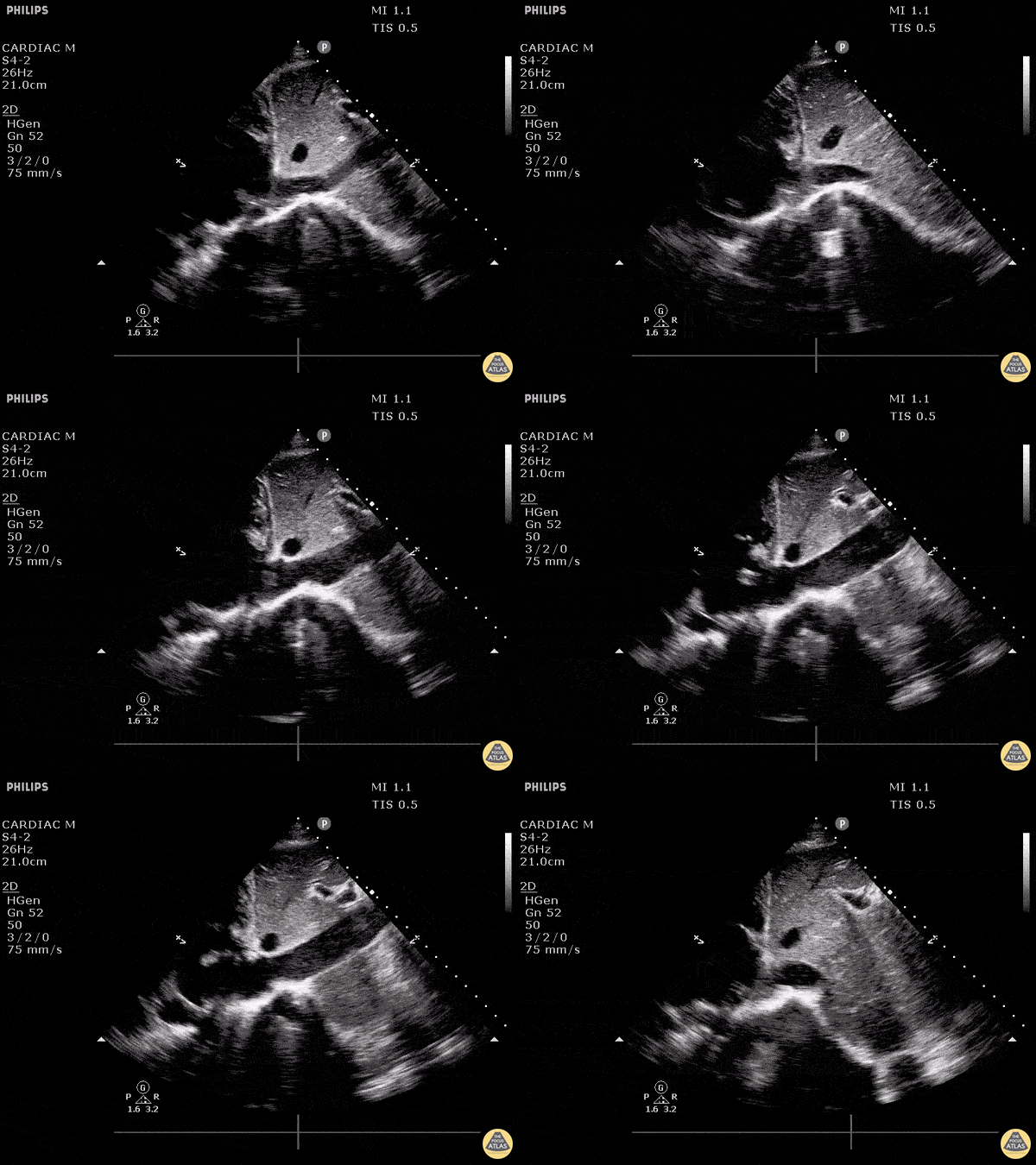
This patient initially presented post-operatively to the emergency department with complaints of dyspnea. As we fan through this saggital view of the IVC as it enters the right atrium, we can see hyperechoic structures suggestive of clot formation. An alternative view of this clot from a subxiphoid view can be seen here. The patient was subsequently diagnosed with a DVT that extended into their central femoral vein, at the same leg that was recently operated on.

Image courtesy of Robert Jones DO, FACEP @RJonesSonoEM
Director, Emergency Ultrasound; MetroHealth Medical Center; Professor, Case Western Reserve Medical School, Cleveland, OH
View his original post here
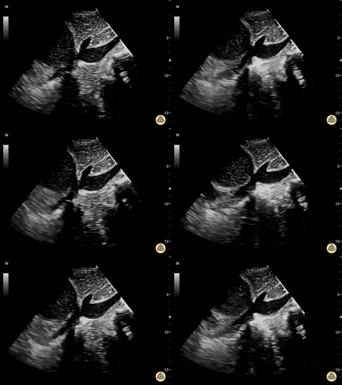
58 y/o F with PMHx of metastatic adenocarcinoma of lung presents with progressive SOB for one week. The patient was tachycardic to 103, normotensive, afebrile, mildly tachypneic and saturating 95% on room air. EKG demonstrated sinus tachycardia without electrical alternans.
POCUS revealed a large, complex, loculated, anterior pericardial effusion. Sonographic findings of right atrial/ventricle collapse and IVC dilatation confirmed cardiac tamponade. In this long-axis subxiphoid view, the IVC is seen enlarged and has minimal respiratory variation.
Common ultrasound findings of cardiac tamponade include: RV end-diastolic collapse, RA systolic collapse, plethoric IVC, septal “bounce”, decrease of mitral valve inflow velocity >25% with inspiration. Echocardiography is the modality of choice to evaluate for pericardial effusion and to assess for cardiovascular compromise (right chamber collapse and IVC). Accurate determination of this patient’s tamponade allowed for rapid surgical intervention. Patient underwent pericardial window with partial pericardiectomy a few hours after presenting to the ED.
Dr. Pumarejo, Dr. Tran and Dr. Patel. Aventura Hospital and Medical Center Emergency Medicine.
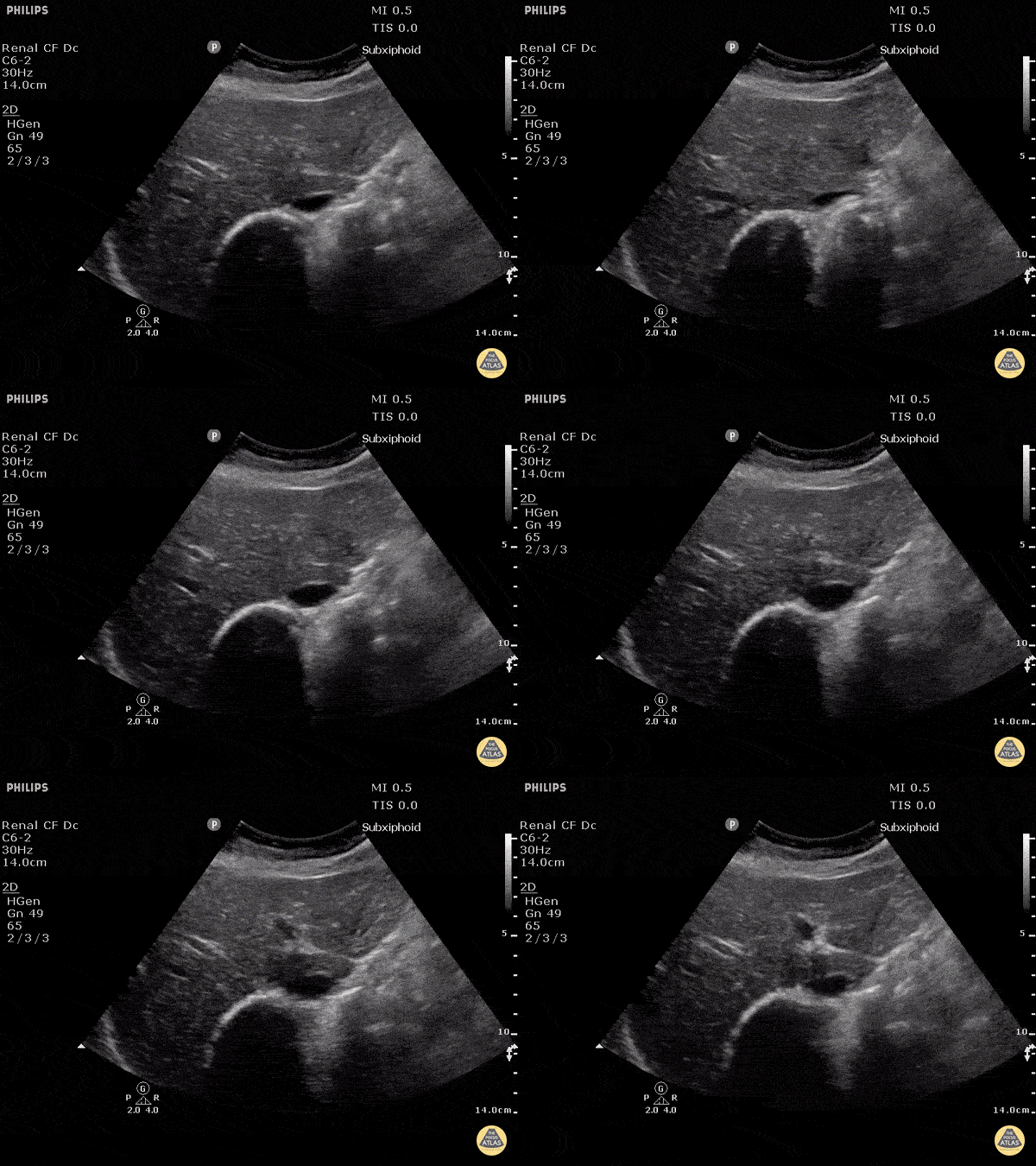
Cardiac Tumors
TEE
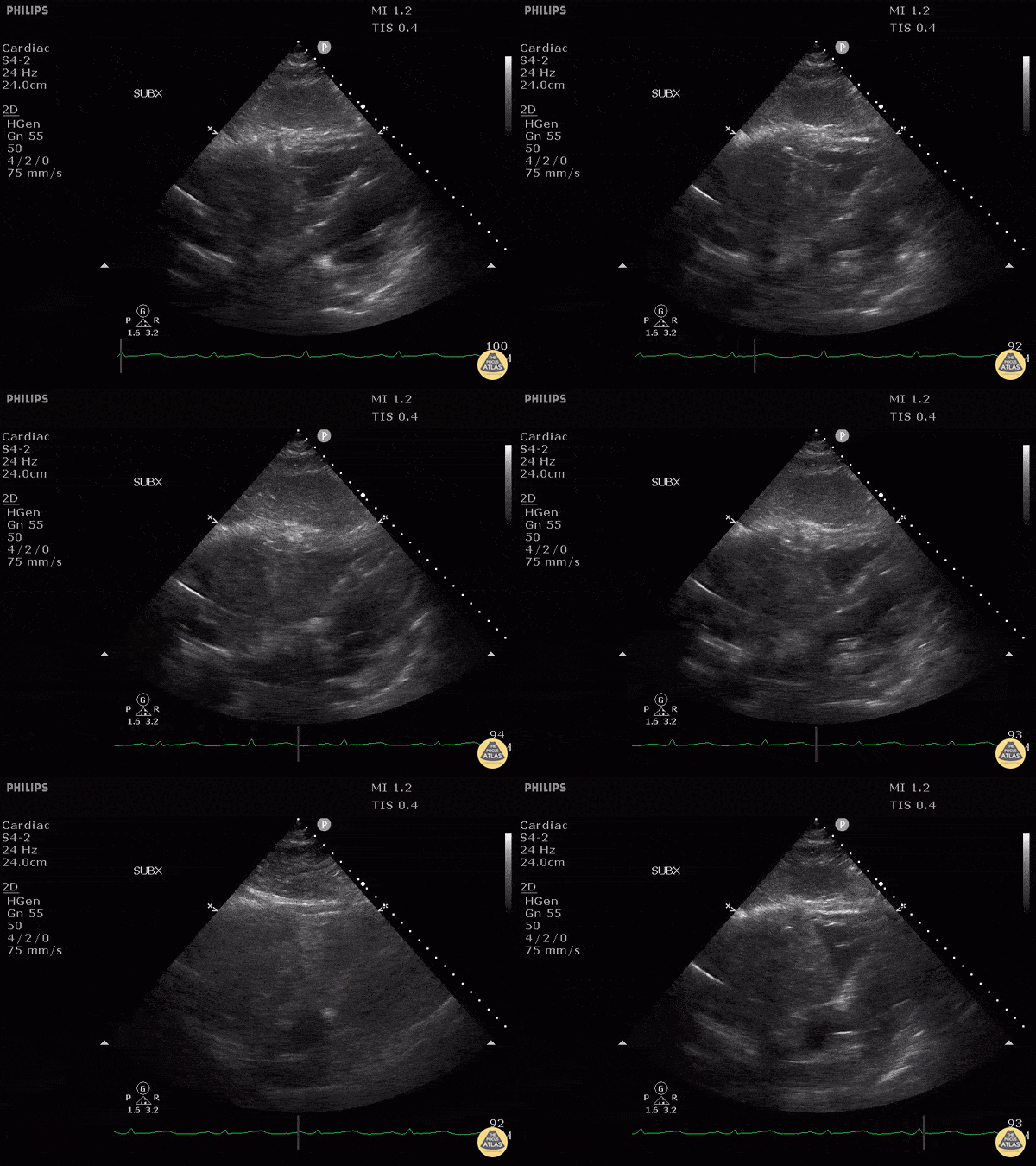
This patient presented to the emergency department after a syncopal episode upon exertion. From this subxiphoid view of their heart, we can see isoechoic structures within their right atrium. This patient was ultimately diagnosed with a cardiac sarcoma.
Image courtesy of Robert Jones DO, FACEP @RJonesSonoEM
Director, Emergency Ultrasound; MetroHealth Medical Center; Professor, Case Western Reserve Medical School, Cleveland, OH
View his original post here
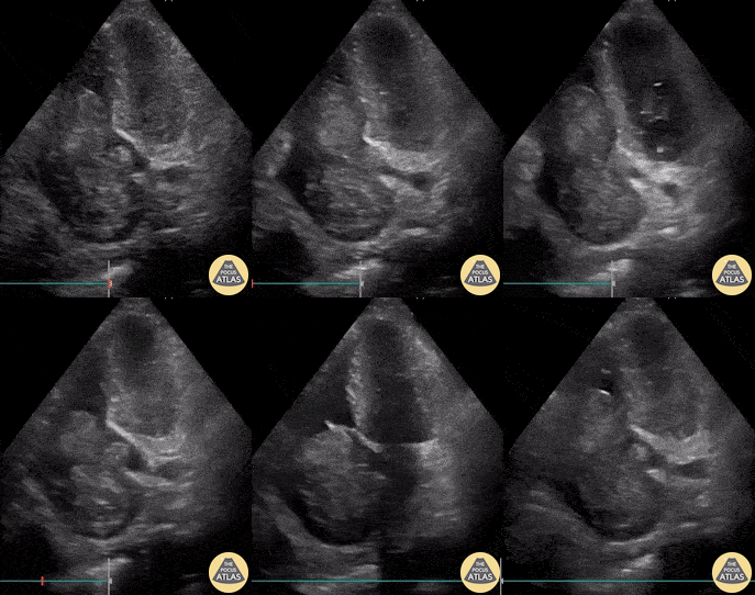
64 yo man with history of hepatic cirrhosis presented to ED with hypotension. During the RUSH exam, we incidentally identified this large hyperechoic mass occupying a large part of the right heart. Cardiac myxomas are the most common primary cardiac tumor in adults, though only 15–20% originate within the right atrium as does this one. The etiology of hypotension in our patient ended up being septic shock secondary to SBP. However, bedside ultrasound sometimes surprises you with stunning and unexpected images!
Renato Tambelli, @JediPocus
Emergency Physician (HCFAMEMA /Sao Paulo, Brazil)
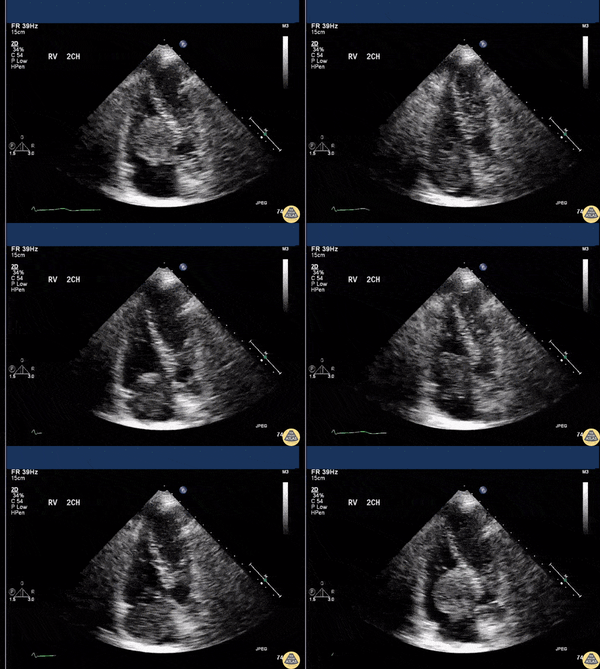
An apical 4 chamber view on a patient with CP, SOB, and palpitations revealed a right sided atrial myxoma.
Image courtesy of Robert Jones DO, FACEP @RJonesSonoEM
Director, Emergency Ultrasound; MetroHealth Medical Center; Professor, Case Western Reserve Medical School, Cleveland, OH
View his original post here
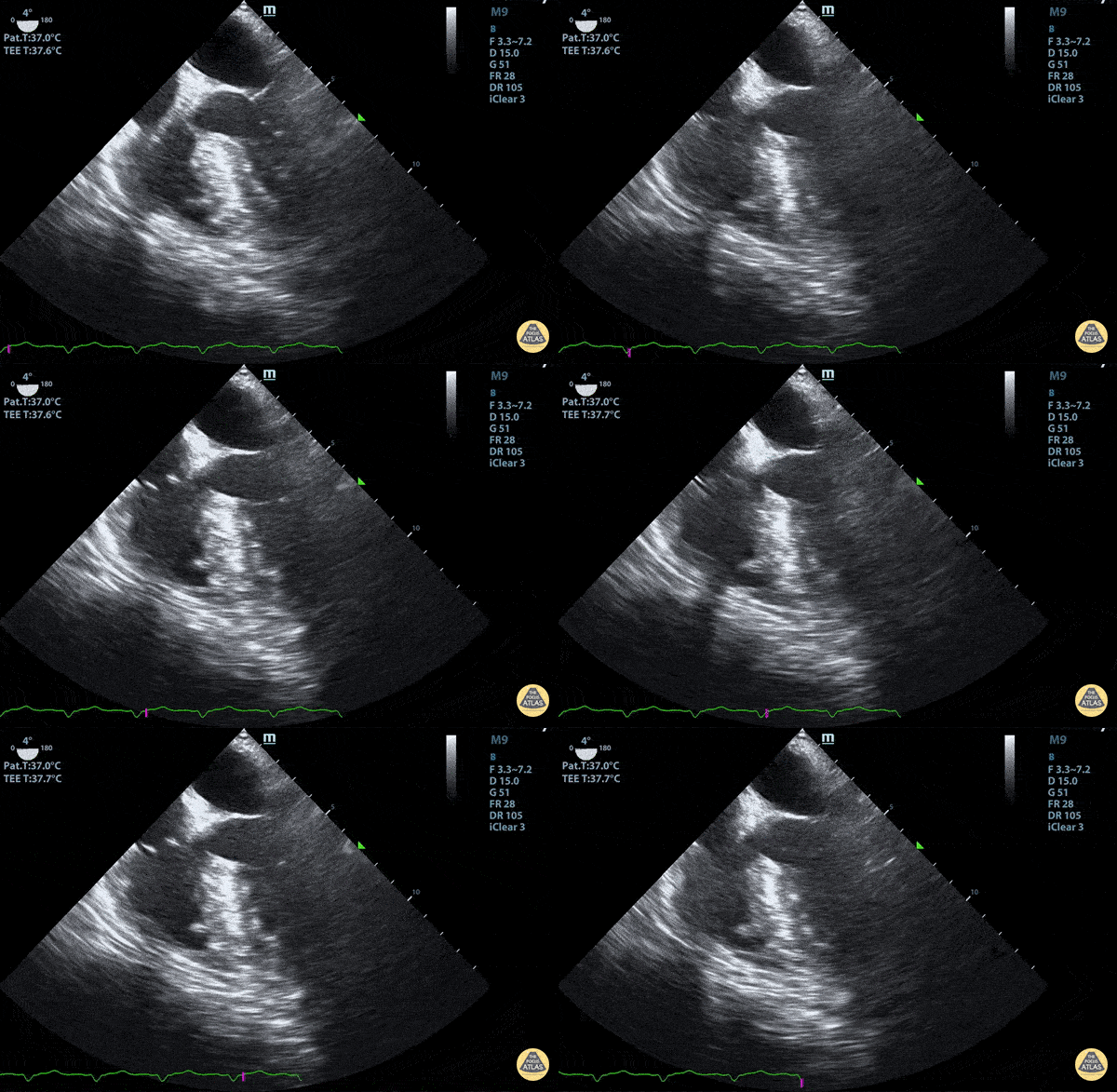
Mid-Esophageal 4 Chamber View with a trace pleural effusion
Duncan McGuire, DO, Emergency Medicine, Beaumont Health
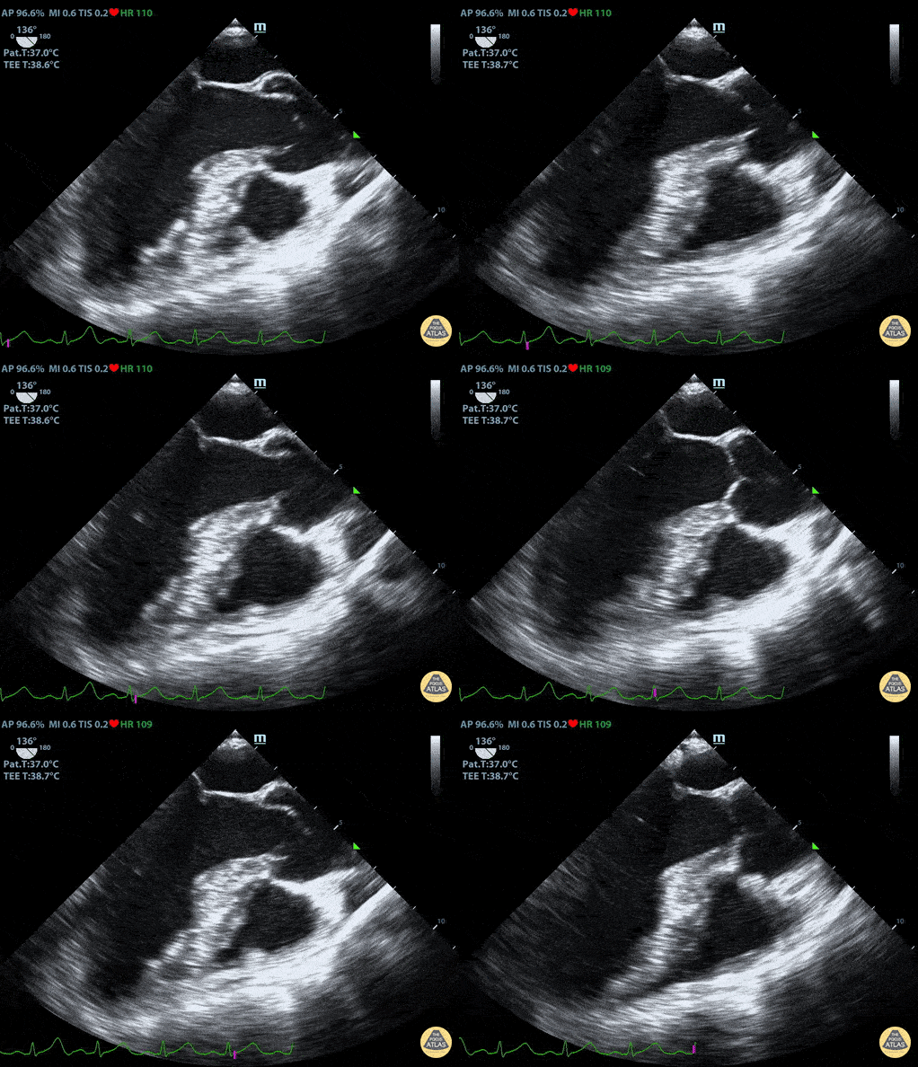
Normal Mid-Esophageal Long Axis View
Duncan McGuire, DO, Emergency Medicine, Beaumont Health
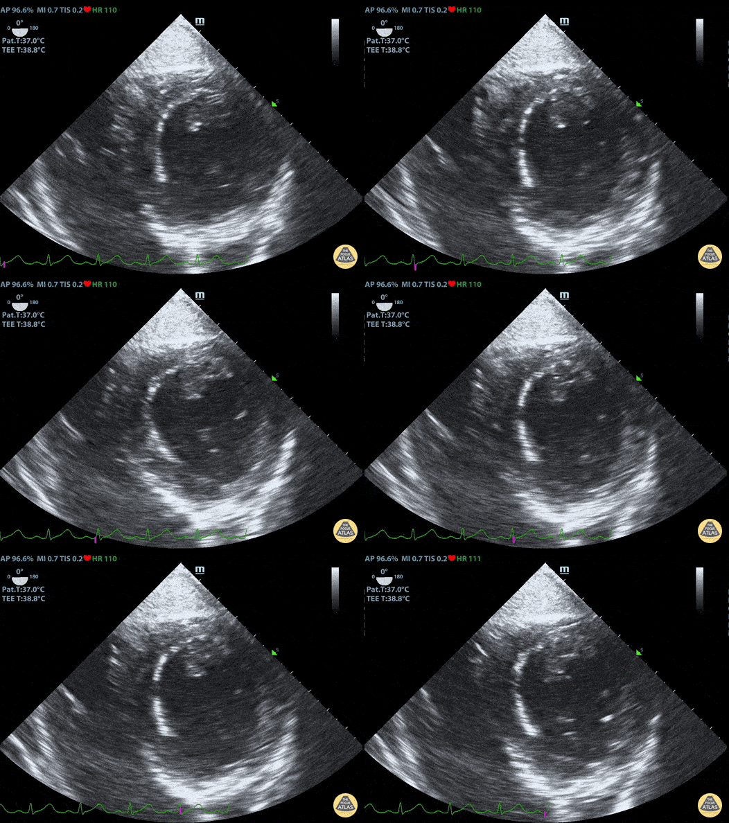
Normal Trans-Gastric Short Axis View
Duncan McGuire, DO, Emergency Medicine, Beaumont Health
Other Cardiac Pathology
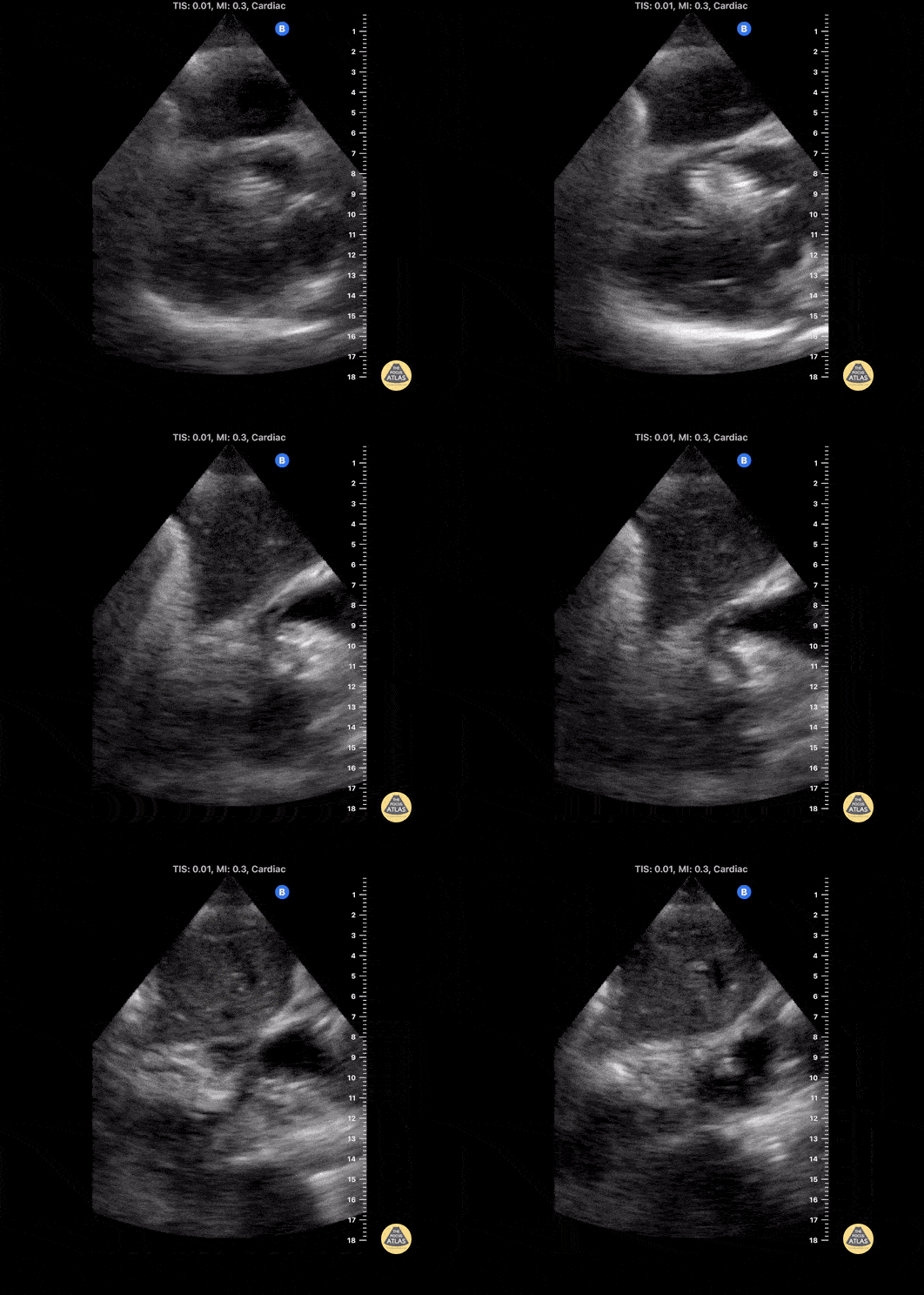
Middle aged male with recent right iliac DVT s/p thrombectomy and venous stent placement is found to have asymptomatic migration of stent into the right heart. In the subxiphoid view, the hyperechoic coiled-appearing stent is seen traversing the tricuspid valve. Percutaneous attempt at removal was unsuccessful, so he ultimately underwent open-heart surgery.
Contributed by: Eric Reid, MD
This patient initially presented post-operatively to the emergency department with complaints of dyspnea. From this subxiphoid view of the heart, we can see a hyperechoic structure suggestive of clot formation. An alternative view of this clot viewed from the right atrium and IVC junction can be seen here, termed “clot-in-transit”. The patient was subsequently diagnosed with a DVT that extended into their central femoral vein.
Image courtesy of Robert Jones DO, FACEP @RJonesSonoEM
Director, Emergency Ultrasound; MetroHealth Medical Center; Professor, Case Western Reserve Medical School, Cleveland, OH
View his original post here
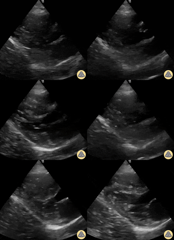
This is a PLAX view obtained during CPR . It reveals adequate compressions with the LV “squashing” and obliterating the LV lumen; then subsequently the LV lumen reappearing and recoiling, which allows the LV to refill.
Renato Tambelli, @JediPocus
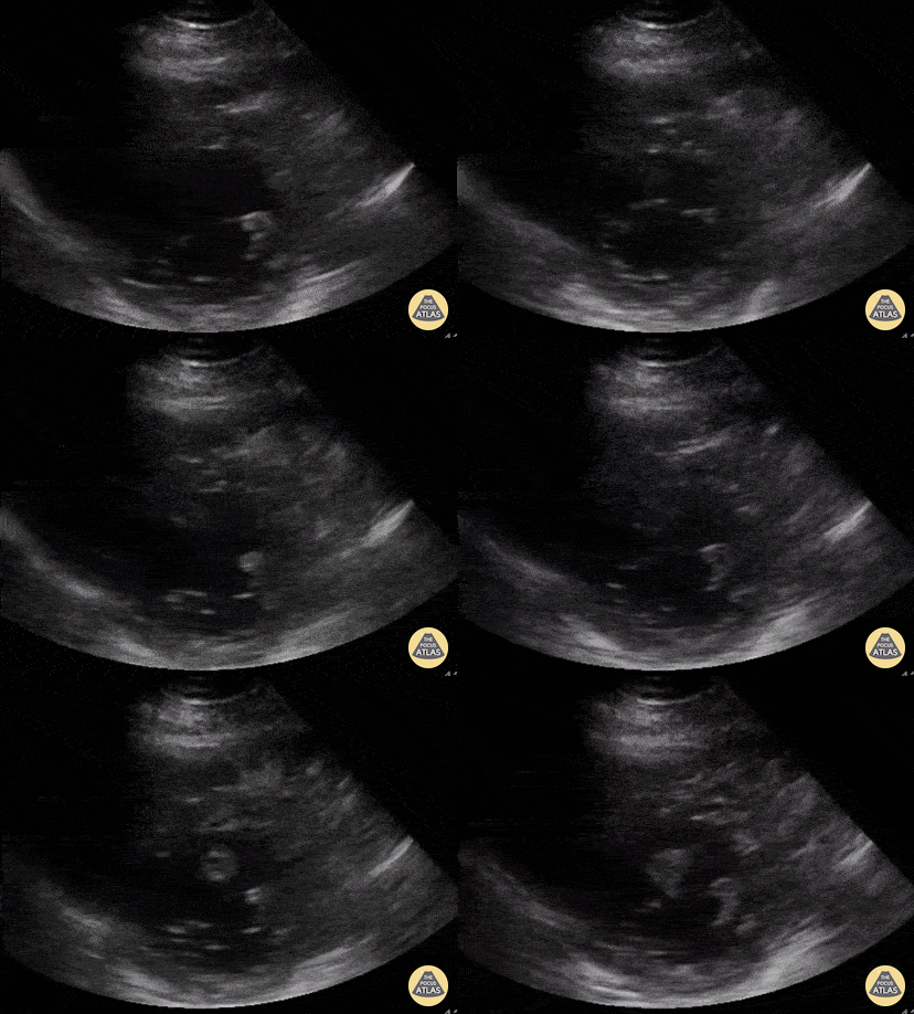
Parasternal short axis view of a 47yo with PMH non-obstructive CAD incidentally found to have mildly reduced LV systolic function and an echogenic mass in LV extending to outflow tract. Determined to be a large LV thrombus of unknown etiology.
Andrew Balster, MD
Paul Musgrave, MD (OHSU IM POCUS fellow)
@POCUSaurusDx
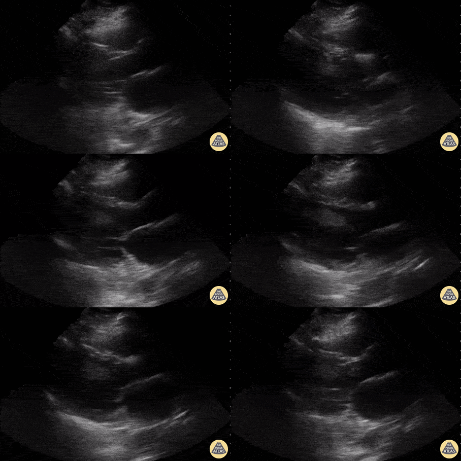
Parasternal long axis view of a 47yo with PMH non-obstructive CAD incidentally found to have mildly reduced LV systolic function and an echogenic mass in LV extending to outflow tract. Determined to be a large LV thrombus of unknown etiology.
Andrew Balstera, MD
Paul Musgrave, MD (OHSU IM POCUS Fellow)
@POCUSaurusDx
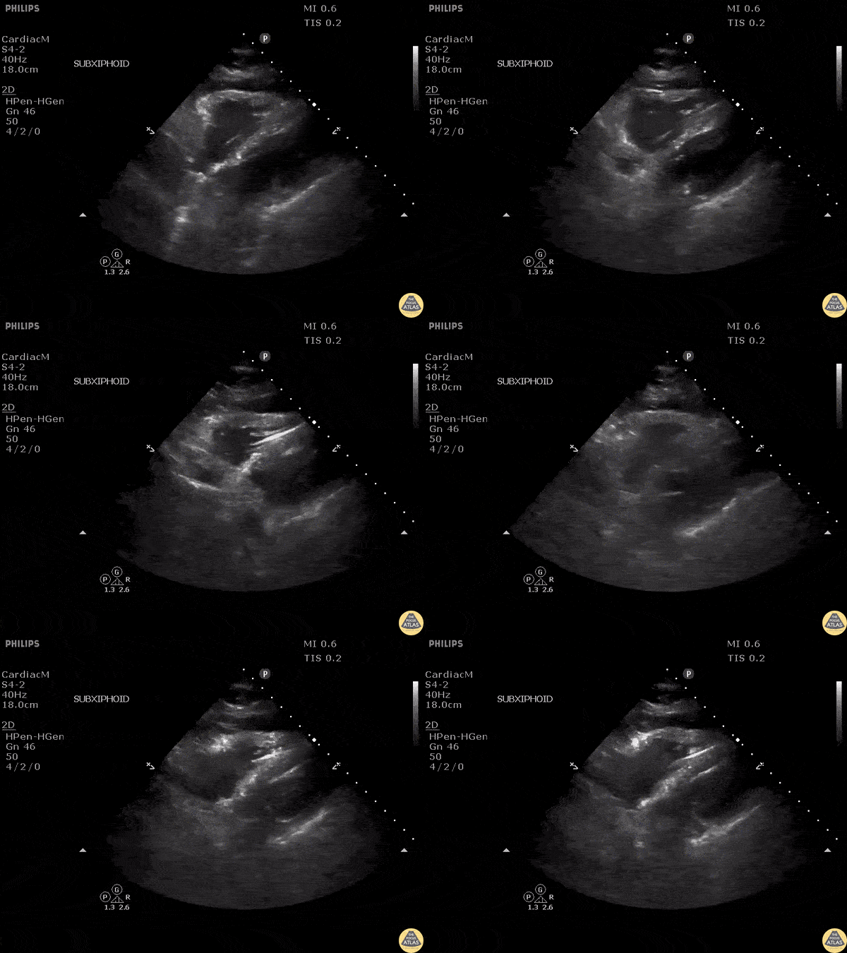
Patient with chest pain with recent history of pacemaker placement. Subxiphoid view reveals the pacer wire in the right ventricle piercing the wall, causing a pericardial effusion.
Image courtesy of Robert Jones DO, FACEP @RJonesSonoEM
Director, Emergency Ultrasound; MetroHealth Medical Center; Professor, Case Western Reserve Medical School, Cleveland, OH
View his original post here

Undergoing Maintenance
We have reorganized our echo section and all images can be accessed below on this page. We are currently working on category pages (buttons below will be functional soon)! Please excuse the mess during this transition!
- The POCUS Atlas Team