
Hydronephrosis
Hydronephrosis and Obstruction
No Hydronephrosis
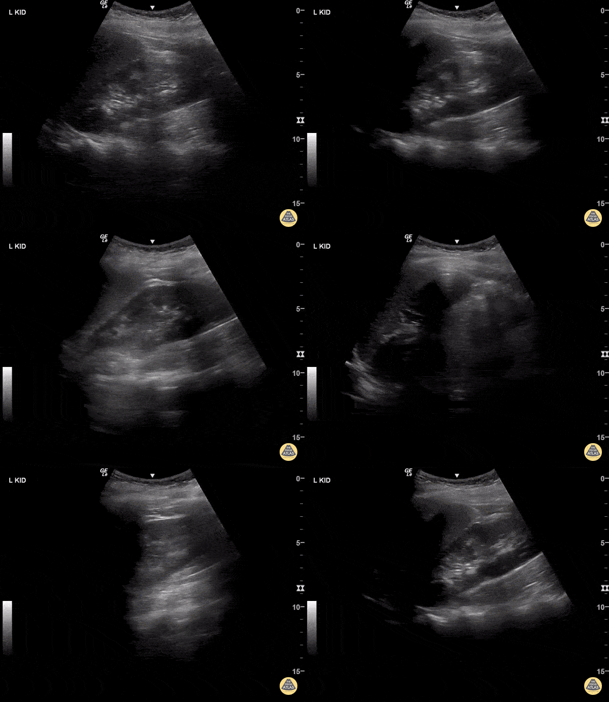
Mild Hydronephrosis

Mild - grade two hydronephrosis with dilation of the renal pelvis and dilation of the some calyces. Grade one would not include dilated calyces.
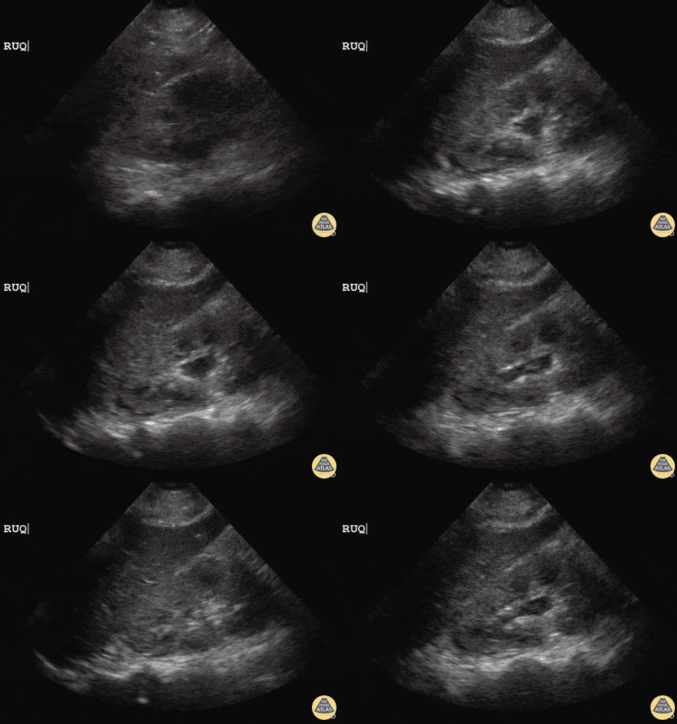
21 y/o female post-op emergency hysterectomy post uterine rupture with rising creatinine in surgical ICU.
POCUS revealed right-sided mild Grade I hydronephrosis with appreciable dilated major calyces and renal pelvis. Initial concern is for obstructive process or ureter injury.
Dr. Sathya Subramaniam, Pediatric EM Fellow - Kings County/SUNY Downstate
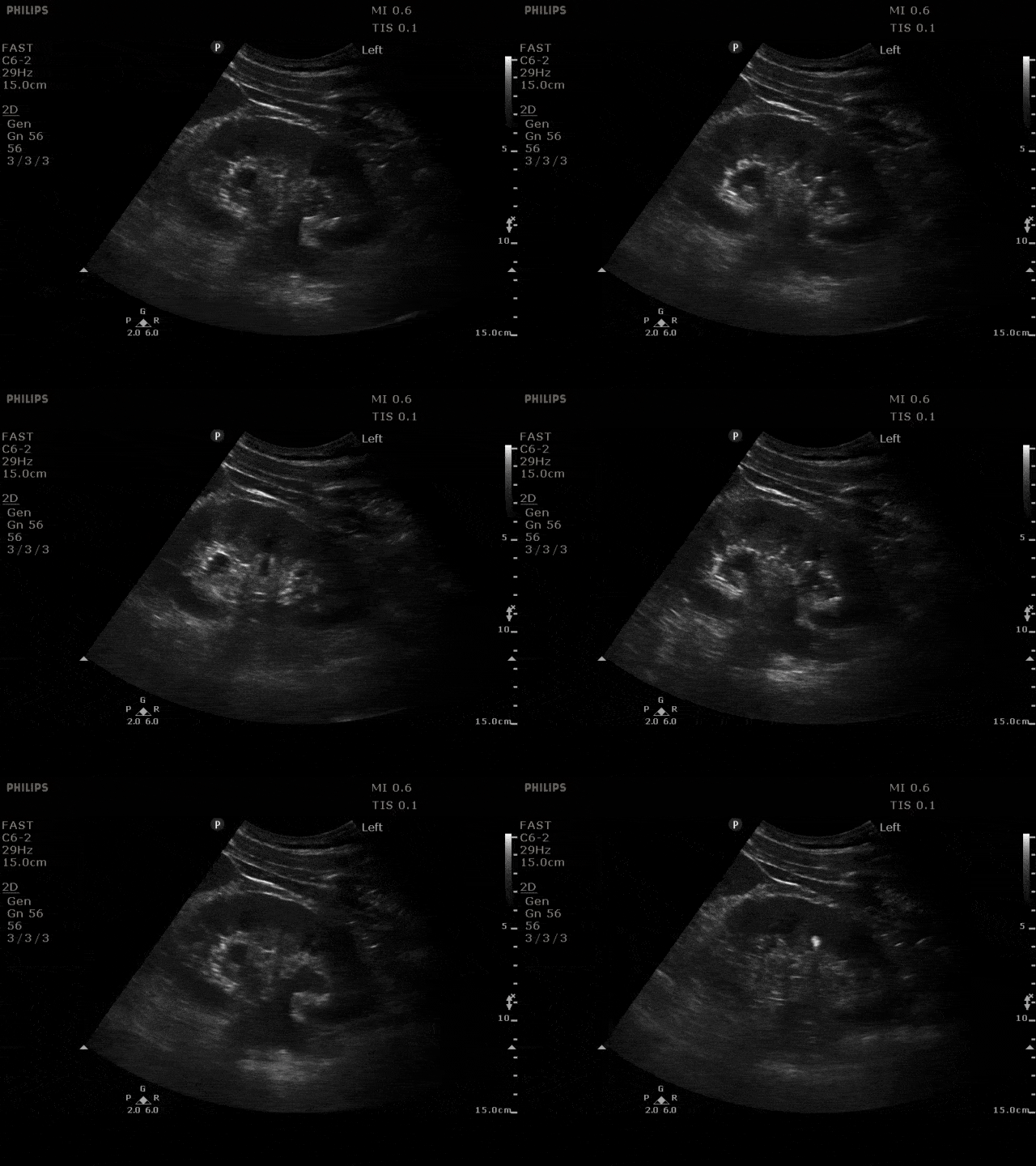
Mild - grade two hydronephrosis with dilation of the renal pelvis and dilation of the calyces. Grade one would not include dilated calyces.
Moderate Hydronephrosis
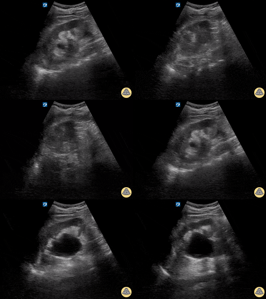
Here is an example of hydronephrosis in the presence of nephrolithasis. This patient presented to the emergency department complaining of new right flank pain after waking up in the morning. She was seen previously for urinary complaints, diagnosed with UTI and started on antibiotic regiment that had been altered twice due to concerns of sub-optimal coverage. She demonstrated right CVA tenderness on physical exam, raising suspicion for pyelonephritis as well as hydronephrosis. Subsequent point of care ultrasound revealed moderate hydronephrosis. CT was obtained to further evaluate and confirmed the presence of an obstructing kidney stone. Note presence of hydronephrosis as the probe is fanned/tilted through the kidney, highlighting the importance of scanning through the entire organ.
Dr. Carlo Zamora, DO, PGY-1
Riverside Regional Medical Center Emergency Medicine Residency (Newport News, VA)
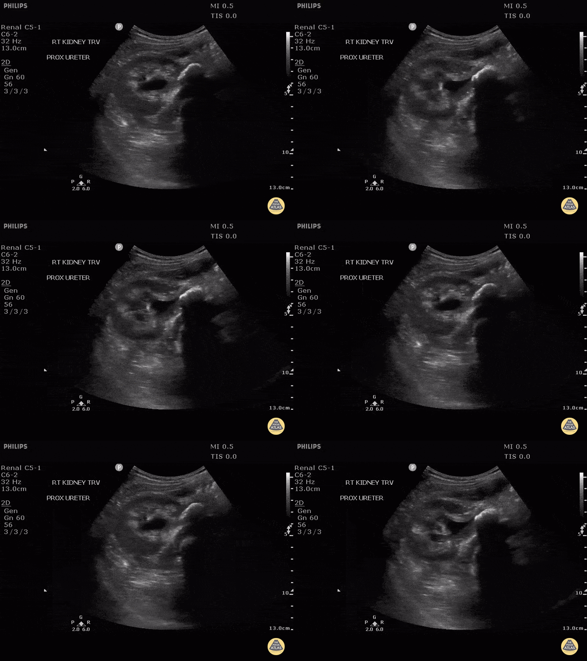
This patient presented with fever and flank pain with a known history of medullary nephrocalcinosis. Here we can see internal echoes within the dilated collecting system consistent with pyonephrosis. A large stone can be seen in the proximal ureter as a hyperechoic line with posterior shadowing.
Image courtesy of Robert Jones DO, FACEP @RJonesSonoEM
Director, Emergency Ultrasound; MetroHealth Medical Center; Professor, Case Western Reserve Medical School, Cleveland, OH
View his original post here
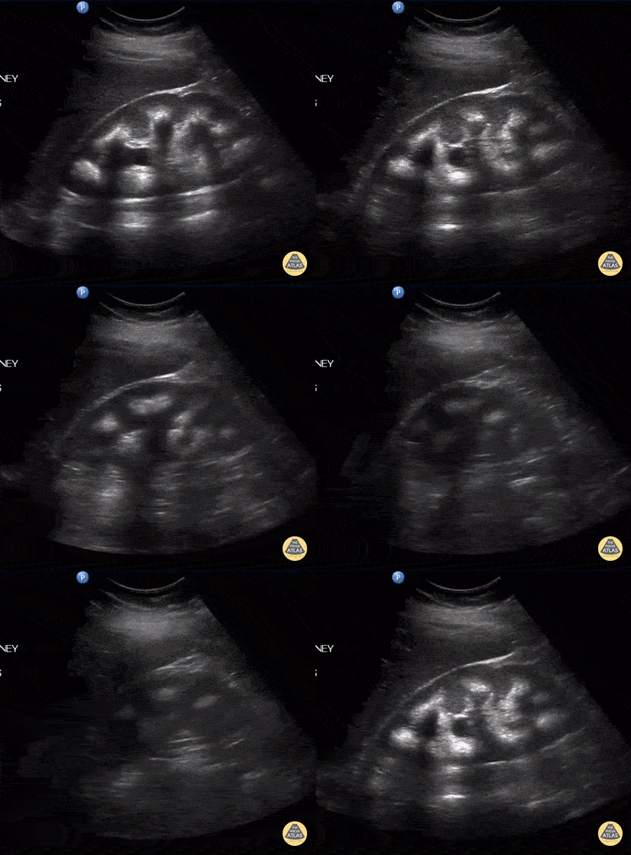
A female patient presented with right flank pain. A longitudinal view of the right kidney indicates medullary nephrocalcinosis with posterior shadowing and moderate hydronephrosis. Note the posterior shadowing partially masks the hydronephrosis. Further ultrasound found a distal ureteral stone.
Image courtesy of Robert Jones DO, FACEP @RJonesSonoEM
Director, Emergency Ultrasound; MetroHealth Medical Center; Professor, Case Western Reserve Medical School, Cleveland, OH
View his original post here
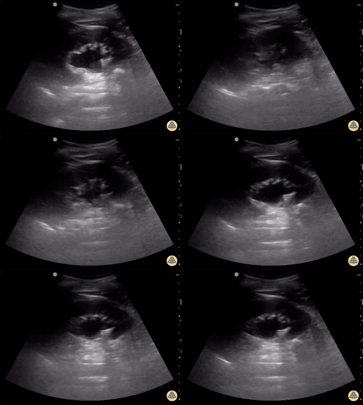
The degree of hydronephrosis is determined by a grading system. Grade 0 (none) means there's no dilation of the renal pelvis.
Grade 1 (mild) means there's mild dilation of the renal pelvis without any dilation of the calyces. Grade 2 means there's moderate dilation of the renal pelvis that extends to a few calyces.
Grade 3 (moderate) means the renal pelvis dilation extends to all the calyces. Grade 4 (severe) means there's extension of dilation to all the calyces with the addition of thinning of the renal parenchyma.
In this clip, the renal pelvis calyces are dilated but there is no thinning of the renal parenchyma making this mild to moderate, grade 2.
Sukh Singh, MD
Caption by Matthew Riscinti, MD
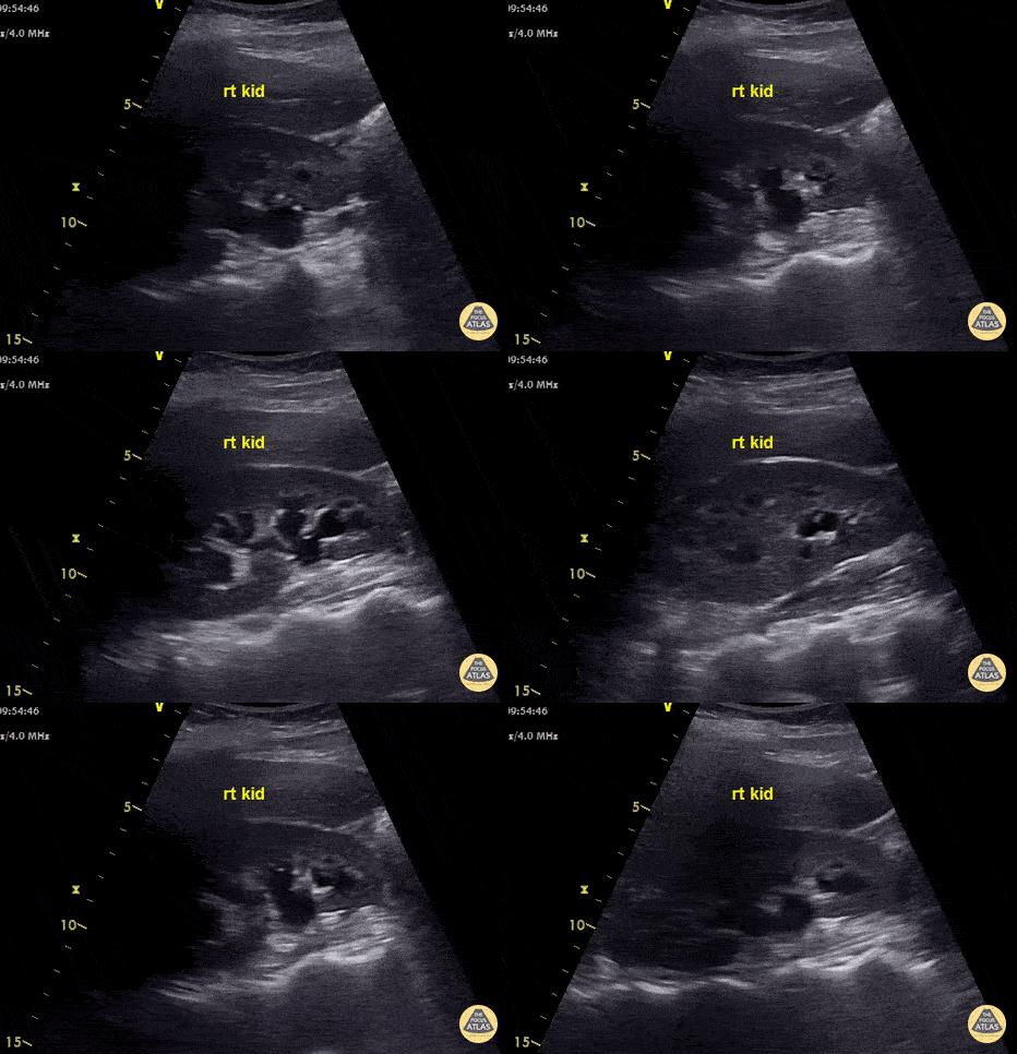
This young lady presented with clinical features of pyelonephritis - fever, rigors and right flank pain. Renal US shows moderate hydronephrosis and hydroureter. CT showed a 5.7mm right mid ureteric stone. Nephrostomy tube was placed to decompress obstructive uropathy.
Ultrasound is insensitive for pyelonephritis - most patients have normal scans. POCUS can be used to check for hydronephrosis, renal abscess, pyonephrosis or emphysematous pyelonephritis as these findings will alter management.
Images recorded by Dr. Khaled Taha
Submitted by Dr Cian McDermott
Mater University Hospital, Dublin, Ireland
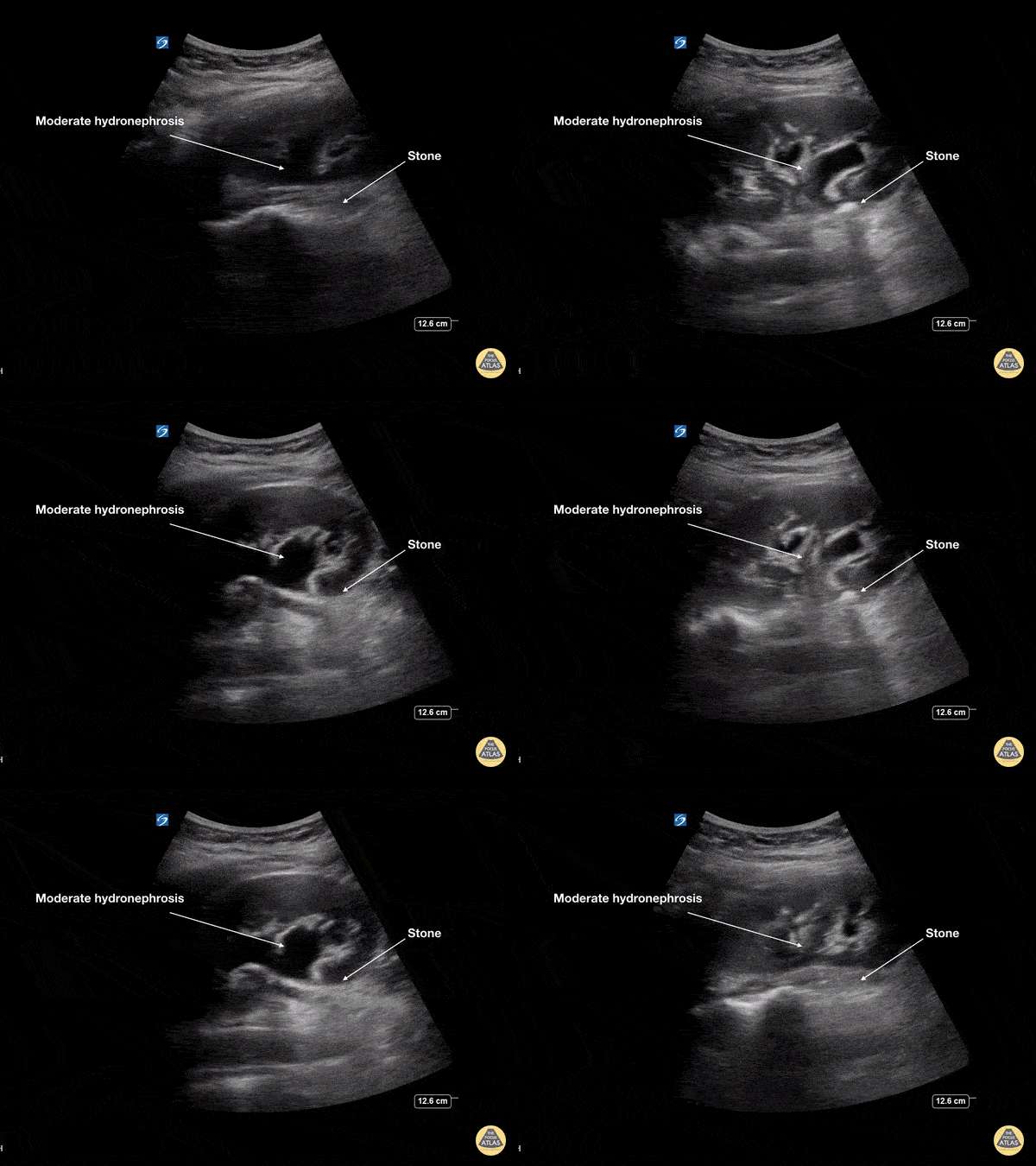
This young female presented with colicky left flank pain worsening over the previous 24 hours. POCUS showed moderate hydronephrosis with rounding of the calyces of the left kidney collecting system. The stone is seen at the ureteropelvic junction (UPJ) as a hyperechoic structure with posterior acoustic shadowing measuring 7mm on CT. At ureteroscopy, the stone was retrieved, fragmented and a double J stent was placed
Dr Cian McDermott, Mater University Hospital, Dublin, Ireland
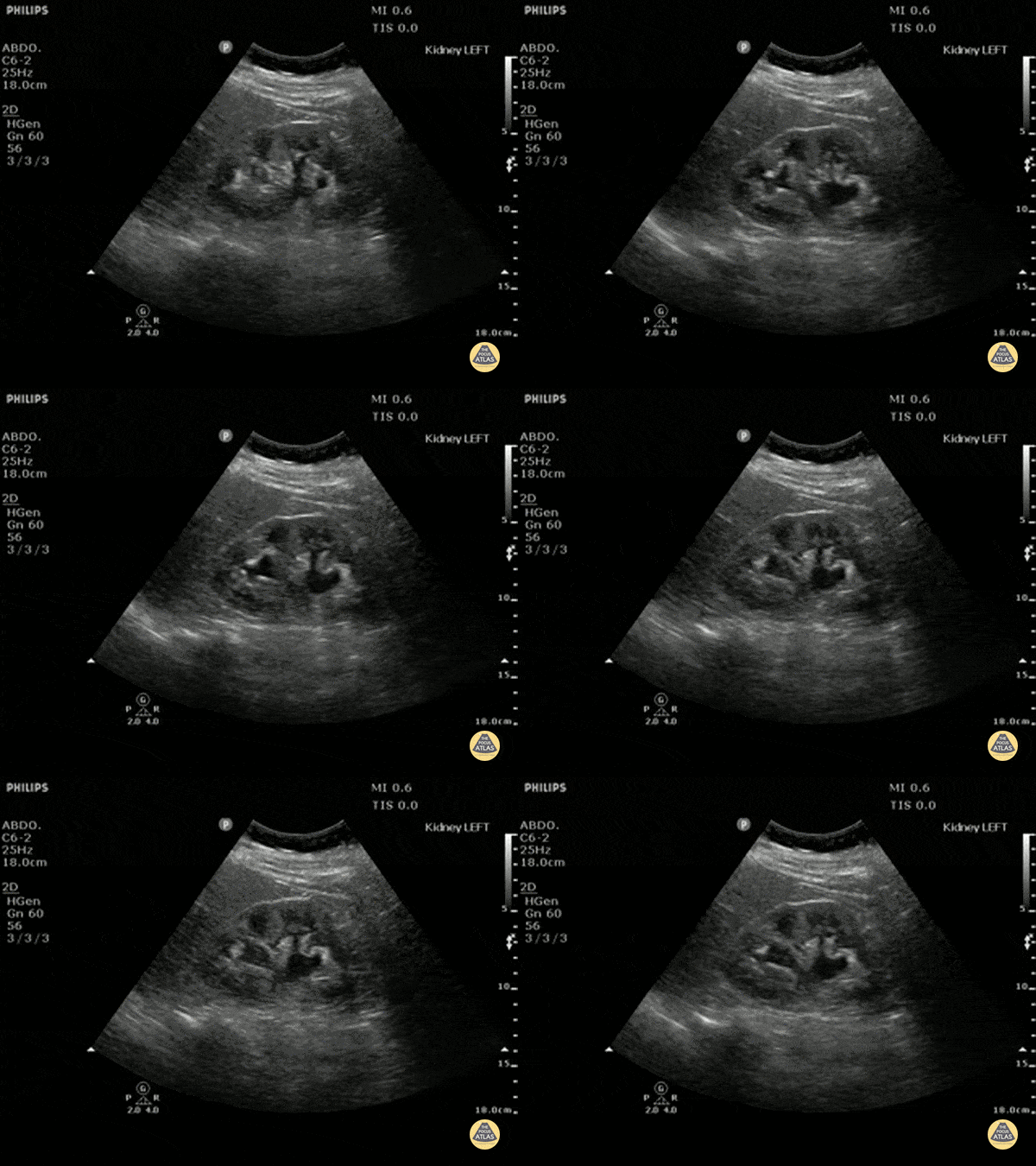
Moderate (grade three) hydronephrosis can be appreciated here with dilation of both the renal pelvis and calcyces. The renal cortex is also thinned. There is not gross atrophy.
Dr. Justin Bowra et al. (Dr. Yogi)
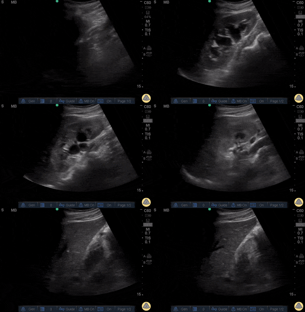
66 yo M with hx of congenital single kidney and prostate cancer presents with suprapubic discomfort x 1 week. Found to be in urinary retention.
POCUS allows grading of hydronephrosis based off of the severity of the dilation of the renal pelvis and calyces. Here we see dilated pelvis, ballooning calyces, and cortical thinning. This represents Grade 3, moderate hydronephrosis. Grade 4, severe, would demonstrate further atrophy and loss of borders, occurs with severe hydronephrosis.
Rushabh Shah, MD, MBA and Maria-Pamela Janairo, MD - Kings County/SUNY Downstate Emergency Medicine
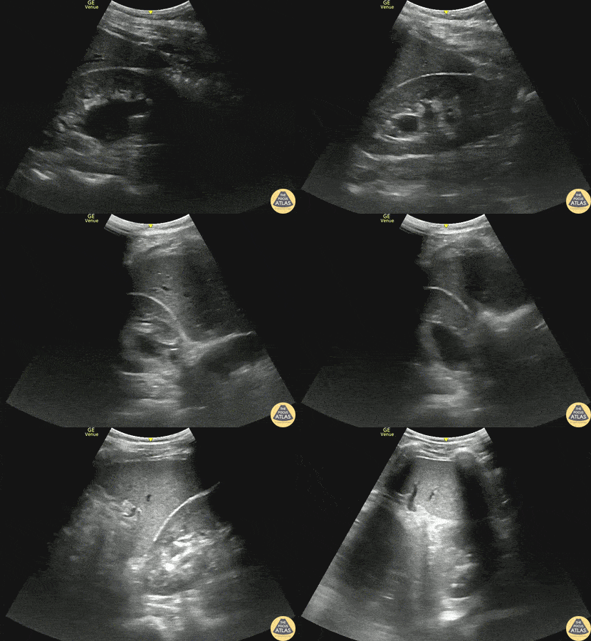
30s M with no past medical history presented with acute onset right sided flank pain. POCUS demonstrated moderate hydronephrosis of the right kidney with evidence of hydroureter as well. Moderate hydronephrosis is seen here with distension of the renal pelvis as well as distension of most of the renal calyces, with intact renal cortical thickness. This patient had his symptoms controlled and was able to be discharged.
Dr. Mark Serpico, PGY3
Denver Health Residency in Emergency Medicine
Severe Hydronephrosis
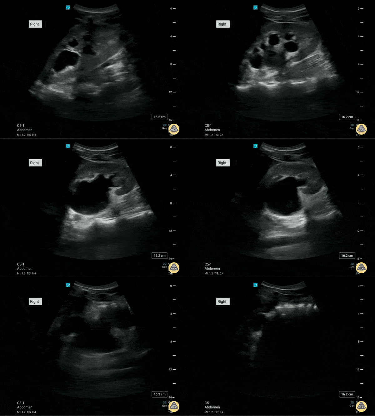
This is the right renal ultrasound of a 20 year old male with a past medical history of recurrent obstructing nephrolithiasis. There is a moderate to severe hydronephrosis present. with what appears to be some thinning of the renal cortex.
Brittany Ladson, DO; Sara Schumbach, MD
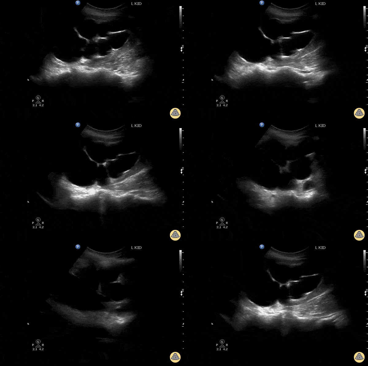
This is a long axis image of severe hydronephrosis that mimics a cystic kidney.
Image courtesy of Robert Jones DO, FACEP @RJonesSonoEM
Director, Emergency Ultrasound; MetroHealth Medical Center; Professor, Case Western Reserve Medical School, Cleveland, OH
View his original post here
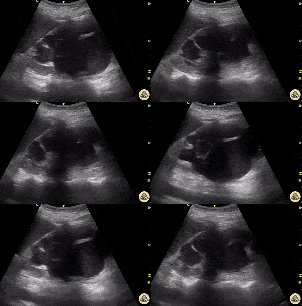
A 43-year-old female presented to the ED reporting fever and left-sided flank and low back pain. HPI was notable for recurrent urinary tract infections. POCUS performed on the Left Upper Quadrant revealed severe hydronephrosis, with hypdronephrotic collections in the region of the calyces resembling the outline of a bear’s paw (referred to as “bear paw sign”). Subsequent abdominal CT confirmed severe hydronephrosis secondary to stenosis of the ureteroplevic junction (UPJ).
Josiane Almeida
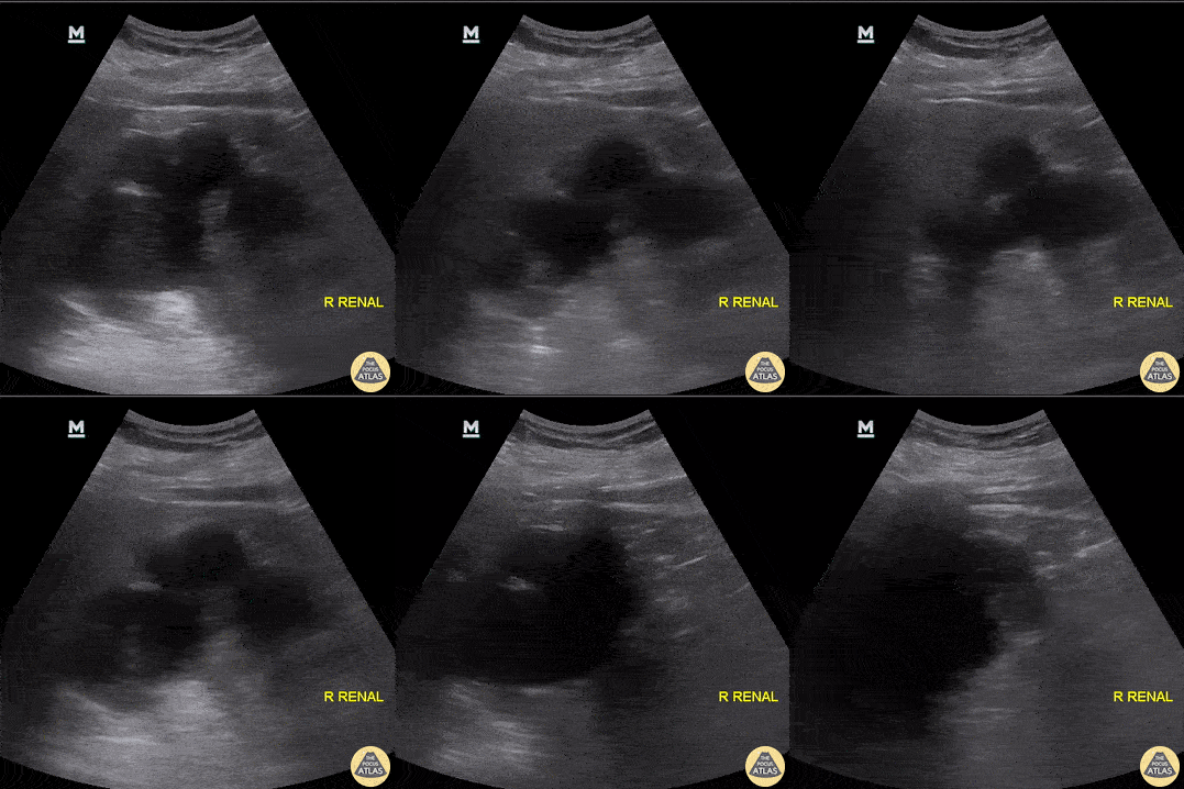
POCUS evidence of severe right renal hydronephrosis, as identified in a patient who had an ipsilateral 2.5cm mid-ureteral calculus.
Aaron Inouye, PA-C, North Canyon Medical Center
@PAintheED
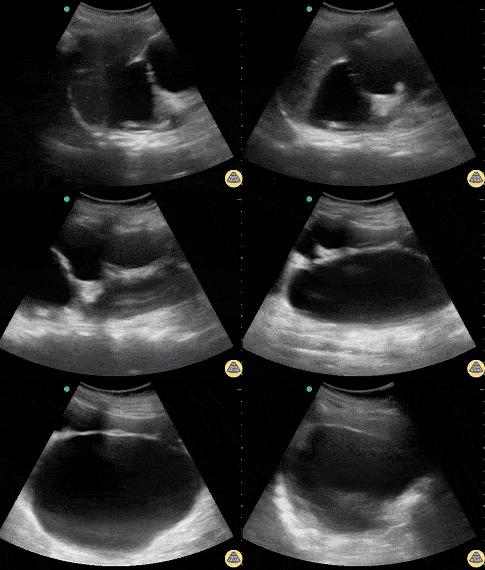
Patient with urinary obstruction with severe hydronephrosis and absolutely massive hydroureter (CT confirmed).
Dr. Stephen Alerhand - US Fellow - Mt Sinai Hospital, NYC