
Recent
Recent Images
Aorta
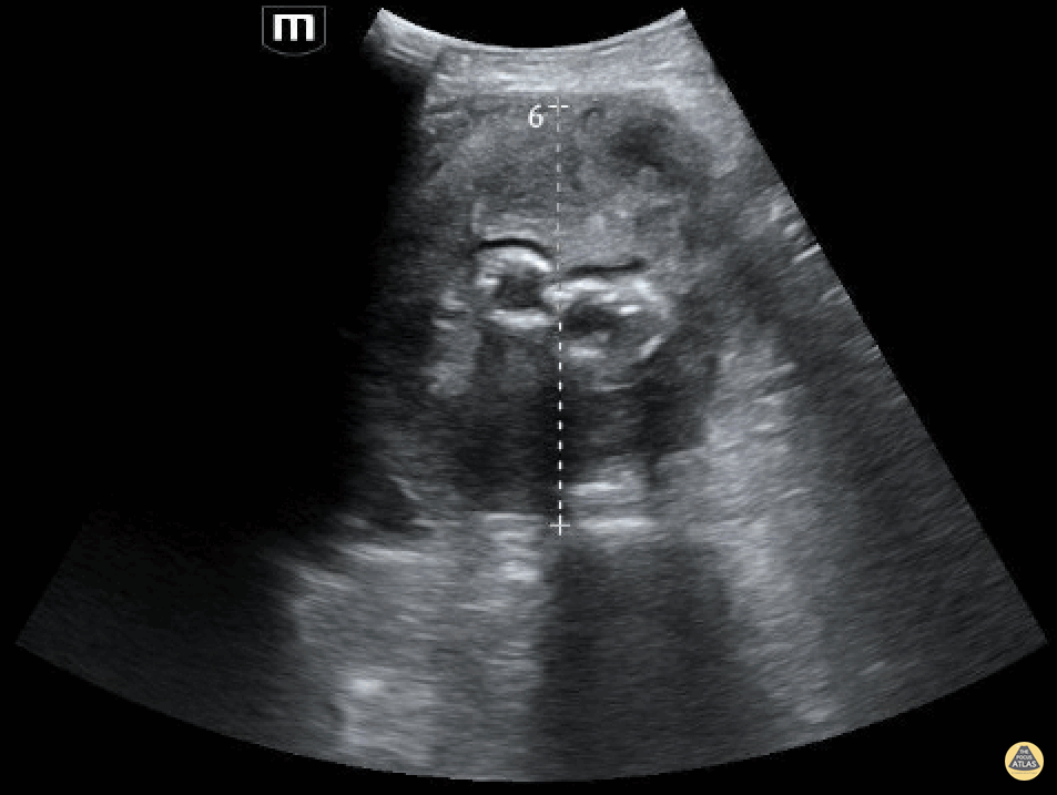
A patient in their 80s with PMH of abdominal aortic aneurysm s/p grafting with type 2 endoleak presented to the ED for dizziness. Bedside ultrasound demonstrated an 8.55 cm aortic aneurism with large thrombus. Endoleak is the hypoechoic area seen above the hyperechoic endograft.
Mehtab Galeh, MD; Bayley Espinoza, MD
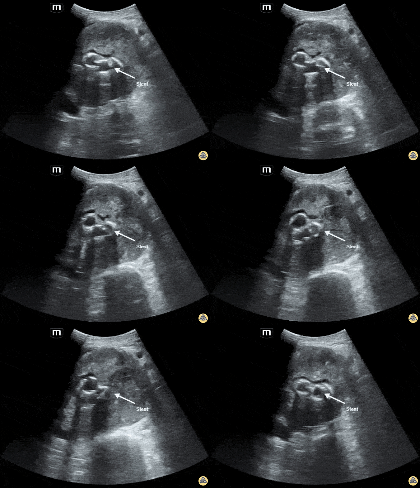
A patient in their 80s with PMH of abdominal aortic aneurysm s/p grafting with type 2 endoleak presented to the ED for dizziness. Bedside ultrasound demonstrated an 8.55 cm aortic aneurism with large thrombus. Endoleak is the hypoechoic area seen above the hyperechoic endograft.
Mehtab Galeh, MD; Bayley Espinoza, MD
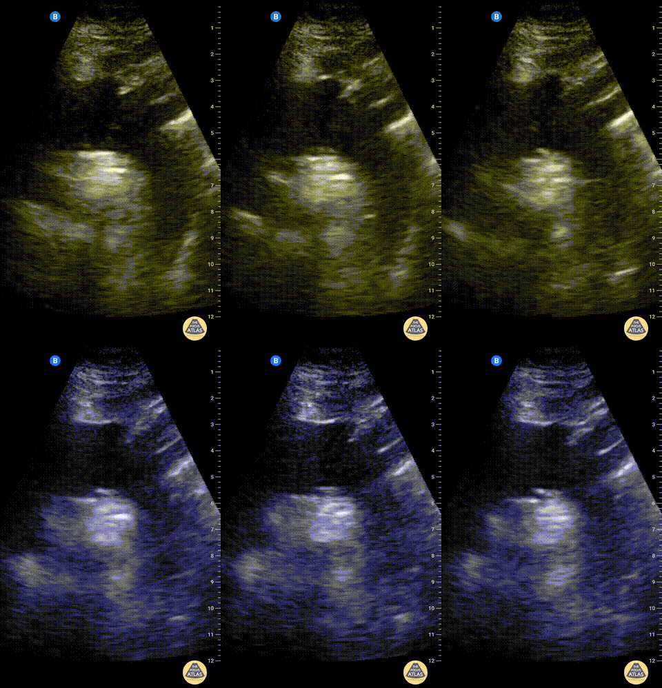
Suprasternal notch view of the aortic arch with brachiocephalic artery (BCA), left common carotid artery (LCA), and left subclavian artery (LSA) branching off. Right pulmonary artery is also visible adjacent to the ascending aorta.
Charles Jang, EM PGY-3
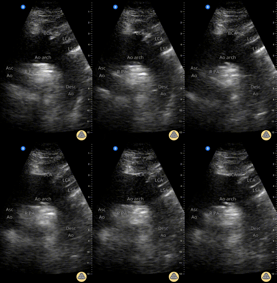
Suprasternal notch view of the aortic arch with brachiocephalic artery (BCA), left common carotid artery (LCA), and left subclavian artery (LSA) branching off. Right pulmonary artery is also visible adjacent to the ascending aorta.
Charles Jang, EM PGY-3
Bowel
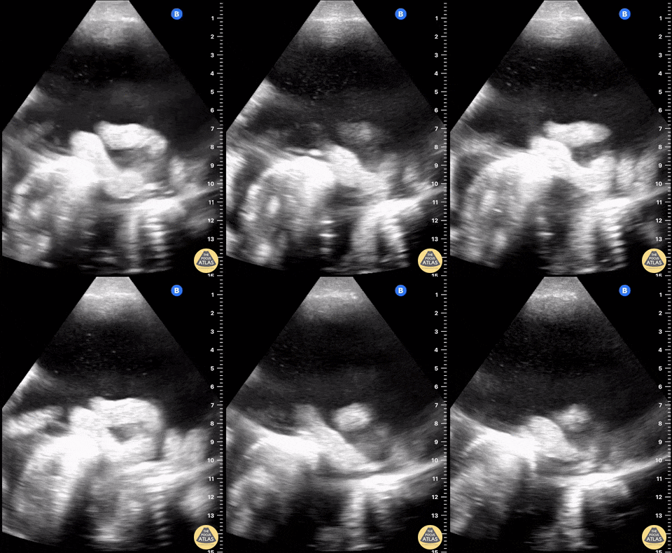
Malignant ascites with plankton sign in woman with metastatic ovarian cancer.
Samuel Eglin, MD
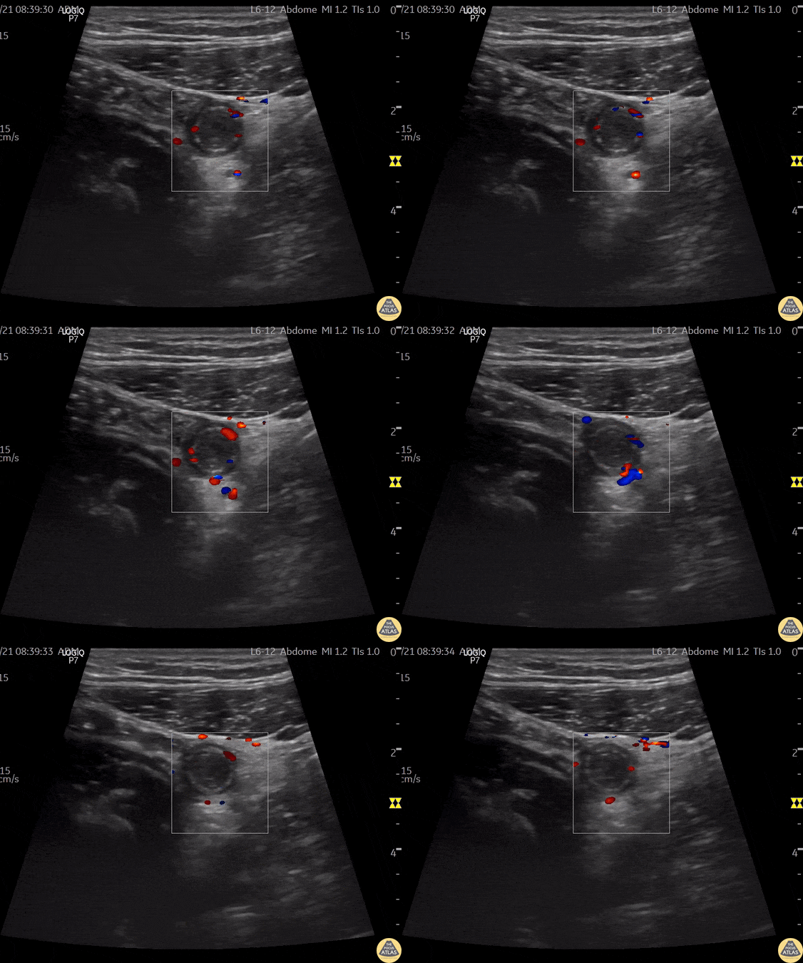
A 24 year old male comes to ER with abdominal pain, vomiting, and fever.
A directed scan with a high-frequency linear transducer in the right iliac fossa shows a tubular, non-compressible structure, a "target like" image, with increased echogenicity of the fat around it. When applied Color Doppler - we can observe increased vascular flow around the appendix - a sign known as "The Ring of Fire". This finding suggests the diagnosis of acute appendicitis.
Contributor: Renato Tambelli (@R_Tambelli @Jedipocus)
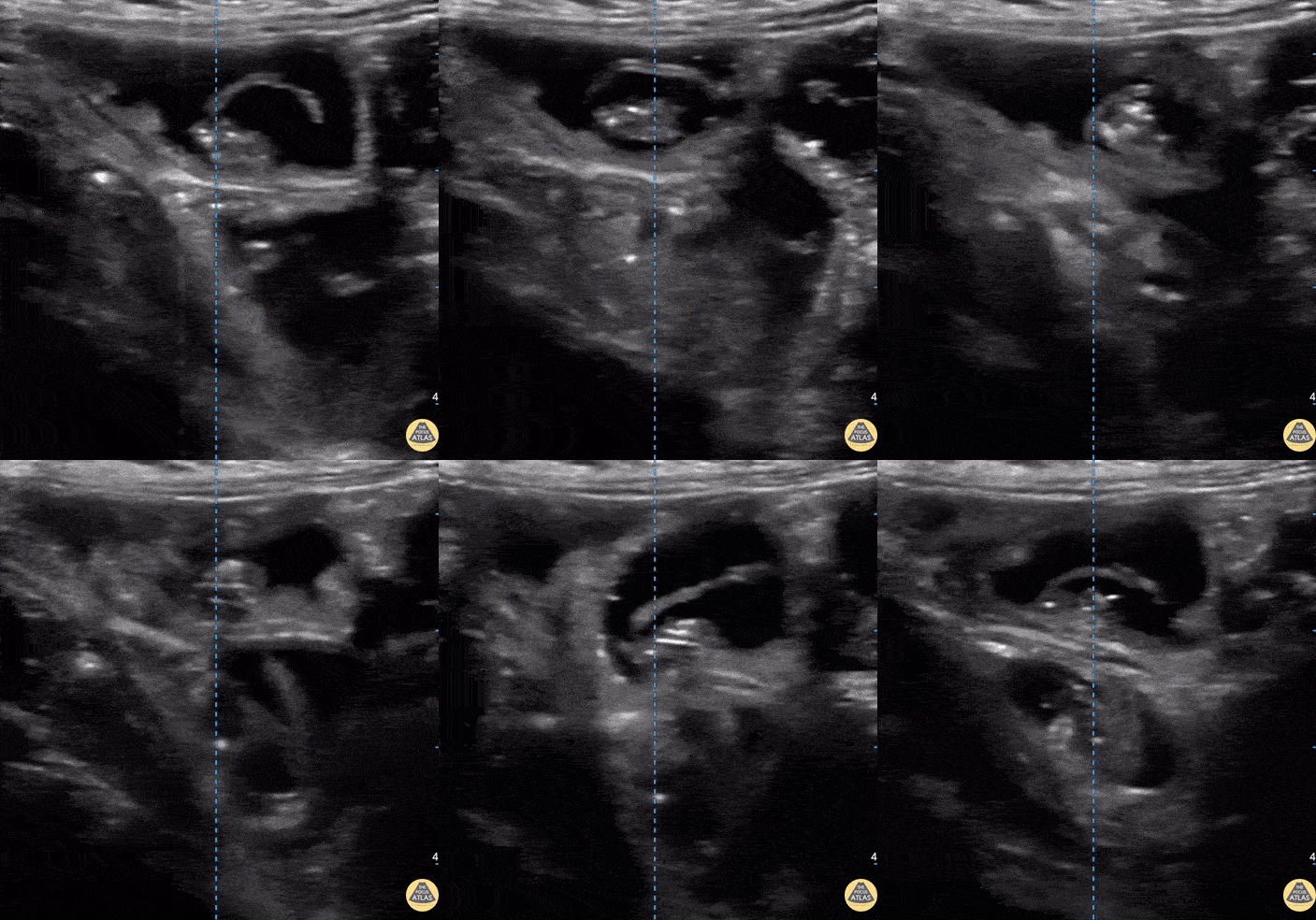
Fatigue for the past 3–4 months with mild disturbance of bowel habits. The patient reported visualizing proglottids in the stool.
POCUS findings: Ultrasound performed in the left lower quadrant revealed a structure consistent with parasitic segments.
Diagnosis: Laboratory confirmation a few days later identified Taenia saginata.
Dr. Guillaume Schramme
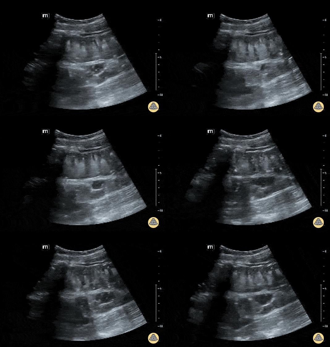
Fecal material can be seen moving forward and backwards through dilated bowels in the patient with a bowel obstruction.
Contributed by: Brittany Garza, DO and Saleem Nasseh, MD and Sadie Ellenson, MS4
Echo
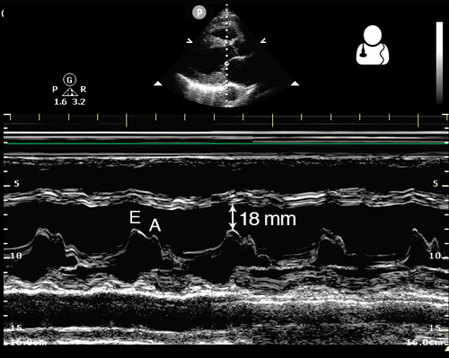
One way to evaluate cardiac function is to measure the distance from the anterior leaflet of the mitral valve to the septum during diastole. This is known as end point septal separation (EPSS).
An EPSS < 7 mm is considered normal, while EPSS > 10 mm suggests decreased cardiac function.
This measurement should be obtained in a parasternal long axis view, using M-mode with the cursor placed through the tip of the mitral valve.
Image credit: Ultrasound of the Week
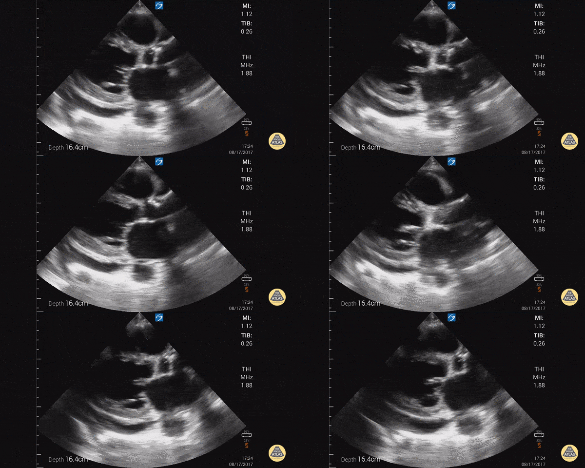
Parasternal long axis with cardiomyopathy, pericardial effusion, and decreased EF.
Dr. Gordon Johnson MD
Internist
Portland Oregon & Uganda
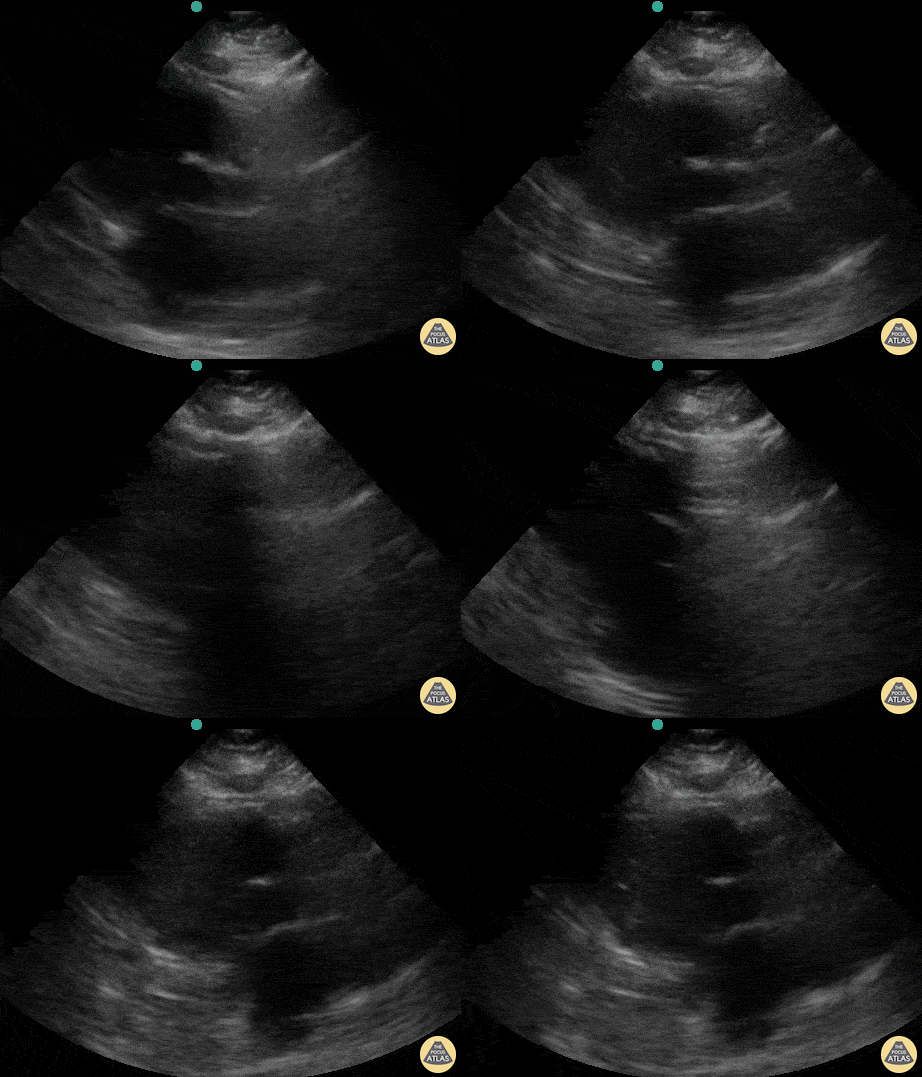
A patient presented with out-of-hospital cardiac arrest. POCUS was used to confirm presence of cardiac standstill. Note the absence of movement of the left ventricular free wall.
Melissa Myers, MD. Emergency Medicine in Texas. Ultrasound Fellowship Program Director.
@melissamyersmd
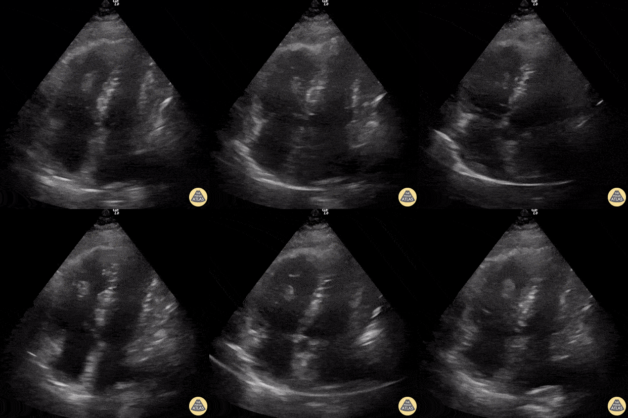
60 year-old smoker presents with dyspnea and chest pain. Apical 4 chamber view is notable for a thrombus within the RV and associated evidence of RV strain including increased RV size and impaired systolic function with sparing of the apex (known as McConnell’s sign).
Renato Tambelli, @R_Tambelli
Emergency Physician Hospital das Clínicas de Marília, Sao Paulo/Brazil
Hepatobiliary
Drainage tube within a located liver fluid collection.
Contributors: Dimitri Livshits, DO; Jane Belyavskaya, MD; Chris Hanuscin, MD
Kings County/SUNY Downstate
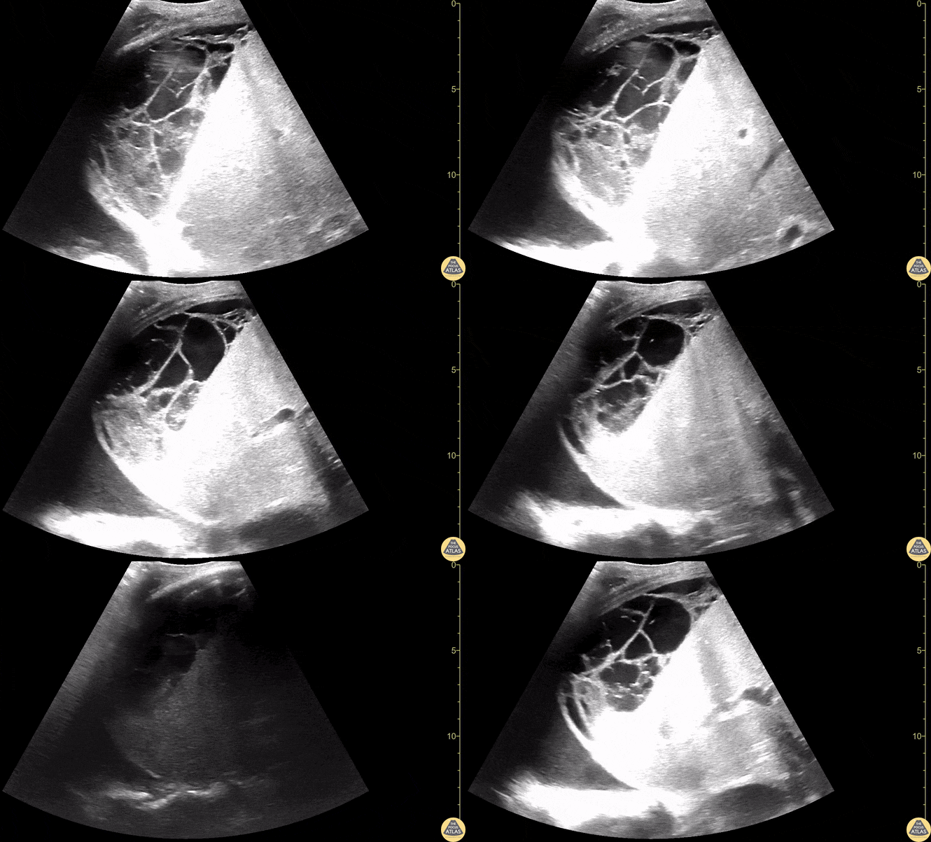
Located collection within the liver consistent with likely liver abscess.
Contributors: Dimitri Livshits, DO; Jane Belyavskaya, MD; Chris Hanuscin, MD
Kings County/SUNY Downstate
![Thick Walled Gallbladder in Dengue Fever (Short Axis) [2/2]](https://images.squarespace-cdn.com/content/v1/58118909e3df282037abfad7/1728883282342-87F9ZQFLQ314C6WAFAAC/image-asset.gif)
A 28 year old female comes to the ER with fever, myalgia, hypotension, and abdominal pain.
This case illustrates one of the ultrasound findings in Dengue Fever in the Leakage Syndrome Stage. We can see a large, thick-walled gallbladder and the presence of pericholecystic fluid. This clip demonstrates the gallbladder in short-axis view.
Contributor: Renato Tambelli (@R_Tambelli @JediPocus)
![Thick Walled Gallbladder in Dengue Fever (Long Axis) [1/2]](https://images.squarespace-cdn.com/content/v1/58118909e3df282037abfad7/1728882762911-WS6YZE9IZ5WVP4XD8BX1/image-asset.gif)
A 28 year old female comes to the ER with fever, myalgia, hypotension, and abdominal pain.
This case illustrates one of the ultrasound findings in Dengue Fever in the Leakage Syndrome Stage. We can see a large, thick-walled gallbladder and the presence of pericholecystic fluid. This clip demonstrates the gallbladder in long-axis view.
Contributor: Renato Tambelli (@R_Tambelli @JediPocus)
Musculoskeletal
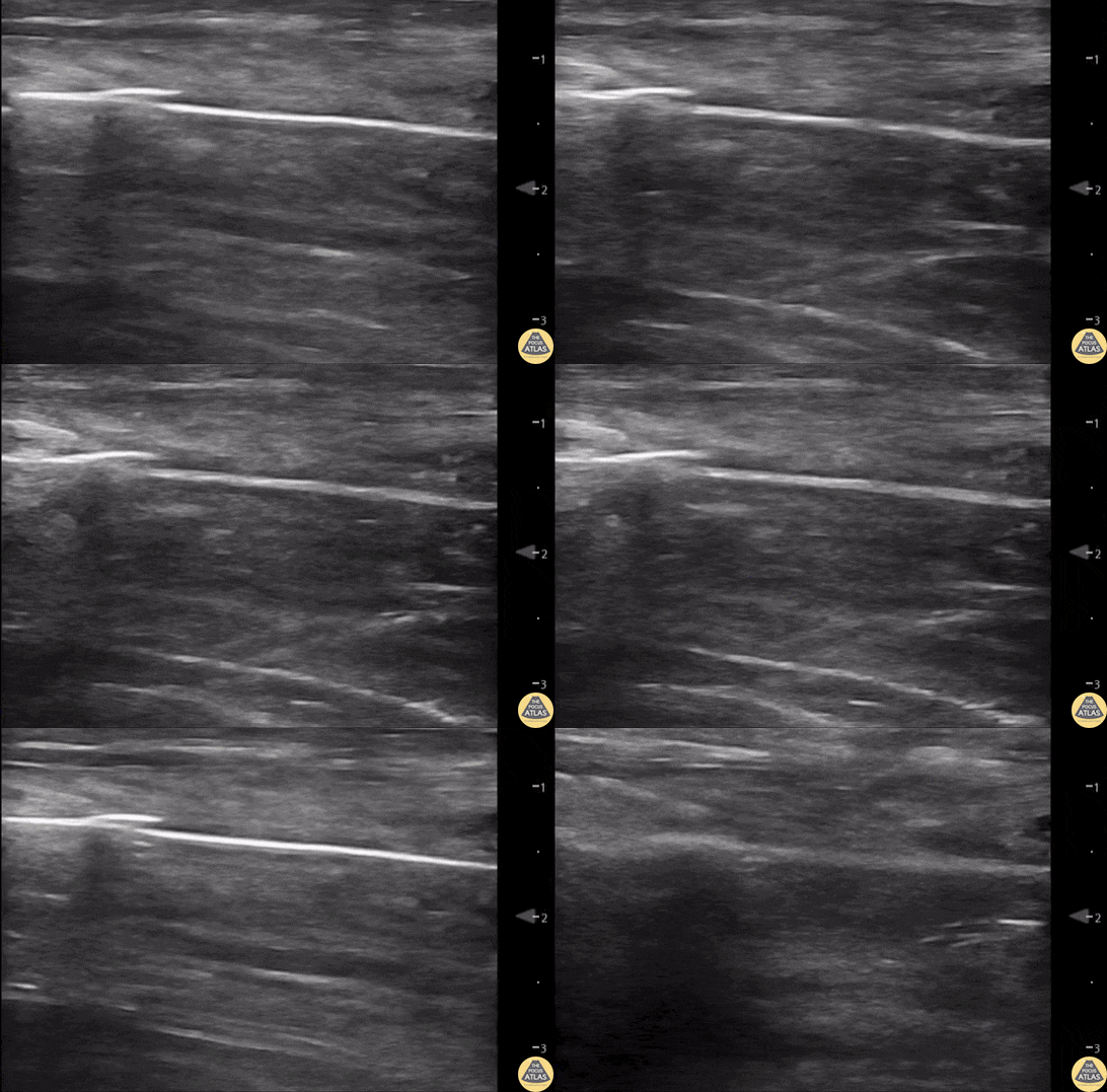
55 yo F s/p RT TKA w/ revision presents after a fall. POCUS was w/o tendinous/bony abnormality. RT knee x-ray was w/o definite acute fracture.
The American College of Radiology (ACR) promotes US evaluation of “pain after...(TKA with) suspect(ed) periprosthetic soft-tissue abnormality unrelated to infection” after radiographic evaluation as “usually appropriate”.
Although ACEP US Guidelines boast 96.8% sensitivity and 99.7% specificity for diagnosing fractures, POCUS evaluation of traumatic arthroplasty complications does not exist. This creates an area of interest within POCUS.
Contributors: Lauren Lowes, DO; Justin Morin, DO; Garrett Richardson, MS4; Nava Kendall, MD
Central Michigan University Residency of Emergency Medicine
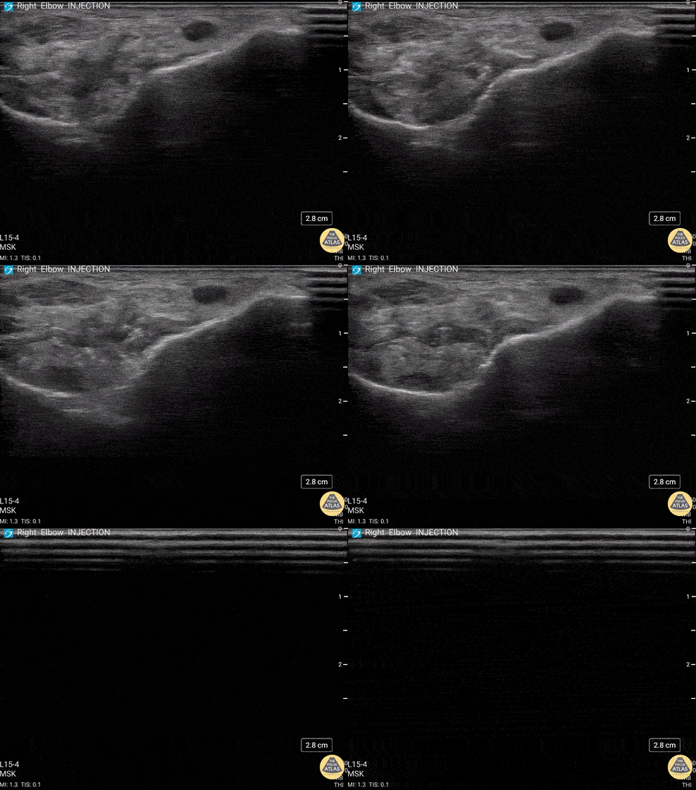
92 y/o M presented with moderate to severe osteoarthritis on elbow x-ray who was treated with ultrasound-guided corticosteroid joint injection.
Video shows transverse view of the posterior elbow as corticosteroid is injected with a posterolateral approach deep to the posterior elbow fat pad located in the olecranon fossa. This area is continuous with the elbow joint capsule. Posterolateral approach is advantageous due to decreased risk of disrupting neurovascular structures or the articular cartilage from other approaches to the joint space. Posterior approach is also advantageous compared to lateral approach in moderate to severe arthritis due to limited access to the radiocapitellar joint space.
Eben Alexander, DO
Devesh Patel, MD
Eastern Virginia Medical School
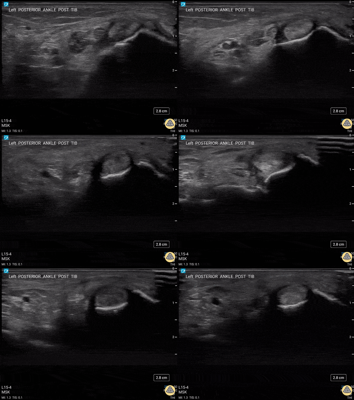
40 y/o F presented with 9 month history insidious onset L medial ankle pain and was found to have tibialis posterior tendon partial tear.
Video shows transverse view scanning proximal to distal at the posteromedial ankle (left is posterior). There is thickening of the posterior tibialis tendon with an anechoic tendon sheath effusion. At the level of the medial malleolus there are two moderate interstitial tears extending distally. Flexor digitorum and hallicus longus appear unaffected.
Eben Alexander, DO
Devesh Patel, MD
Eastern Virginia Medical School
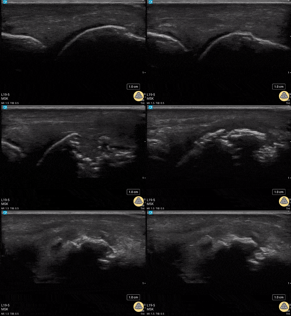
34 y/o M presented with fall while skiing injuring closed fist against ice and was found to have a displaced/impact fracture of his third metacarpal head.
Video shows sagittal view scanning in the ulnar to radial direction at the third metacarpophalangeal joint (right is proximal). There is diffuse disruption of the cortex of the radial aspect of the metacarpal head and displaced/impaction fracture of the metacarpal head. Usually this surface should appear smooth. Articular cartilage can be seen as anechoic at the interface between the two bones. This defect was not visualized on initial or 2 week post x-ray, so ultrasound was able to guide the clinical team to appropriately splint and manage as a fracture.
Eben Alexander, DO
Devesh Patel, MD
Eastern Virginia Medical School
OB/Gyne
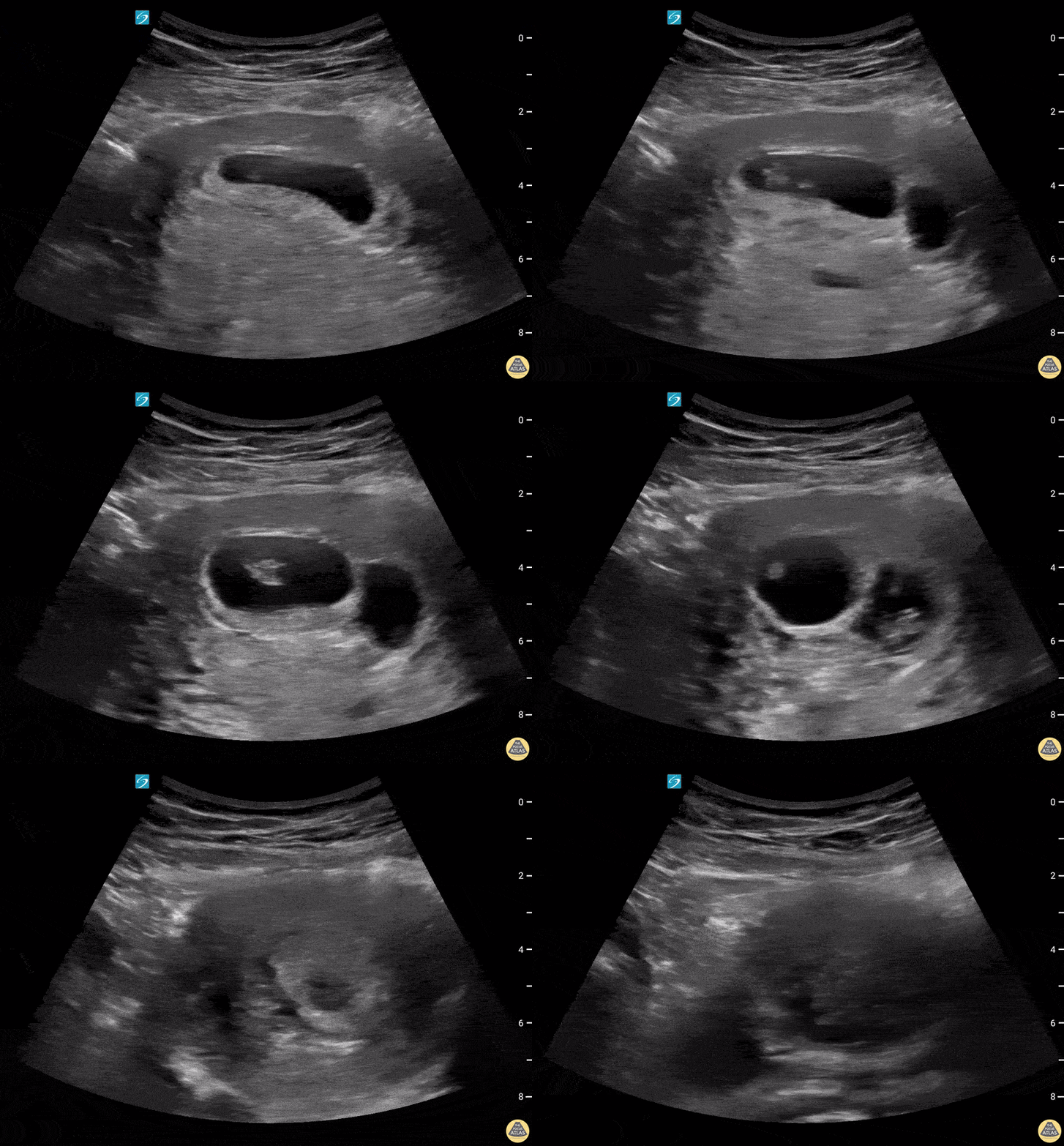
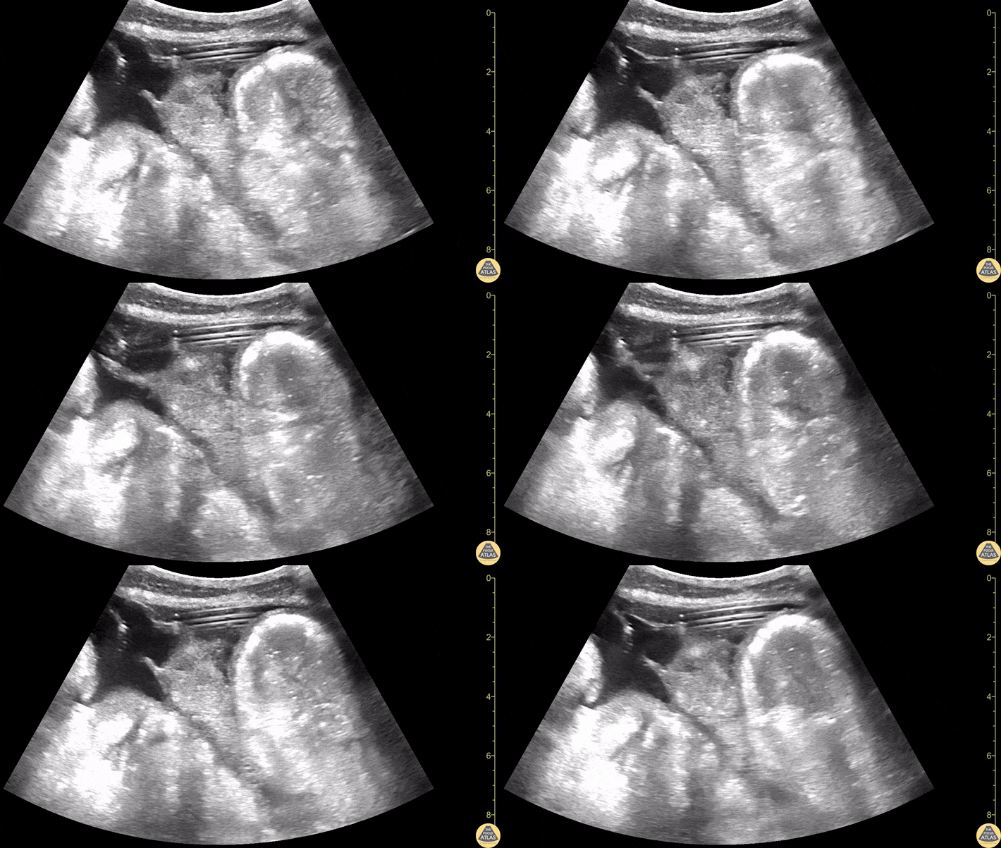
A 27-year-old female presented for evaluation of cough, congestion, decreased appetite, muscle aches, and fatigue. Patient was unsure of the date of her last menstrual period.
This image is a transabdominal ultrasound of the uterus demonstrating a dichorionic diamniotic twin intrauterine pregnancy. The pregnancy was dated at approximately 8 weeks and 2 days, with fetal heart rates of 167 bpm and 174 bpm.
Arthur Sieron BMBS, PGY-1 Emergency Medicine, Central Michigan University; Eric Spencer DO, PGY-3 Emergency Medicine, Central Michigan University; Halimah Hamidu, MS4 Central Michigan University College of Medicine.
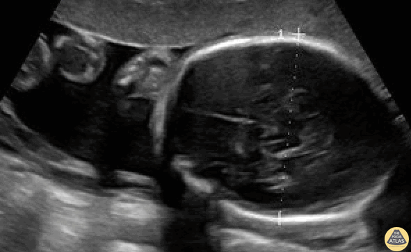
BPD should be measured in the axial plane, perpendicular to the falx cerebri, and from the outer edge of the near calvarial wall to the inner edge of the far calvarial wall.
Contributors:
Stephen Holihan (MD); Dillon Nerland (MD); Madison Waddell (MS4)
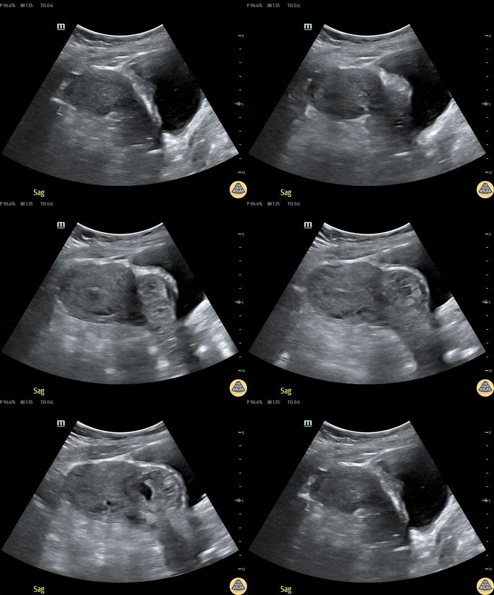
32 year old female at 8-9 weeks gestation by LMP referred to the ED for pregnancy of unknown location. She reported 1-2 weeks of mild pelvic cramping and vaginal spotting.
Bedside trans-abdominal imaging demonstrated a round, heterogenous structure without a clear yolk sack or fetal pole, but with several hallmarks of caesarean scar ectopic pregnancy: position in the anterior uterine wall; in the lower (closer to the cervix) half of the uterus; bulging into the neighboring bladder; with thin interposed myometrium. After confirmatory formal ultrasound, she was admitted for methotrexate termination, which proved successful.
Contributors: Nicholas Maurer, MD, MPH (PGY-1); Megan Chenworth (PGY-3)
Emergency Medicine, Northwestern McGaw Medical Center
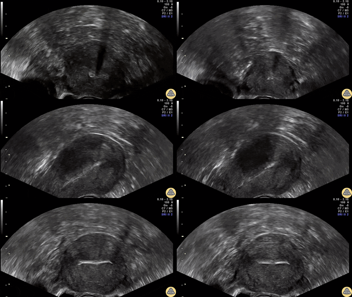
Transvaginal ultrasound showing the IUD correctly placed in both sagittal and transverse plane.
Contributor: Dr. Nicolay B. Werner
Akershus University Hospital, Norway
Ocular
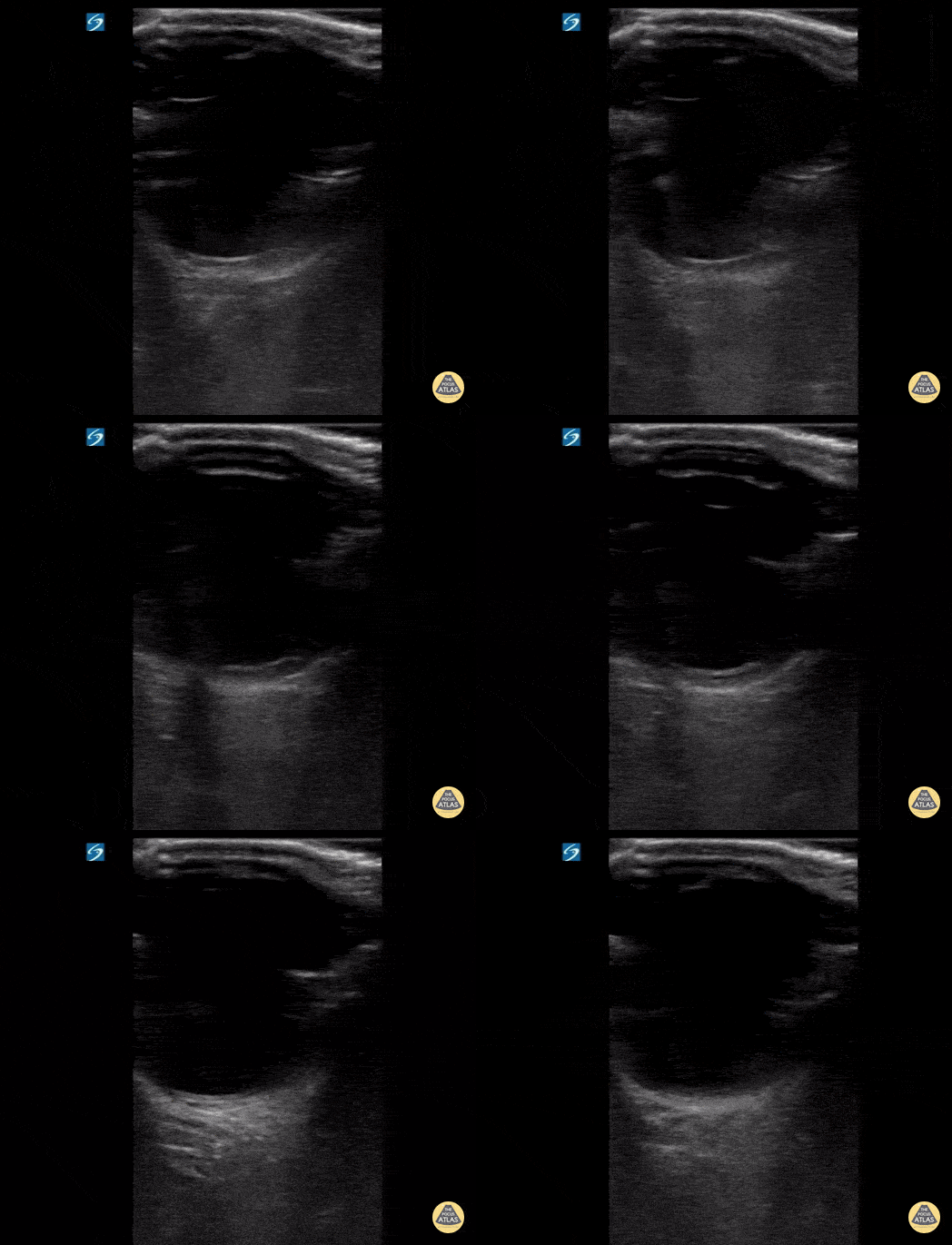
17 y/o female with sudden onset bilateral blurry vision after prolonged indoor cat exposure. Fundoscopy showed retinal pigment epithelium loss consistent with bilateral neuroretinitis from cat scratch disease (Bartonella henselae).
The hyperechoic retina, normally flush with the posterior aspect of the globe, is seen separated by a hypoechoic band of edema.
Retinal edema from cat scratch disease is rare (Gen Opth 19th ed. 2017:378-422), but can be diagnosed by POCUS.
Contributed by: John Hipskind, MD, Ultrasound Director, Kaweah Health EM Residency
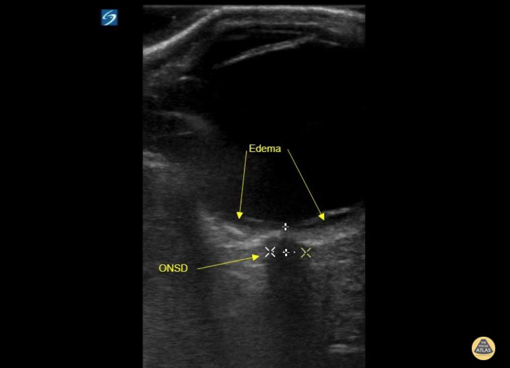
17 y/o female with sudden onset bilateral blurry vision after prolonged indoor cat exposure. Fundoscopy showed retinal pigment epithelium loss consistent with bilateral neuroretinitis from cat scratch disease (Bartonella henselae).
The hyperechoic retina, normally flush with the posterior aspect of the globe, is seen separated by a hypoechoic band of edema.
Retinal edema from cat scratch disease is rare (Gen Opth 19th ed. 2017:378-422), but can be diagnosed by POCUS.
Contributed by: John Hipskind, MD, Ultrasound Director, Kaweah Health EM Residency
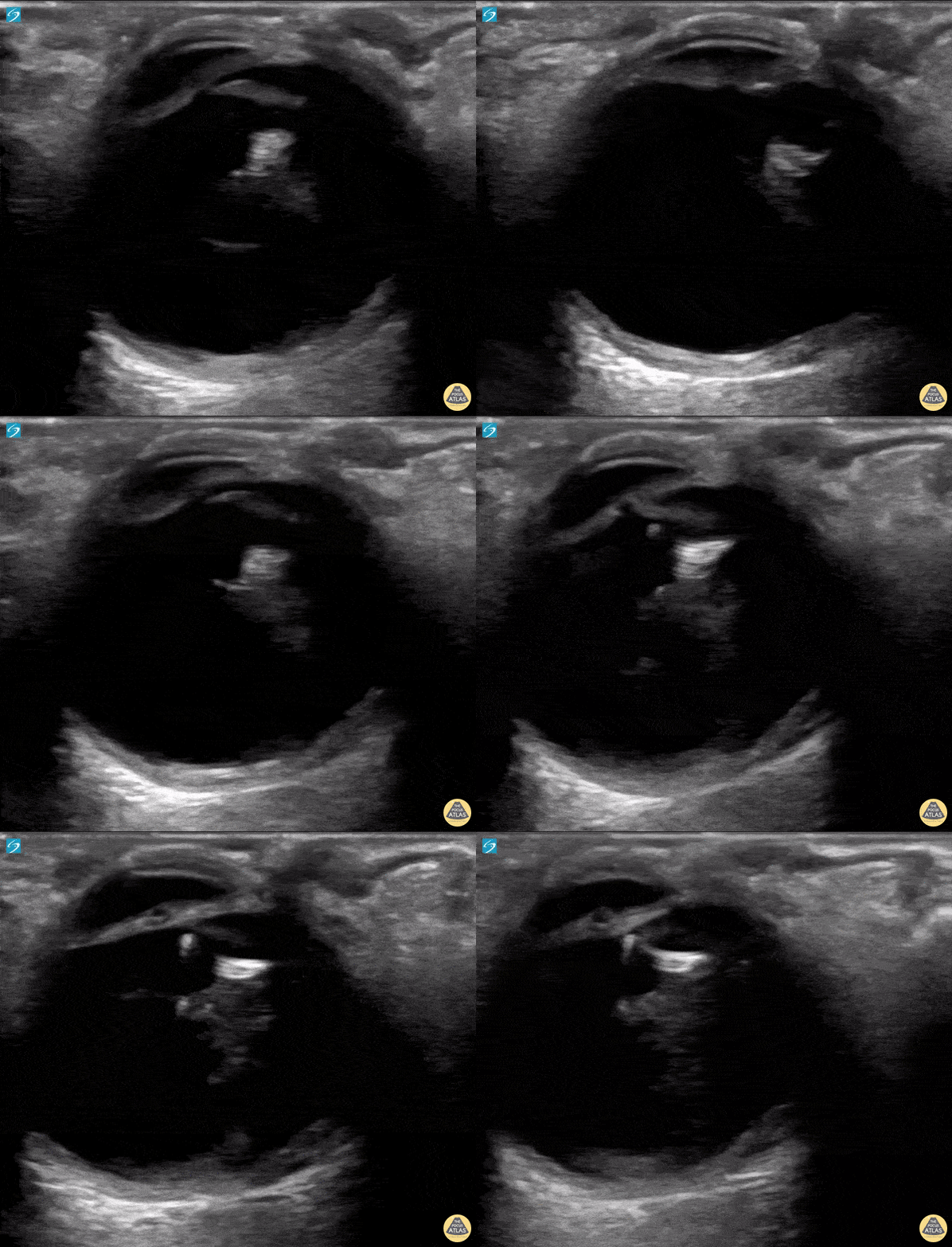
A 50 year old male with a prior history of bilateral intraoccular lens (IOL) transplants presented to our ED with sudden onset foggy vision in his right eye while getting out of the shower. He was unable to participate in visual acuity due to the extent of his blurred. POCUS demonstrated “iridodonesis” and a provisional diagnosis of IOL subluxation/dislocation was made. Ophthalmology was consulted and confirmed the diagnosis.
Dr. Piaseczny, PGY4 Emergency Medicine, Queen's University, Kingston, Ontario, Canada
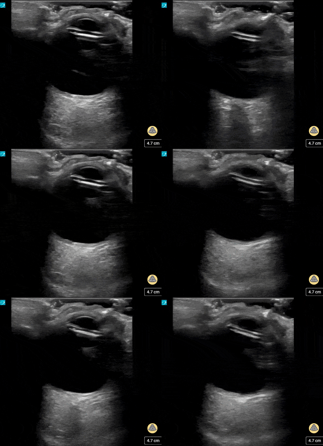
This patient presented with decreased vision on a background of advanced macular degeneration. VA in the eye had decreased from 20/150 to 20/400 on presentation.
Interestingly the patient stated "I think my ocular lens has displaced."
Contributed by: Colin Bell, FRCPC, DPSPC
Pulmonary
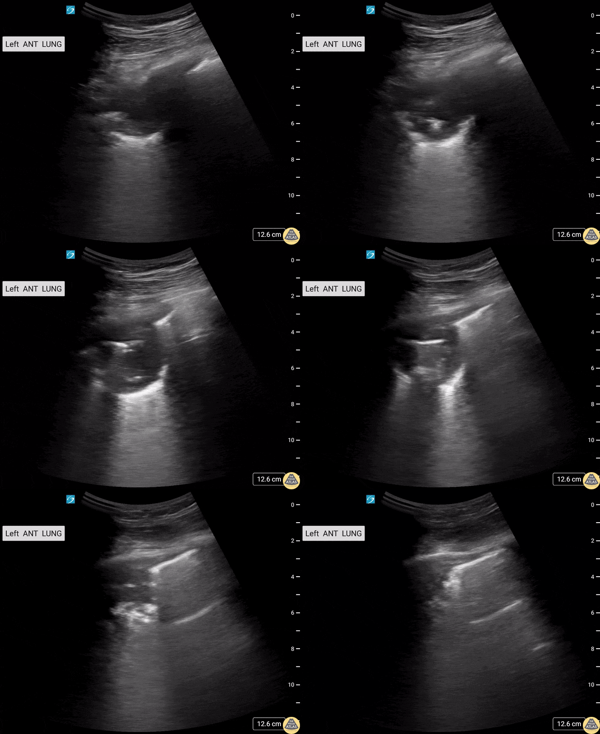
HIV+ male patient presents with cough and shortness of breath. Thoracic US demonstrated a left upper lobe cavitary lesion with calcifications.
Differentials included TB, aspergilloma, lung abscess.
Contributors: Sara Schambach, MD; Garrett Mason, MD
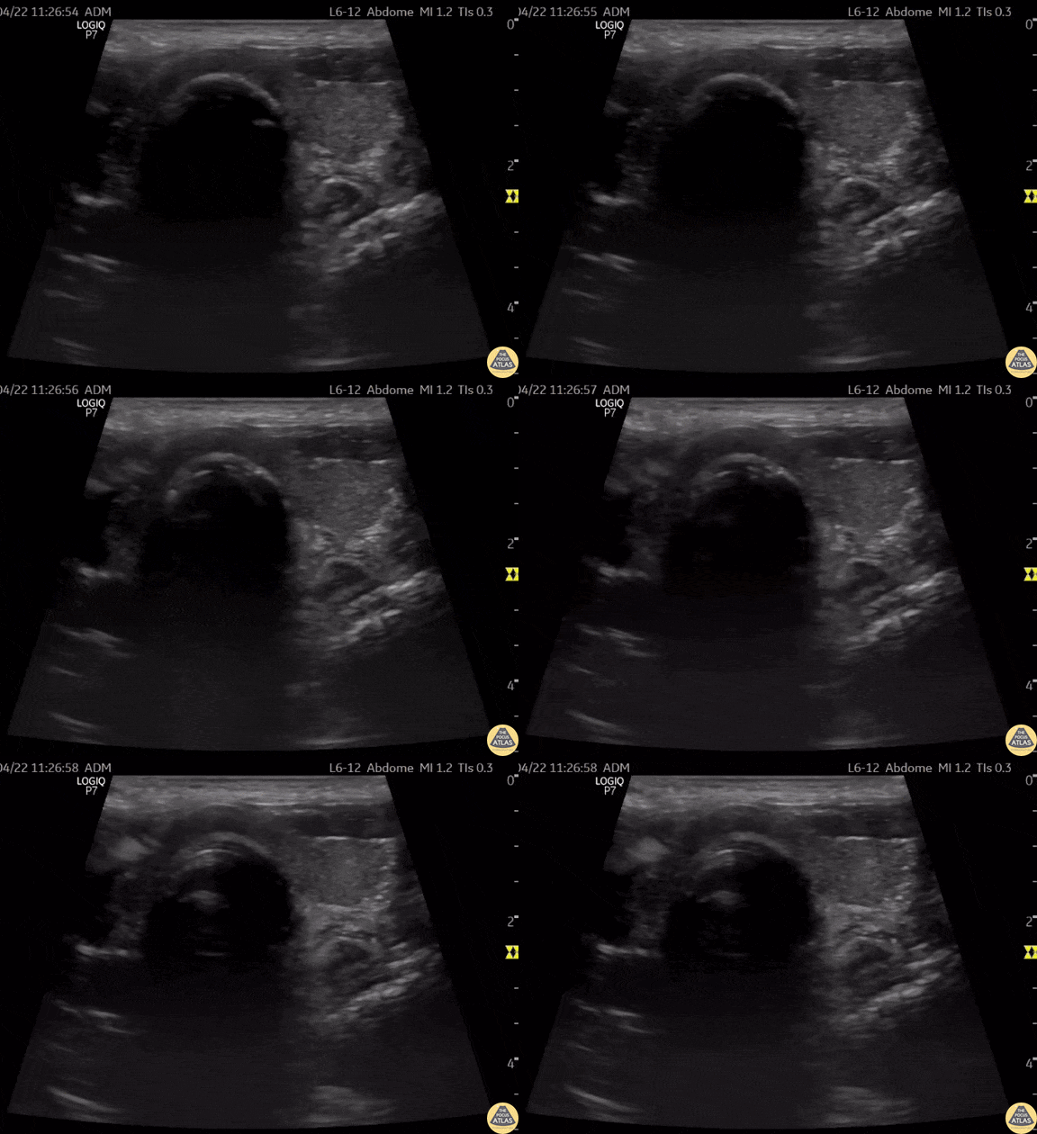
This is a clip of a cervical airway view during intubation attempt.
Some artifact is visible in the trachea when passing the tube demonstrating a "Snowstorm Sign" which can be used to confirm the position of an endotracheal tube. In this view it is possible to see the esophagus next to the trachea. As demonstrated there is no motion detected in esophagus during intubation therefore confirming tracheal intubation.
Contributor: Renato Tambelli (@R_Tambelli @Jedipocus)
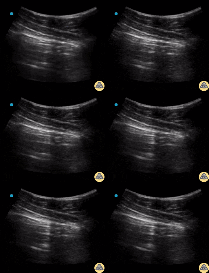
Lung point is a pathognomonic finding on US for pneumothorax. It refers to the junction between healthy lung and collapsed lung.
This is represented in the US recording as lung sliding seen on the left of the pleural line but no lung sliding seen on the right of the pleural line.
Contributors: Dimitri Livshits, DO; Jane Belyavskaya, MD; Chris Hanuscin, MD
Kings County/SUNY Downstate
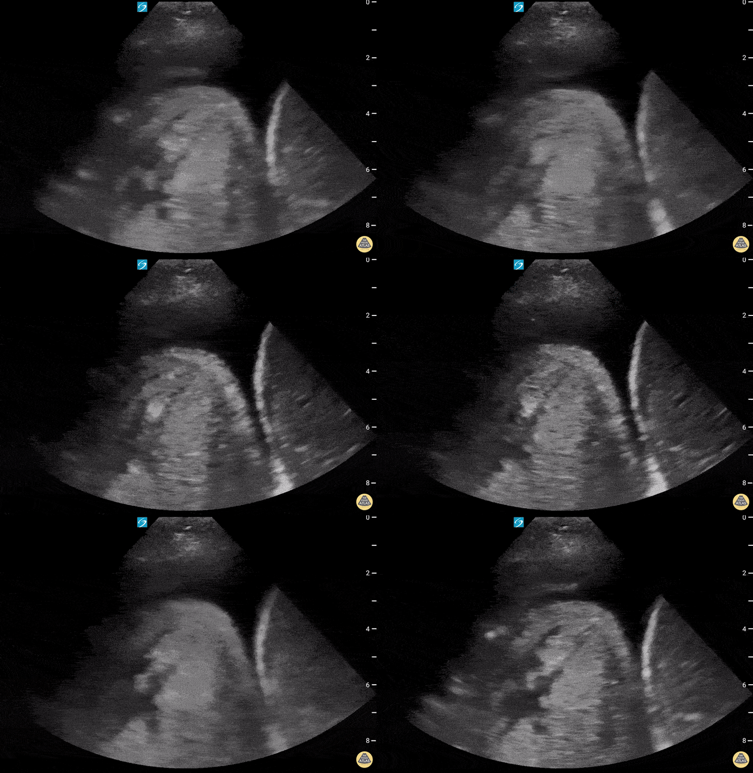
72 year old with past medical history of hiatal hernia presenting to the ED with new onset shortness of breath requiring rescue BPAP. She was diagnosed and treated with CHF exacerbation. Lung ultrasound showed a large pleural effusion with uncertain mass-like object with heterogenous fluid contents on the right (shown here), along with dense B-lines. Correlation with prior CT suggested that the structure on the right chest was a large hiatal hernia.
Contributed by: William McGill, PA-C
Pediatrics
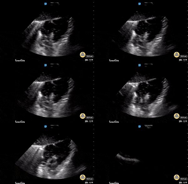
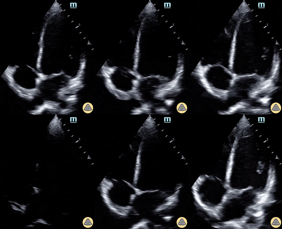
14 year-old male presents to the ED with chest pain two weeks after being having been diagnosed with COVID-19. Labs were notable for elevated CBC, CRP, ESR, and troponin. POCUS revealed moderately decreased function and LV dilation, consistent with the diagnosis of COVID-19 myocarditis.
Paul Khalil, MD and N. Akwesi Poteh, MD at University of Louisville
@khalil3paul
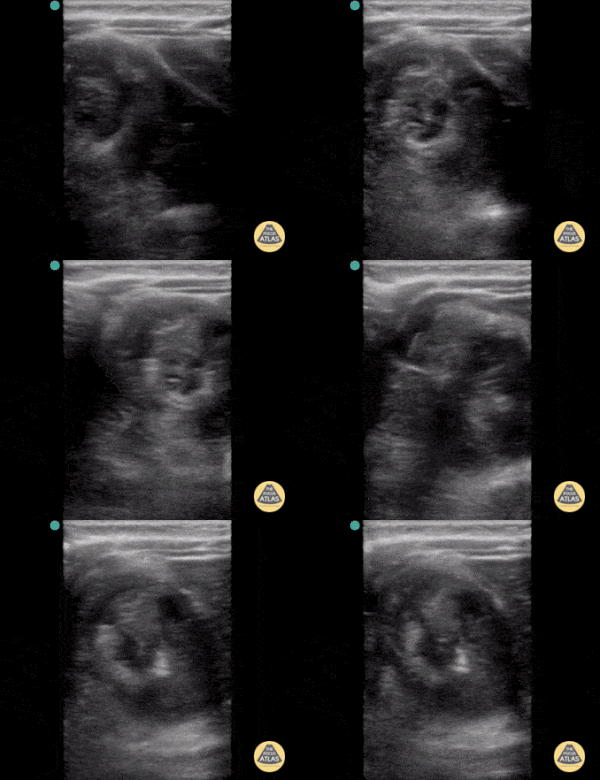
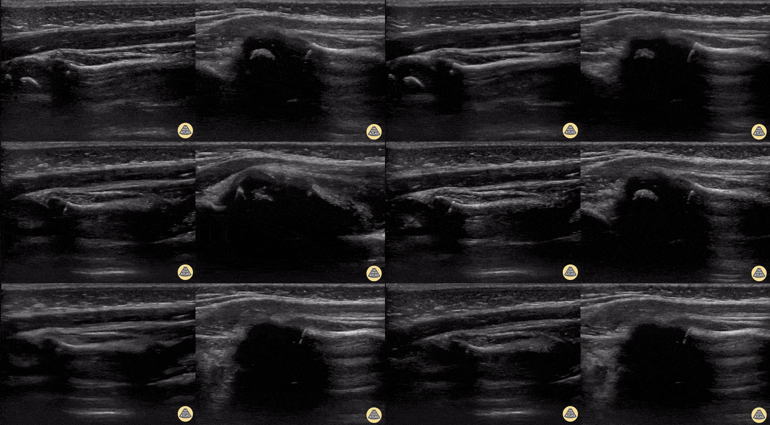
2-year-old female was brought to the ED refusing to use her left arm after her father caught her by the arm to prevent her falling down flight of stairs. POCUS was performed to confirm clinical suspicion of a Nursemaid’s elbow (or radial head subluxation). The left image reveals classic findings of a Nursemaid’s elbow including widening of the synovial fringe and inward extension of the annular ligament. Further, you can appreciate both the supinator muscle, as well as the annular ligament are being pulled into the joint space, a finding known as hook sign. The image on the right is the post-reduction image showing normal alignment and contour of the annular ligament.
Austin Meggitt, MD
Pediatric Emergency Ultrasound Fellow
Denver Health Medical Center @DenverEMed
Renal
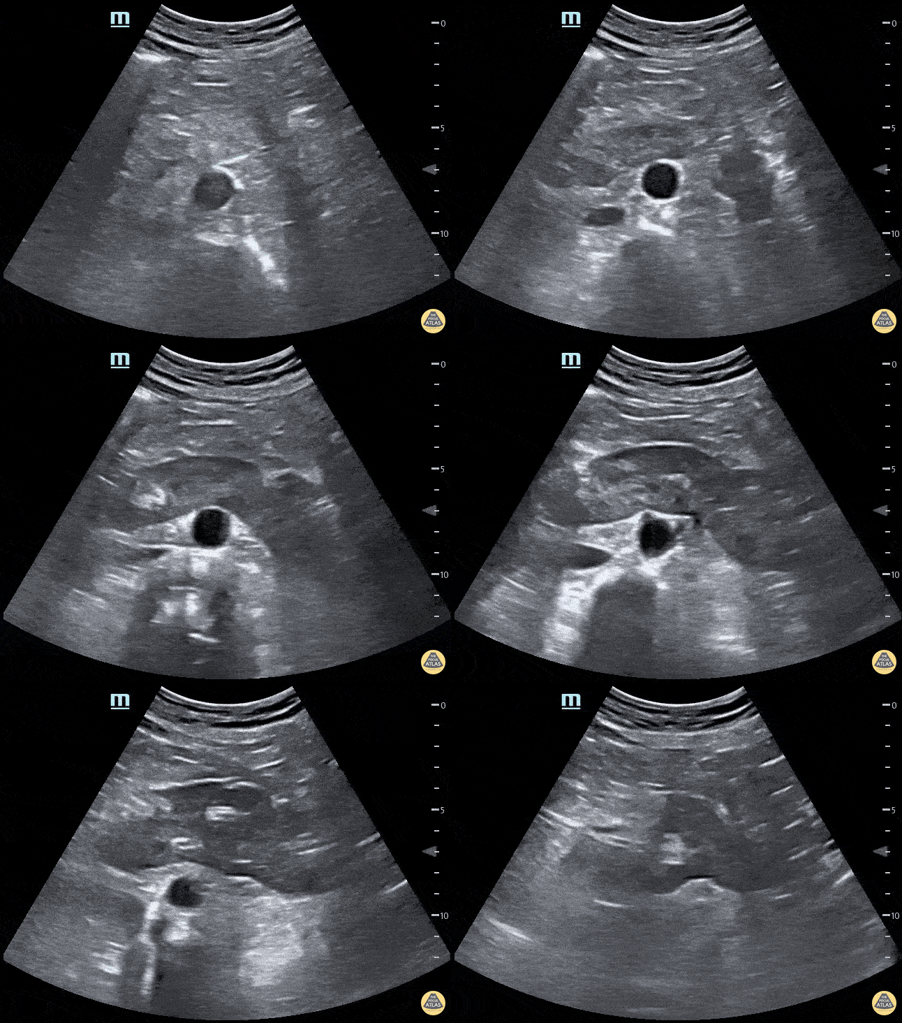
Horseshoe kidney. No kidney was identified in either flank area, this anterior abdominal view shows one large kidney that is anterior to the Aorta.
Contributed by: Marion Memmott, DO; Michael Bernard, DO; Central Michigan University Emergency Medicine Residency
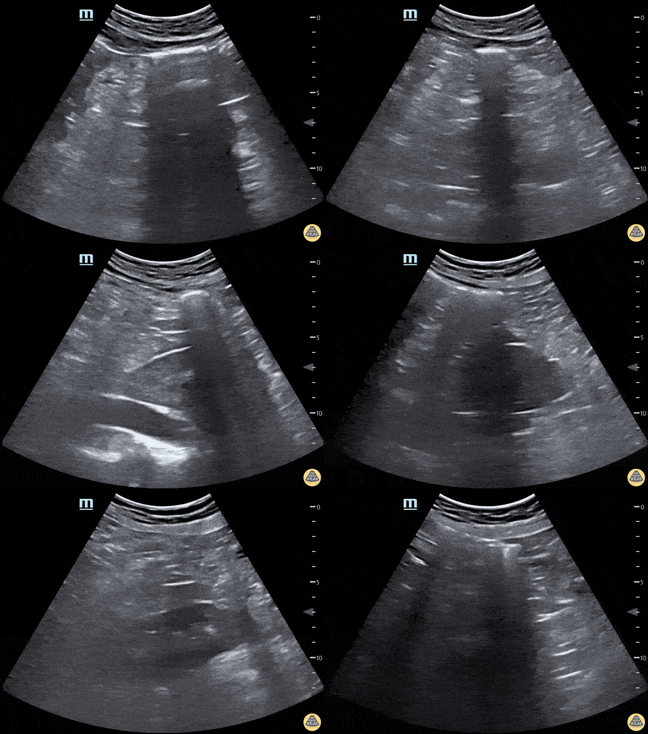
Horseshoe kidney. No kidney was identified in either flank area, this anterior abdominal view shows one large kidney that is anterior to the Aorta.
Marion Memmott, DO; Michael Bernard, DO; Central Michigan University Emergency Medicine Residency

Prominent prostate on ultrasound in a 72 year-old-male with known prostate carcinoma and recent PET scan with confirmation. No known masses or metastasis. This is seen in both transverse and sagittal views.
Lindsay Davis, DO, MPH, PGY3; Matthew Welch, MS4; Alex Schlangen, DO, PGY1
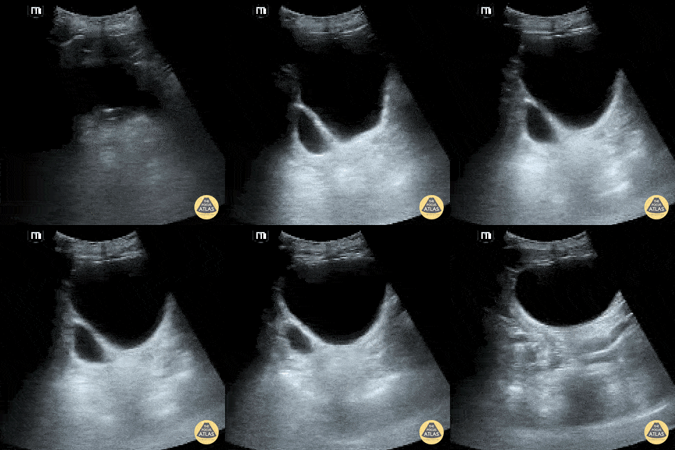
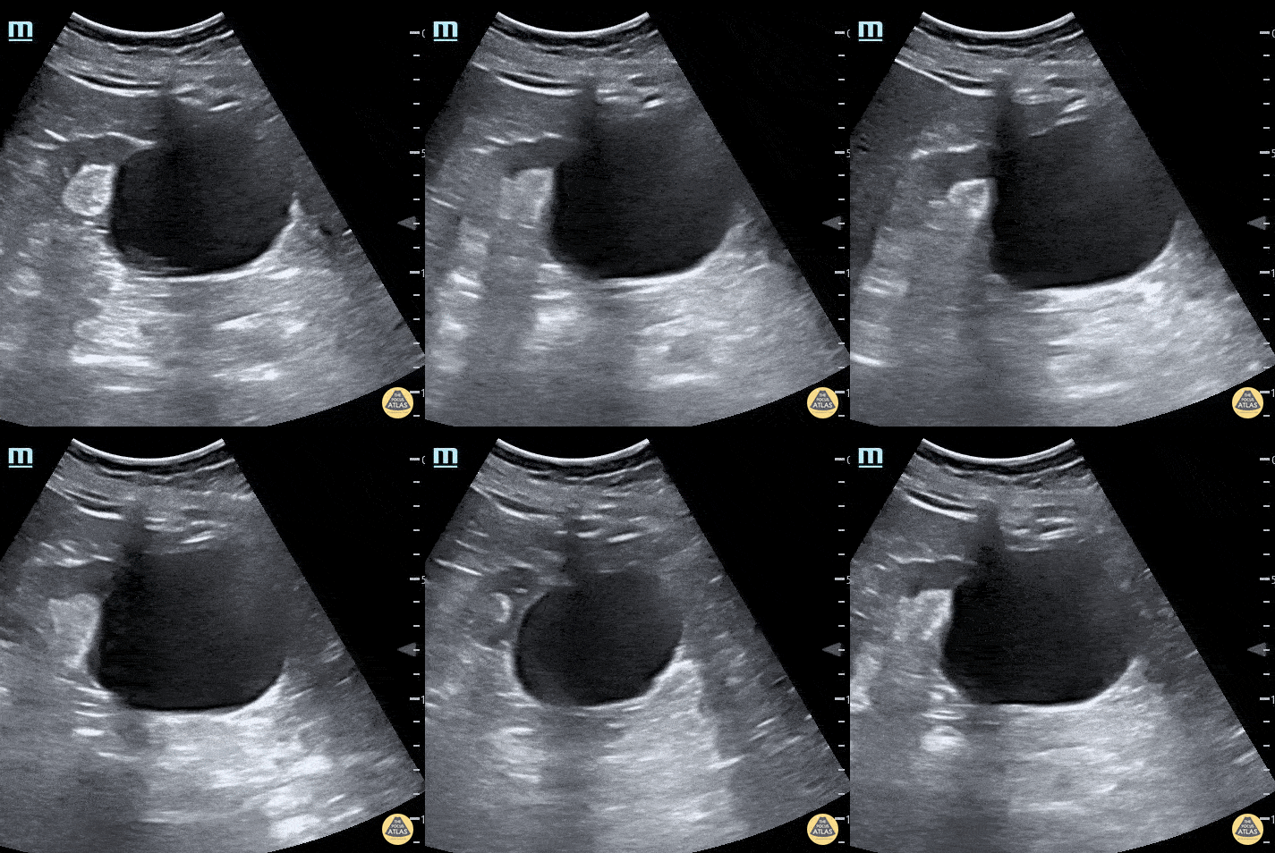
A bladder diverticulum is demonstrated deep to the bladder as the ultrasound probe is fanned.
Contributed by: Brittany Garza, DO; Saleem Nasseh, MD; Sadie Ellerson, MS4
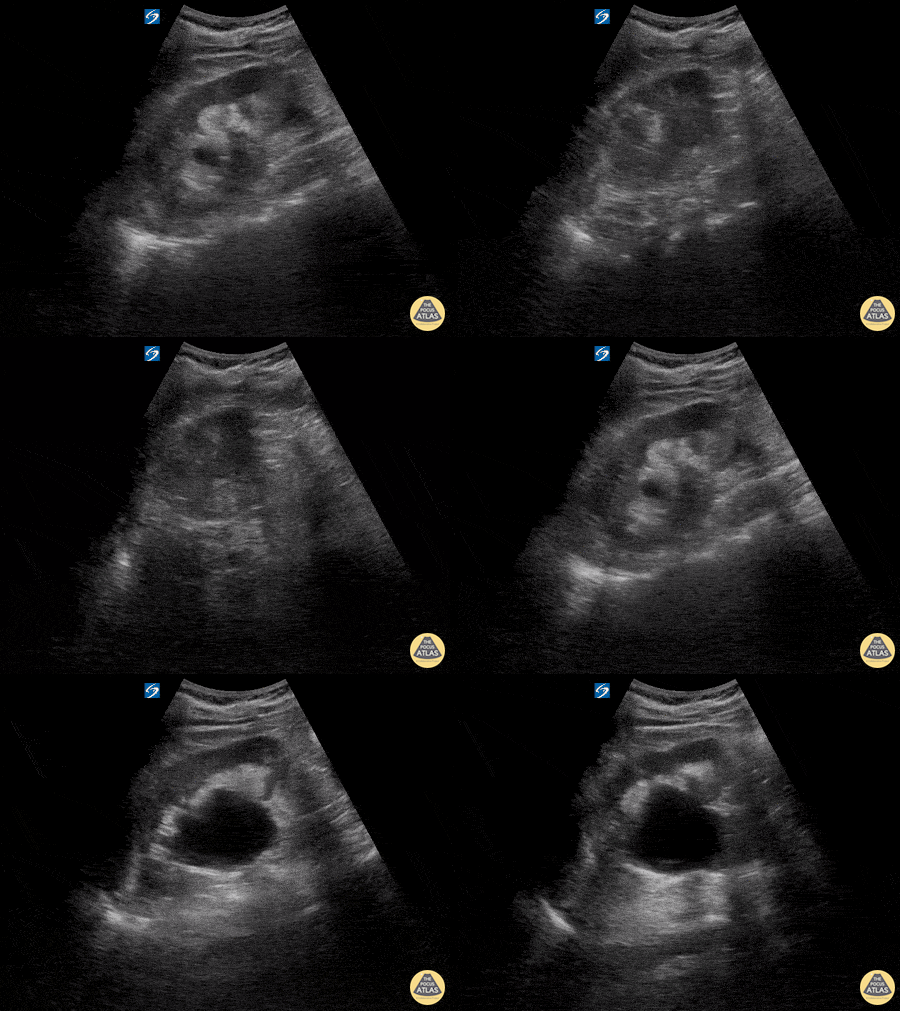
Here is an example of hydronephrosis in the presence of nephrolithasis. This patient presented to the emergency department complaining of new right flank pain after waking up in the morning. She was seen previously for urinary complaints, diagnosed with UTI and started on antibiotic regiment that had been altered twice due to concerns of sub-optimal coverage. She demonstrated right CVA tenderness on physical exam, raising suspicion for pyelonephritis as well as hydronephrosis. Subsequent point of care ultrasound revealed moderate hydronephrosis. CT was obtained to further evaluate and confirmed the presence of an obstructing kidney stone. Note presence of hydronephrosis as the probe is fanned/tilted through the kidney, highlighting the importance of scanning through the entire organ.
Dr. Carlo Zamora, DO, PGY-1
Riverside Regional Medical Center Emergency Medicine Residency (Newport News, VA)

A young female presented with acute RLQ abdominal pain and found to have a 5mm right ureter stone at the UVJ with ureteral dilation. The patient also had moderate hydronephrosis.
Matthew Petruso, DO; Harpreet Grewal, MD; Eric Versluis, MS4; Central Michigan University: Emergency Medicine Residency.
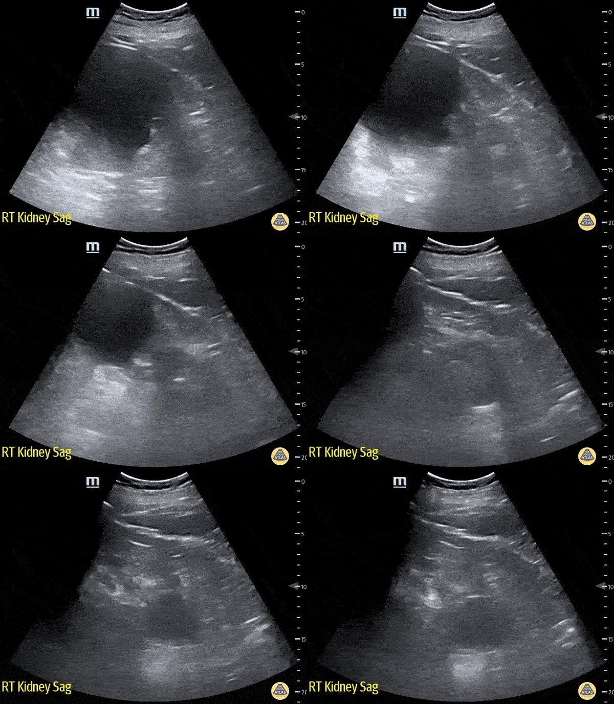
83 year old male presented to the ED for right flank pain.
The renal ultrasound showed two right renal cysts. The superior renal pole cyst measuring 12 x 13cm. The inferior pole cyst measuring 4 x 5cm.
Laura Schroeder, MS, MD; Syeda Zehra, MD; Conner Shea (Medical Student) Central Michigan University Emergency Medicine Residency
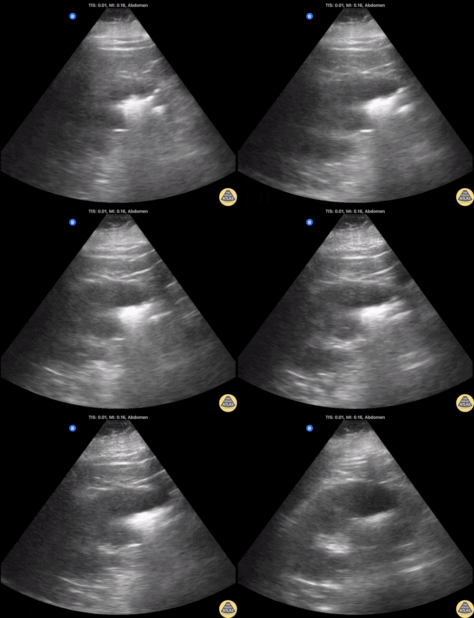
60 year old female with poorly controlled diabetes. Initial CT showing R-sided pyelonephritis, US performed 12 hours later demonstrated development of emphysematous pyelonephritis noted by the hyperechoic areas with dirty shadowing seen within the renal parenchyma (indicative of gas). This was later confirmed on repeat CT imaging.
Contributor: Shane Solger, MD
Kings County/SUNY Downstate EM/IM
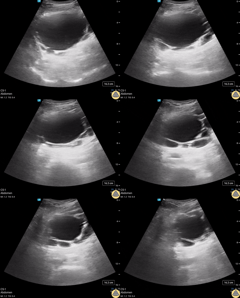
82 year old male with history of subarachnoid bleed presented for evaluation after a fall. During a EFAST exam, bladder US revealed multiple, anechoic, cystic-appearing structures consistent with bladder diverticula (confirmed by CT).
A bladder diverticulum is a protrusion of the mucosa through the muscular wall of the bladder and can be congenital or acquired. Acquired diverticula are more common, with bladder outlet obstruction being the most common cause. Urology follow up is recommended to confirm the cause and screen for other concerning etiologies or complications including carcinoma, recurrent UTIs and bladder calculi.
Contributors: Michael Huber, 4th year Medical Student; Chad Bambrick, MD; Ana Camagay, MD
Central Michigan University College of Medicine
Soft Tissue
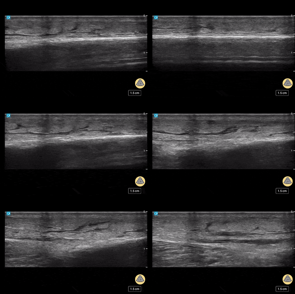
This is an example of “cobblestoning”. This patient initially presented to the emergency department with lower anterior leg pain, especially with walking. Physical exam revealed minor erythema but also exquisite tenderness to palpation as well as with plantar flexion. Although cellulitis is clinically diagnosed, point of care ultrasound can be used to support it. The cobblestone presence is a result of edema between subcutaneous fat.
Dr. Carlo Zamora, DO, PGY-1
Riverside Regional Medical Center Emergency Medicine Residency (Newport News, VA)
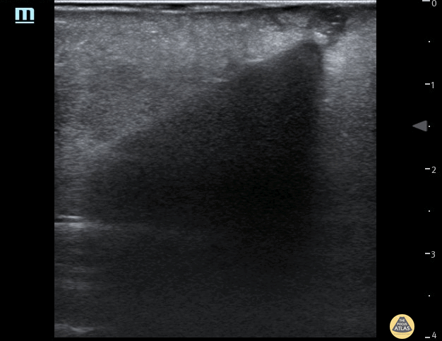
35 year female presented to the Emergncy Department with a self harm retained foreign body in the left forearm. The foreign body was a wooden cocktail stick not seen on x-ray. POCUS easily identified the cocktail stick by it's acoustic shadowing which allowed removal under local anaesthetic. Repeat POCUS scan showed no other foreign bodies.
Dr Pete Hulme @Dr_Pete_EmMed
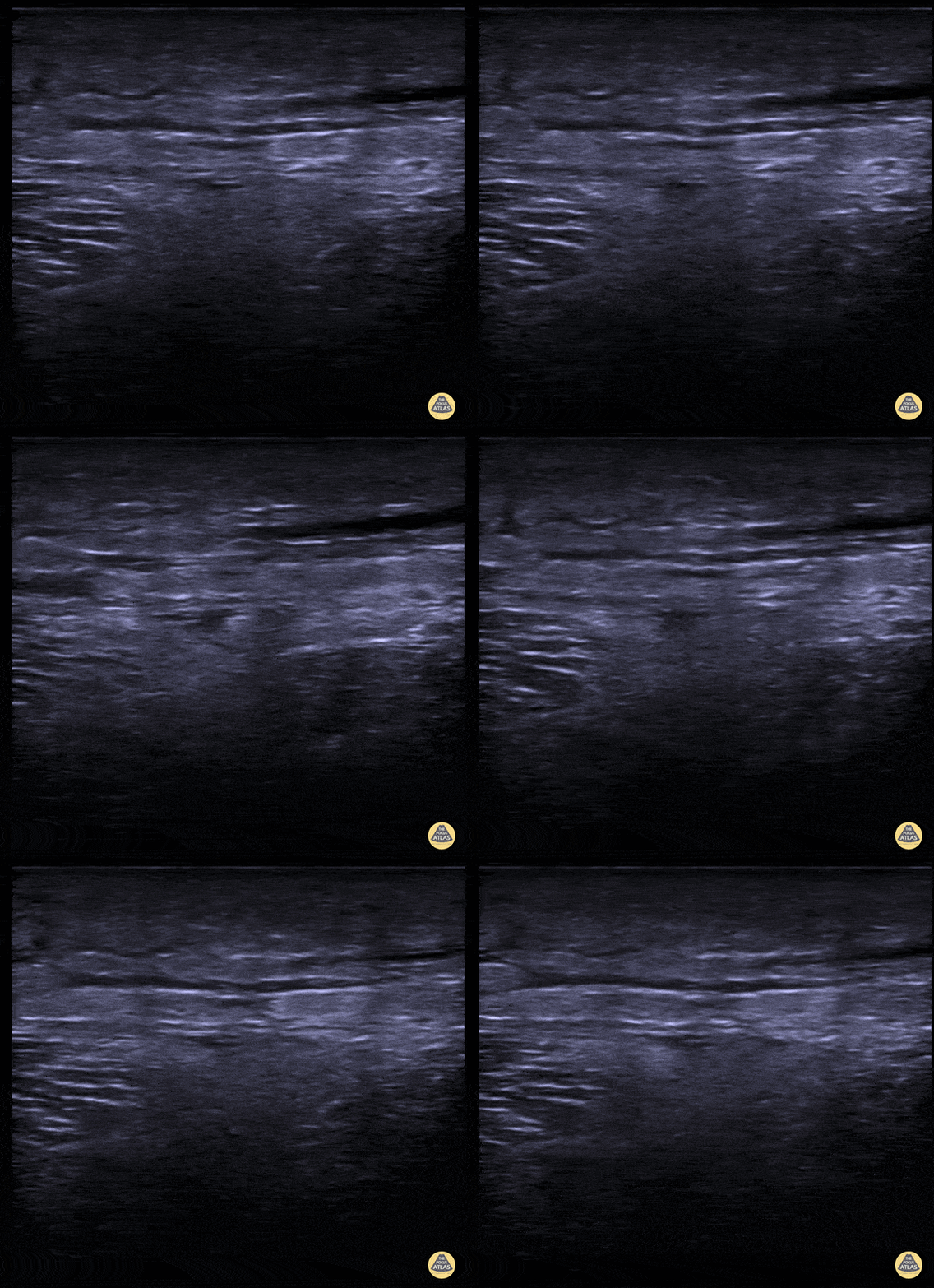
A middle aged male presented to the ED for left leg pain after sustaining puncture wound to the foot 4 hours prior arrival. The ultrasound image demonstrates free air causing dirty shadowing seen deep to the fascial plane obscuring the normal muscle architecture in the middle of the screen.
Nicmarie Maldonado, MD PGY-2; Eddie G. Rodriguez, MD; Miguel Agrait, MD; Michelle Surillo, MD - Division of Emergency Ultrasound, St. Luke's Medical Center/PHSU, Ponce, PR
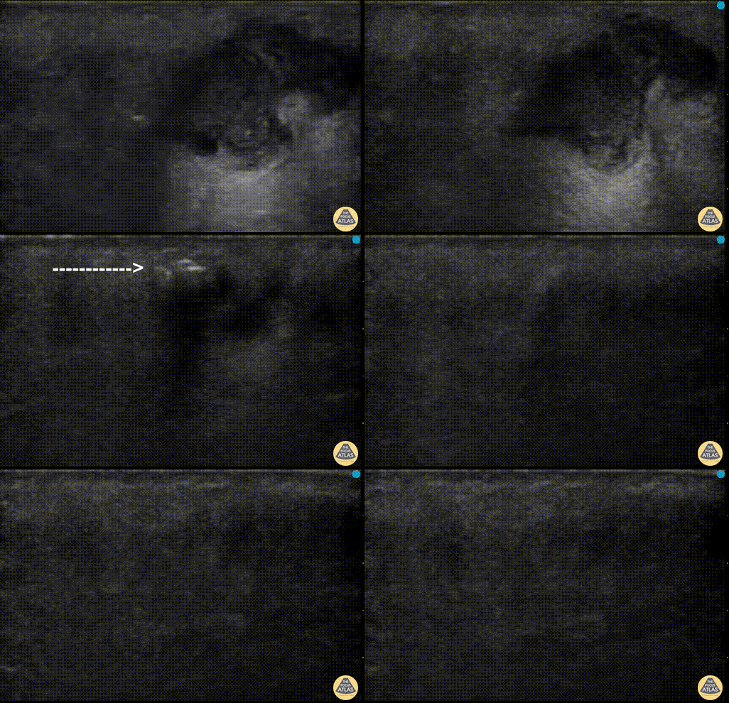
POCUS can help distinguish simple abscess from necrotizing fasciitis. “Dirty air shadowing” visualized in the clip suggests a necrotizing soft tissue infection.
Dr. Farnam Kazi Ultrasound Faculty; Dr. Dimitri Livshits Ultrasound Fellow; Dr. Jane Belyavskaya Ultrasound Fellow (All at Kings County Hospital/SUNY Downstate)