
Normal Anatomy
The best Echocardiography POCUS clips. This is free and open access medical education content. Right click to download.
Echo: Normal Anatomy
Pathology
Normal Echocardiography:
Colorized Echocardiography
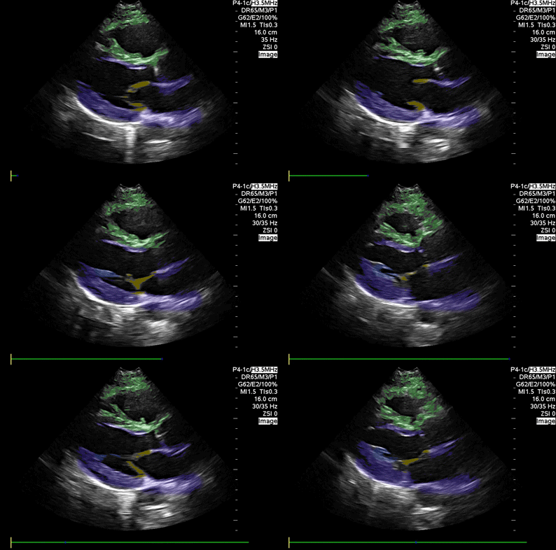
Parasternal Long Axis
Blue : Left atrium and ventricle, Yellow: Mitral valve, Green: Right ventricle
Images: Dr. Lindsay Davis, Dr. Hannah Kopinski. Image Editing: Michael Amador and Dr. Matthew Riscinti
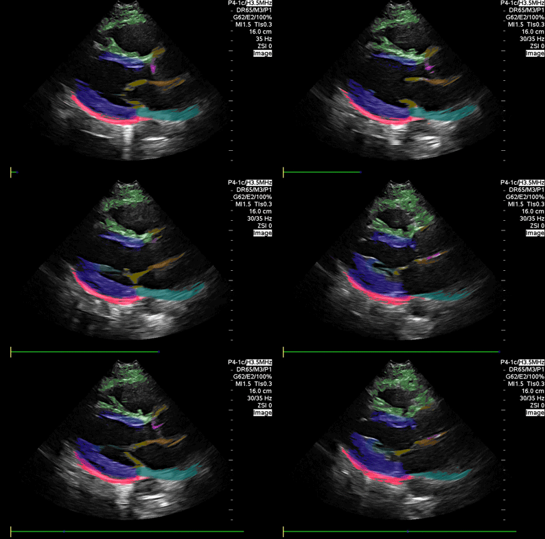
Parasternal Long Axis (detailed)
Blue: Left ventricle, Green: Right ventricle, Orange: Aortic outflow Pink: Aortic valve; Yellow: Mitral valve; Red: Pericardium; Light orange: Left atrium
Images: Dr. Lindsay Davis, Dr. Hannah Kopinski. Image Editing: Michael Amador and Dr. Matthew Riscinti
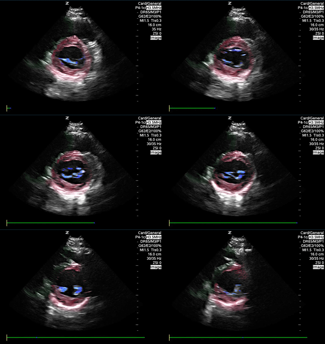
Parasternal short axis
Red: Left ventricle, Blue: Mitral valve, Green: Right ventricle
Images: Dr. Lindsay Davis, Dr. Hannah Kopinski. Image Editing: Michael Amador and Dr. Matthew Riscinti
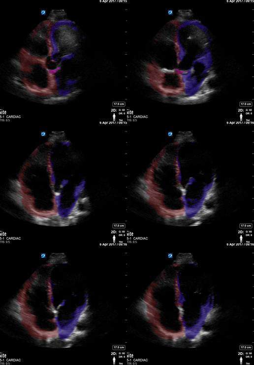
Apical 4
Red: Right atrium and ventricle, Blue: Left atrium and ventricle, Pink: aortic valve
Images: Dr. Lindsay Davis, Dr. Hannah Kopinski. Image Editing: Michael Amador and Dr. Matthew Riscinti
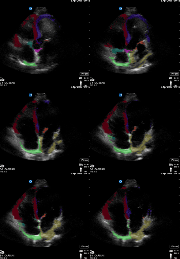
Apical 4 View
Red: Right Ventricle, Green: Right Atrium, Blue: Left Ventricle, Yellow: Left atrium. Pink: Mitral valve. Teal: Tricuspid Valve.
Images: Dr. Lindsay Davis, Dr. Hannah Kopinski. Image Editing: Michael Amador and Dr. Matthew Riscinti
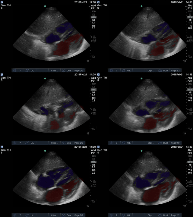
Subxiphoid view
Red: Lumen of left atrium and ventricle, Blue: Lumen of right atrium and ventricle
Images: Dr. Lindsay Davis, Dr. Hannah Kopinski. Image Editing: Michael Amador and Dr. Matthew Riscinti
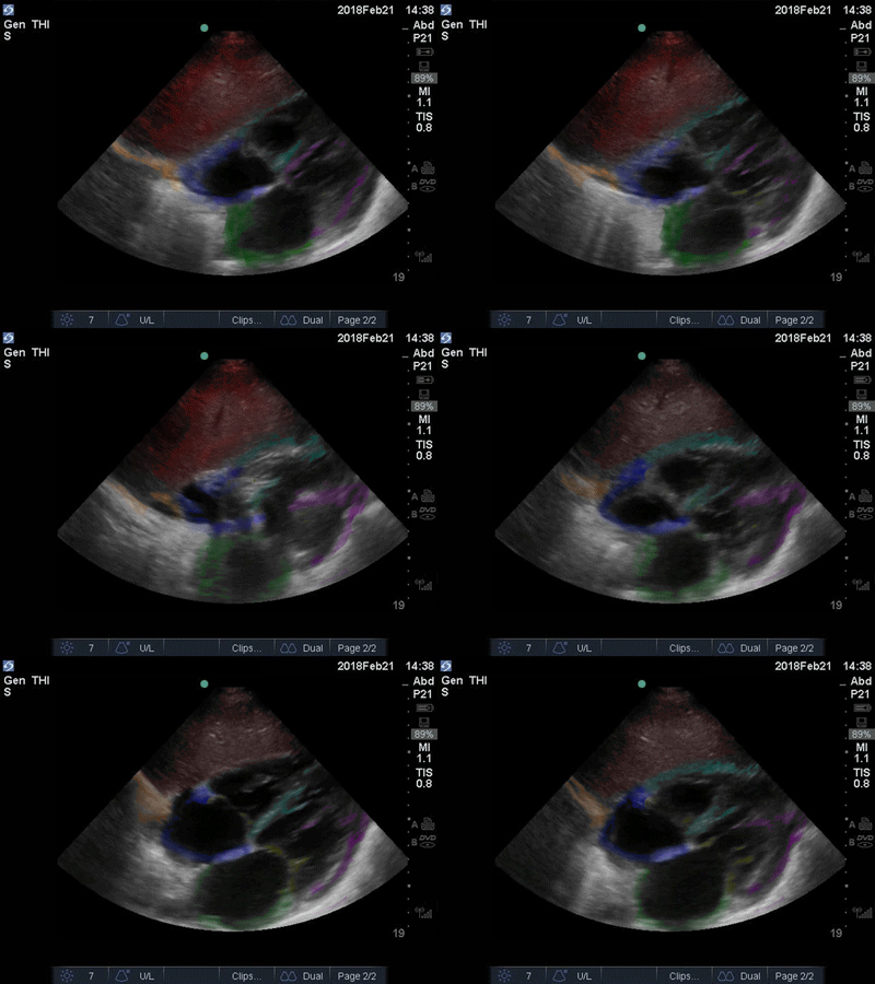
Subxiphoid
Red: Liver. Orange: Diaphragm. Blue: Right Atrium, Teal: Right Ventricle, Green: Left Atrium,Purple: Left Ventricle
Images: Dr. Lindsay Davis, Dr. Hannah Kopinski. Image Editing: Michael Amador and Dr. Matthew Riscinti