
Trauma Teaching
Trauma
The best trauma POCUS clips. Positive and negative fasts. Please see the echo section for pericardial findings. This is free and open access medical education content. Right click to download. See our Evidence Atlas for the evidence behind Trauma POCUS.
Normal FAST Exams
No free fluid is seen around or behind the bladder in this negative FAST.
Dr. Justin Bowra
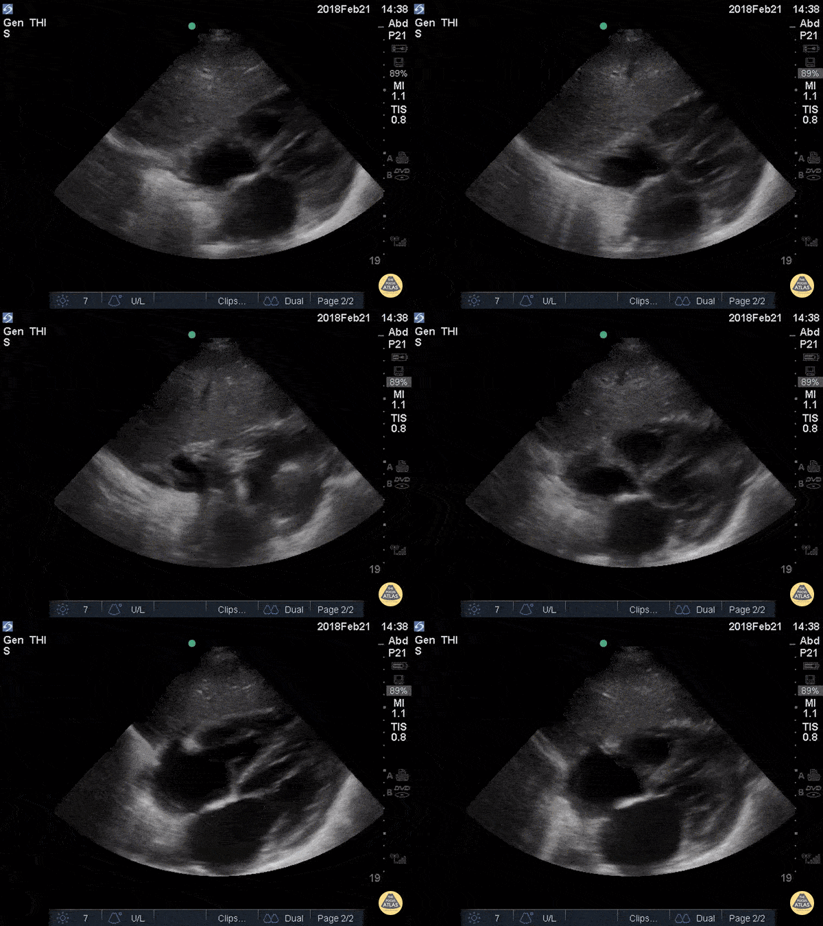
The most superficial structure we see is the liver. Immediately deep to that we see the heart separated from the liver by the diaphragm. Closest to the liver is the right atrium, tricuspid valve, and right ventricle. Deeper to that, we see the left atrium, mitral valve, and left ventricle. There is no anechoic fluid between the bright hyperechoic pericardium and the myocardium, indicating absence of pericardial effusion.
Hannah Kopinksi and Dr. Lindsay Davis - NYU Emergency Medicine
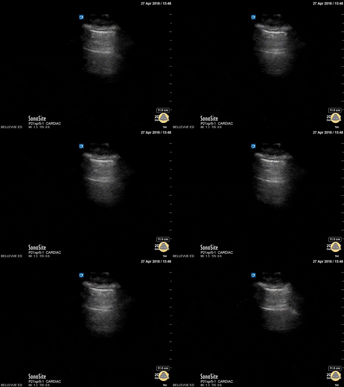
This is a clip demonstrating lung sliding. The most superficial hyperechoic layers are the soft tissue and muscular layers of the chest wall. Immediately deep to that is a bright, thin hyperechoic line which appears to be in motion - this is the pleural line. The parietal pleura rubbing against the visceral pleura as the patient breathes creates this shimmery appearance of lung sliding, also often described as “ants marching”. Lung sliding indicates that there is no pneumothorax.
Hannah Kopinksi and Dr. Lindsay Davis - NYU Emergency Medicine
FAST Exams - Abdominal
A patient presented with LUQ trauma but the perisplenic window was negative for free fluid. Morison’s pouch was positive for free fluid emphasizing the importance of scanning all regions for free fluid in trauma.
Image courtesy of Robert Jones DO, FACEP @RJonesSonoEM
Director, Emergency Ultrasound; MetroHealth Medical Center; Professor, Case Western Reserve Medical School, Cleveland, OH
View his original post here
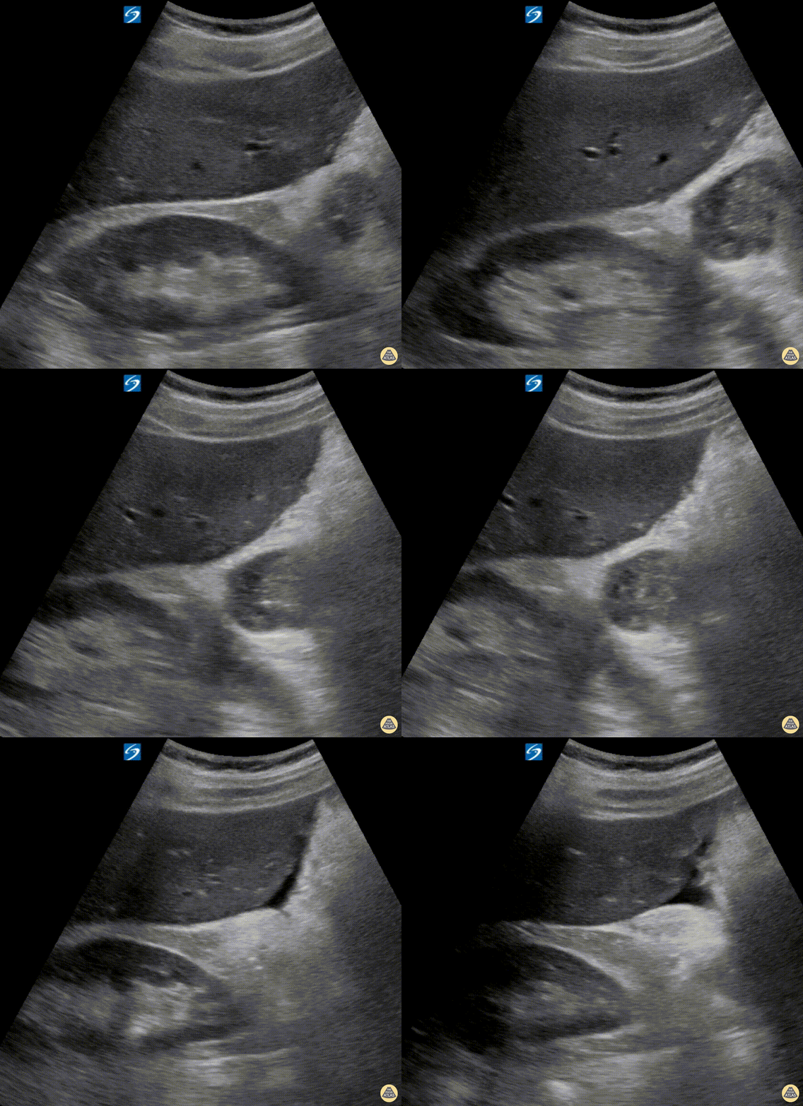
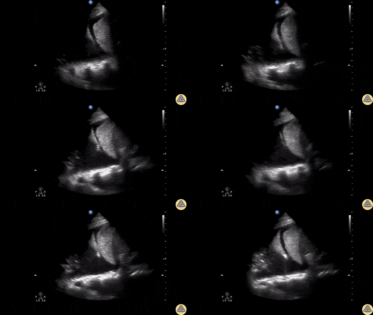
Seen here is a subtle positive RUQ FAST scan in a trauma patient — a pertinent reminder to never stop simply after evaluating Morison's Pouch. Always also trace anteriorly to the lowest part of the liver.
Hjalti Már Björnsson
@hjaltimb
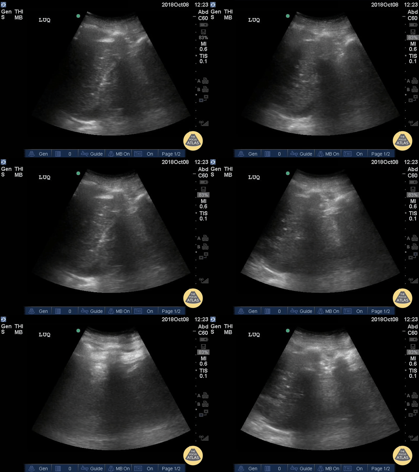
Positive FAST exam demonstrating free fluid in the left upper quadrant. Spontaneous hemoperitoneum in an anticoagulated patient with a left ventricular assist device (LVAD).
Dr. Elias Jaffa, MD
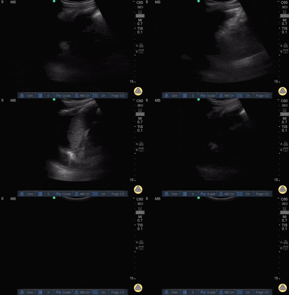
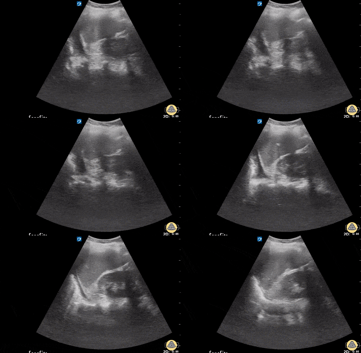
This image demonstrates free fluid in the LUQ. Notice the anechoic fluid seen both superior and inferior to the spleen as the probe is fanned. There is significant rib shadow appreciated obscuring parts of the image which makes the free fluid difficult to appreciate if not evaluated closely.
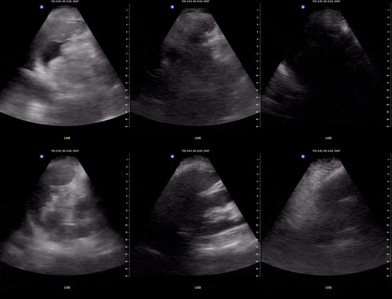
This image demonstrates free fluid present in the left upper quadrant following blunt trauma to the abdomen. Notice the anechoic area present along the the pericolic gutter as well as between the spleen and the diaphragm consistent with free fluid.
Michael Macias, MD
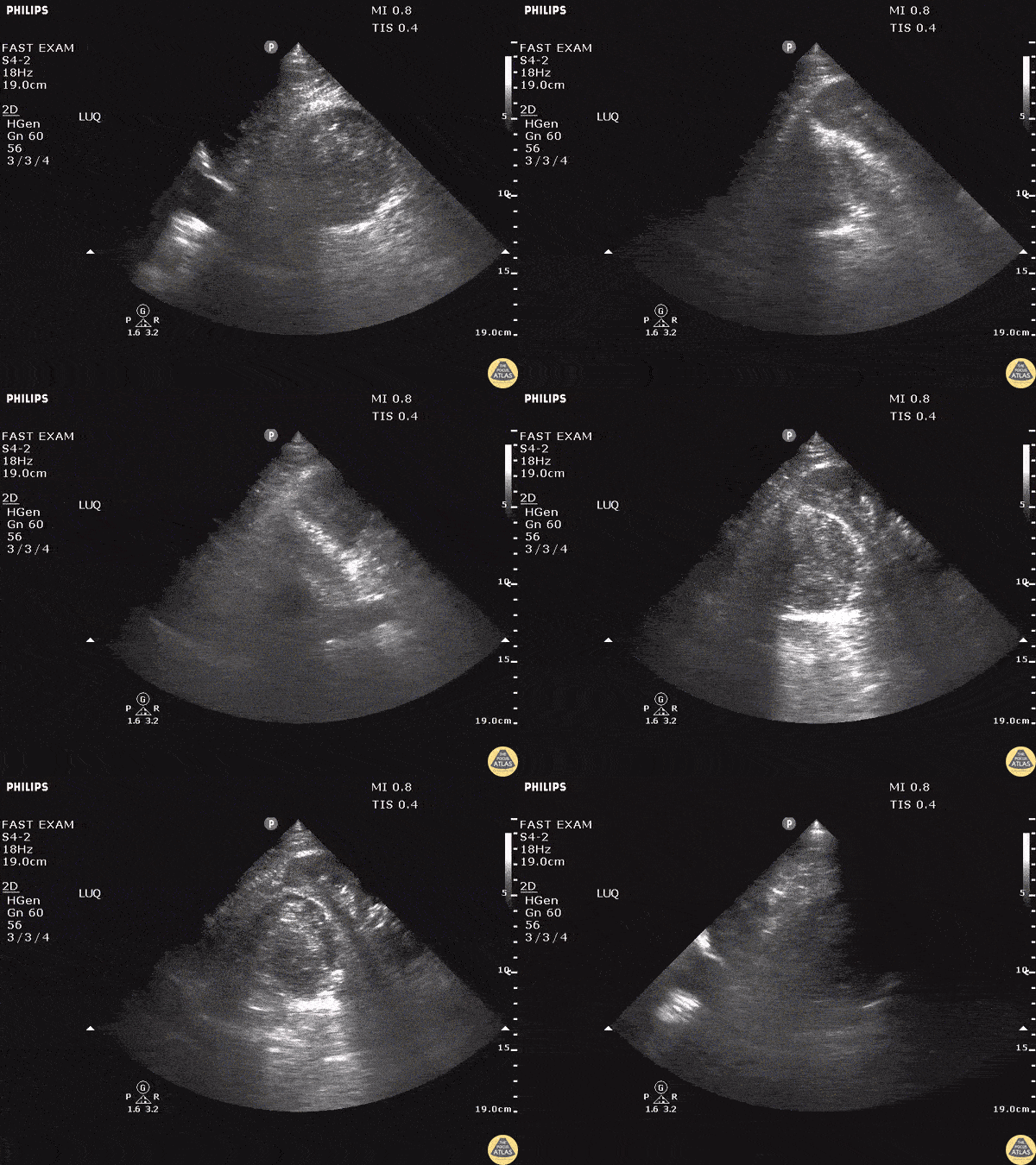
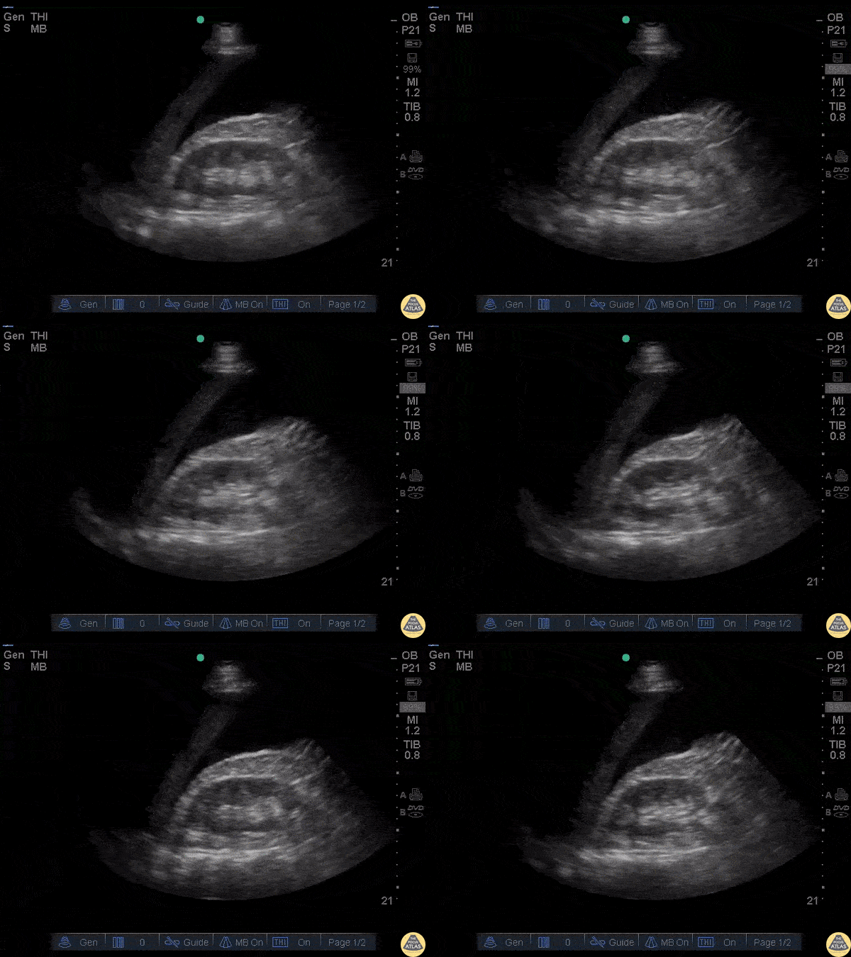
A young male presented to the ED following a 20 foot fall. He presented with LUQ pain, left flank pain, and soft vitals. FAST exam showed no fluid in Morison’s pouch or in the pelvis but revealed a large, left-sided, retroperitoneal hematoma noted by the distortion of the left kidney.
Image courtesy of Robert Jones DO, FACEP @RJonesSonoEM
Director, Emergency Ultrasound; MetroHealth Medical Center; Professor, Case Western Reserve Medical School, Cleveland, OH
View his original post here
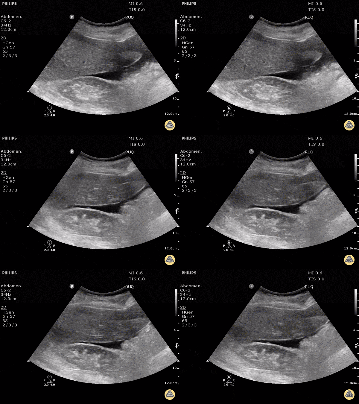
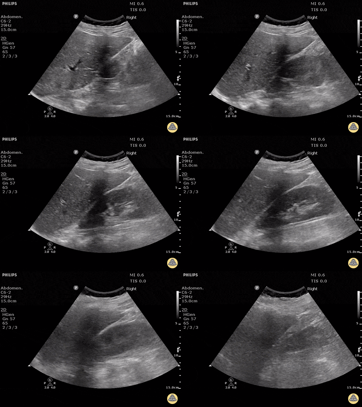
Blunt trauma patient with POSITIVE FAST scan. The liver can been seen floating in free fluid with the kidney posteriorly. The fluid is in Morison's pouch. Be sure to visualize the tip of the liver to complete the evaluation.
Dr. Justin Bowra
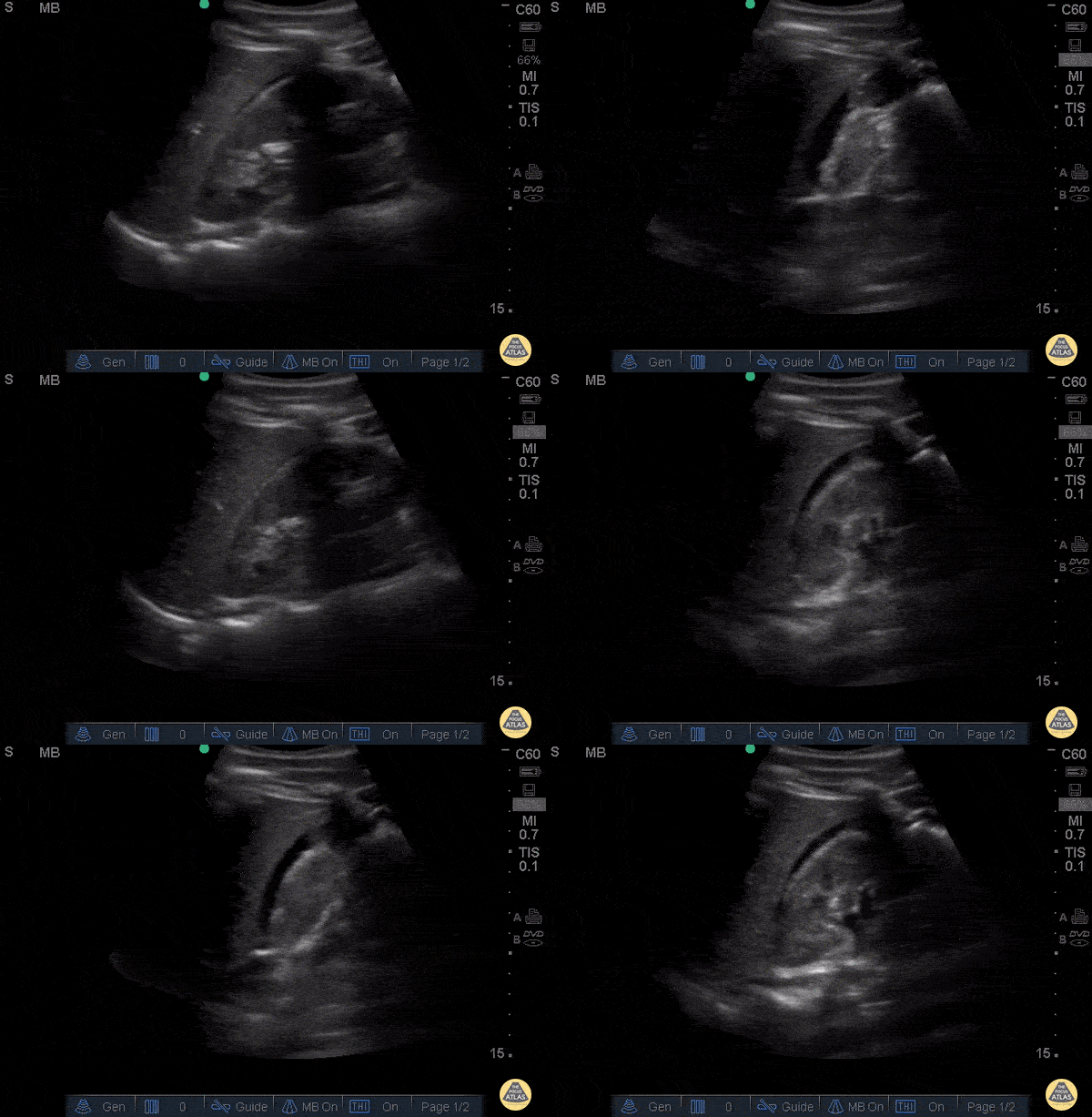
30 y/o pedestrian struck by car, hemodynamically unstable, tachycardic. FAST performed after primary survey revealed free fluid in all four abdominal views of the FAST exam
Free fluid in Morison’s pouch of the RUQ view. This is the most sensitive view to detect free fluid in trauma.
Dr. Catharine Bon - Kings County Emergency Medicine
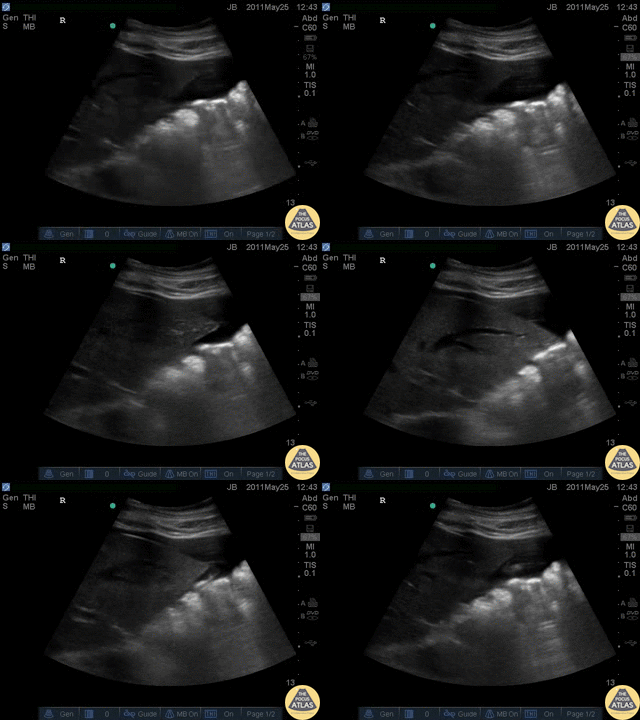
Blunt trauma patient with POSITIVE FAST scan. The liver can been seen floating in fluid with adjacent bowel.
Dr. Justin Bowra
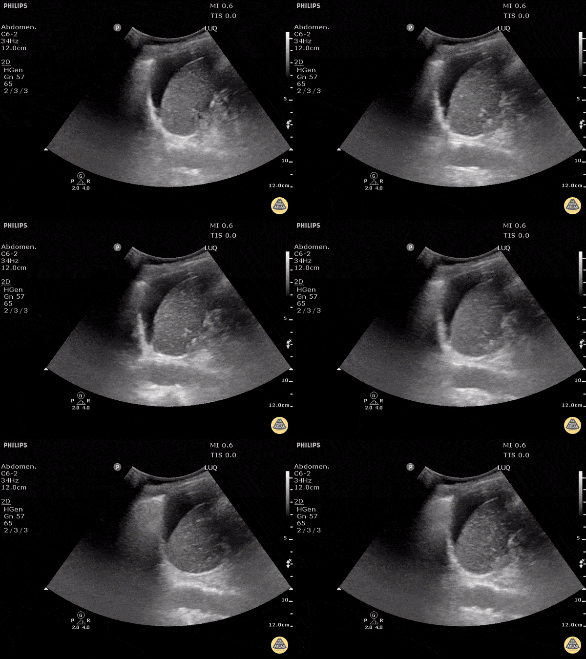
Blunt trauma patient with POSITIVE FAST scan. Free fluid can be seen between the spleen and the diaphragm in this LUQ view.
Dr. Justin Bowra
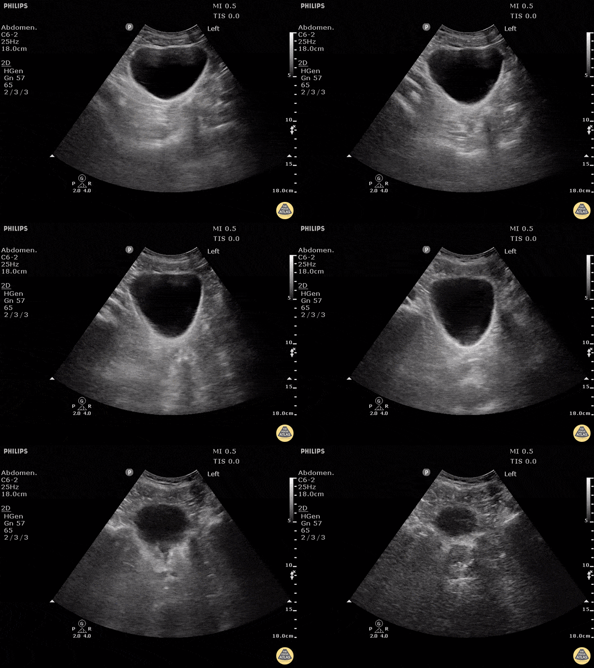
No free fluid is seen around or behind the bladder in this negative FAST.
Dr. Justin Bowra
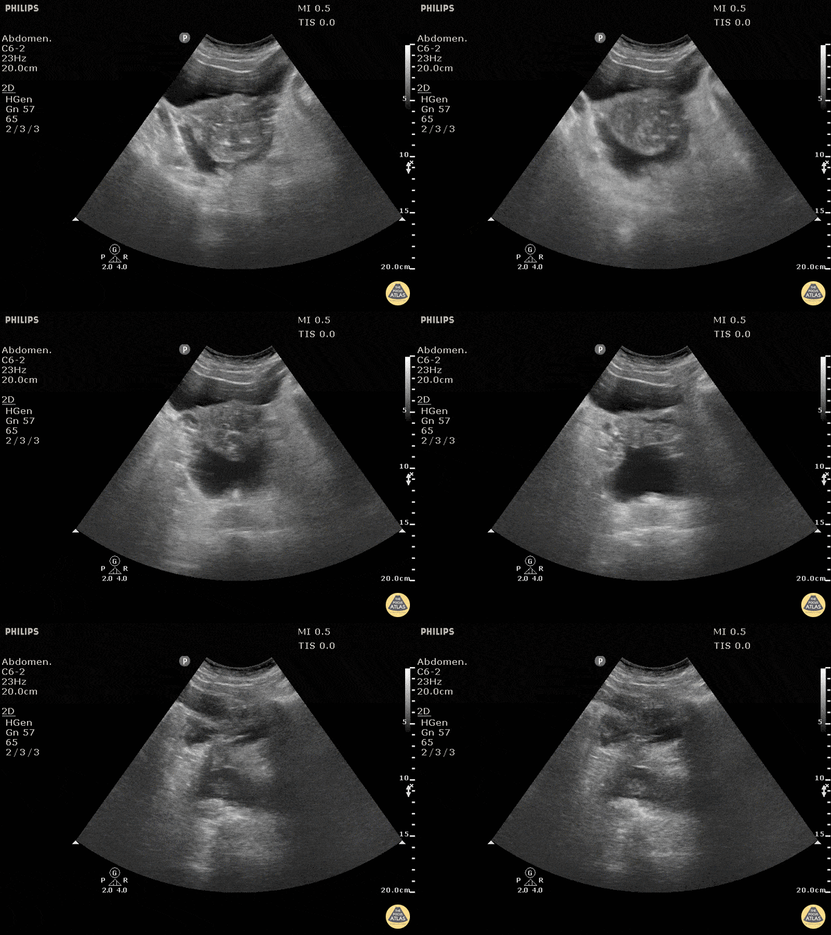
Blunt trauma patient with POSITIVE FAST scan. The uterus can been seen floating in free fluid.
Dr. Justin Bowra
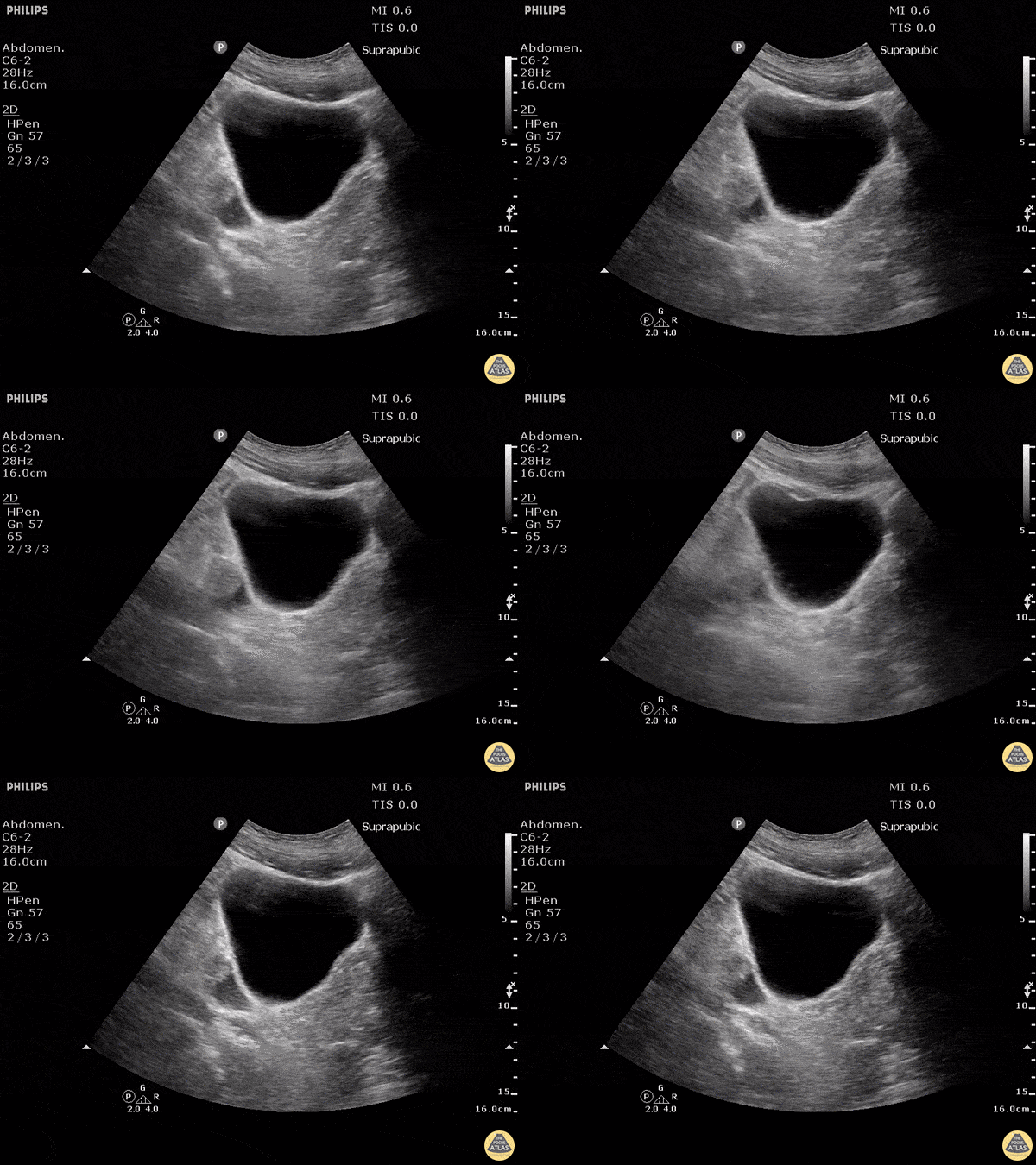
Blunt trauma patient with POSITIVE FAST scan. Free fluid can be seen posterior and lateral to the bladder in this sagittal view.
Dr. Justin Bowra
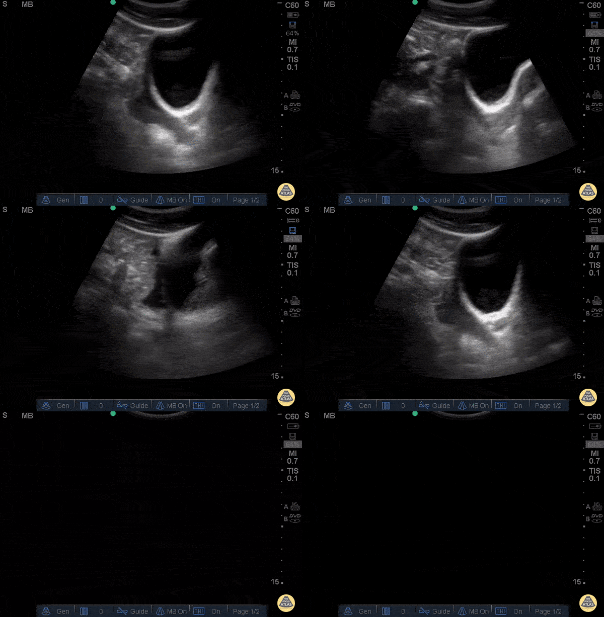
30 y/o pedestrian struck by car, hemodynamically unstable, tachycardic. FAST performed after primary survey revealed free fluid in all four abdominal views of the FAST exam.
Free fluid seen superior and posterior to the bladder in this sagittal view.
Dr. Catharine Bon - Kings County Emergency Medicine
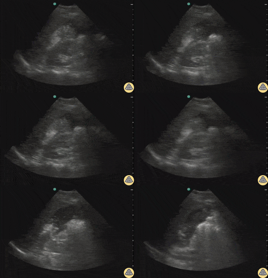
Pt struck by car while riding bike. Perihepatic view on FAST exam revealed a perforated viscus (gastric rupture). Note the free air within the fluid.
Image courtesy of Robert Jones DO, FACEP @RJonesSonoEM
Director, Emergency Ultrasound; MetroHealth Medical Center; Professor, Case Western Reserve Medical School, Cleveland, OH
View his original post here.
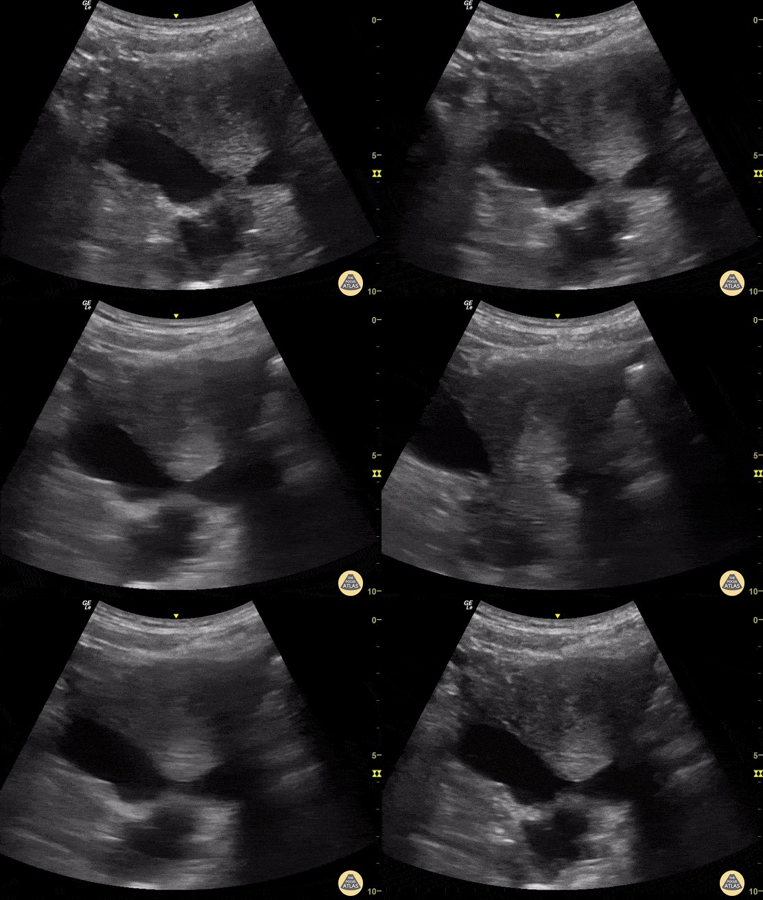
A 35-year-old woman presented to the ED after experiencing blunt abdominal trauma. She was hemodynamically unable. E-FAST Exam performed at bedside was notable for free intra-abdominal fluid (viewed here from suprapubic region). This rapid diagnosis enabled prompt disposition to the operating room.
Josiane Almeida, Emergency Physician; Sao Paulo- Brazil
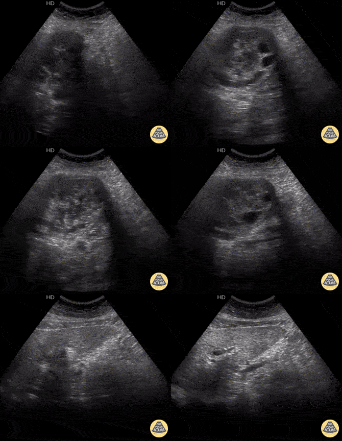
52-yo female presents to ED hypotensive with diffuse urticaria after blunt trauma to abdomen from a fall. FAST exam revealed a small amount of free fluid in RUQ and an abdominal mass. Diagnosis later confirmed to be a ruptured hepatic hydatid cyst.
Image courtesy of Robert Jones DO, FACEP @RJonesSonoEM
Director, Emergency Ultrasound; MetroHealth Medical Center; Professor, Case Western Reserve Medical School, Cleveland, OH
View his original post here
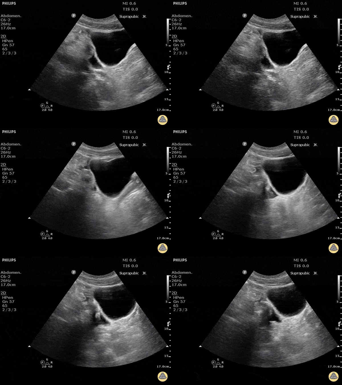
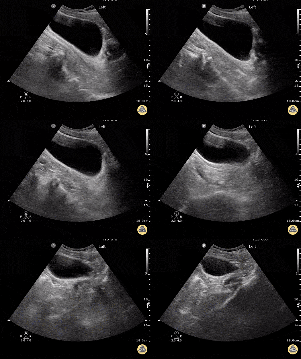
Blunt trauma patient with POSITIVE FAST scan. Free fluid can be seen posterior to the dome of bladder in this sagittal view.
Dr. Justin Bowra
FAST - Cardiac
Pulmonary Trauma and Pneumothorax
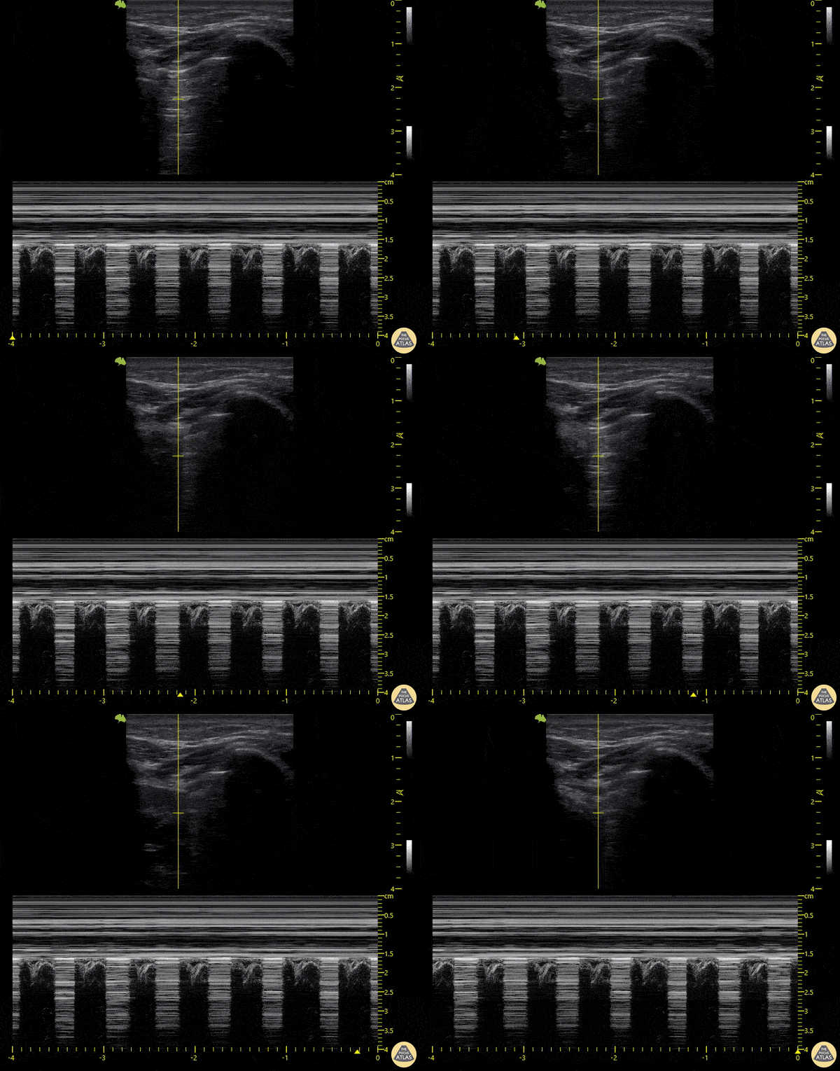
18-year-old patient who presented following a motorcycle accident in which he sustained closed chest trauma with bilateral hemopneumothoraces. In this sequence taken with a linear transducer in left pulmonary zone 1, the "pulmonary point" indicative of pneumothorax can be seen.
Libardo Valencia Chicue
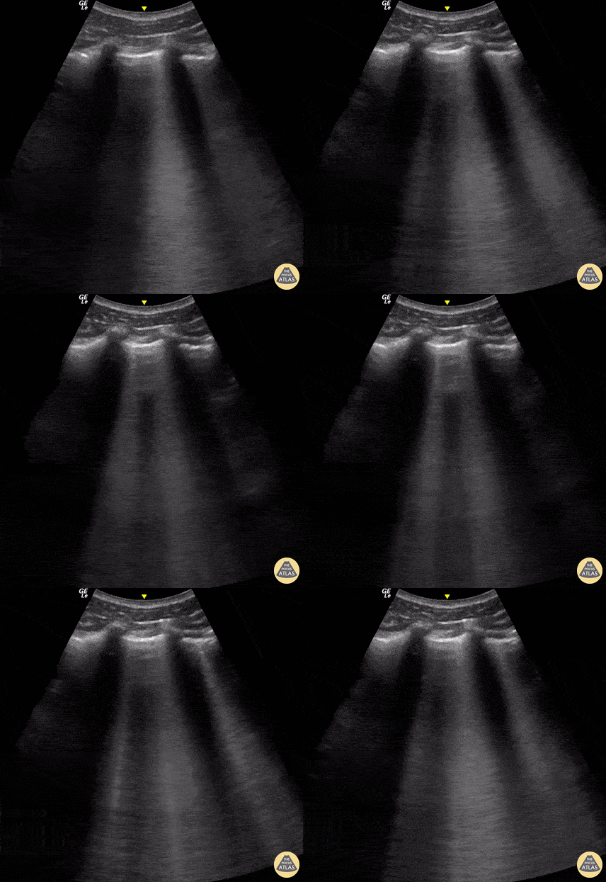
A 23-year-old male was admitted to the ED following a motorcycle accident. He subjectively reported dyspnea; objectively was hypoxic. POCUS seen here (obtained from L3 zone using curvilinear probe) reveals multifocal B-lines consistent with pulmonary contusion. This case illustrates that bedside US is useful beyond diagnosing pneumothorax and hemothorax in trauma patients.
Renato Tambelli, Emergency Physician Hospital das Clínicas de Marília, Brazil.
@R_Tambelli / @JediPocus
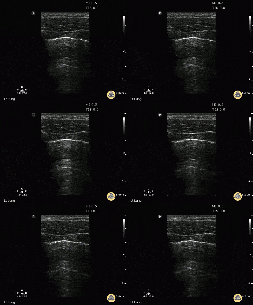
A young male presented to ED with stab wound to chest. Upright CXR was normal. POCUS showed double lung point indicative of small PTX. This was monitored without evidence of progression. No intervention was required.
Image courtesy of Robert Jones DO, FACEP @RJonesSonoEM
Director, Emergency Ultrasound; MetroHealth Medical Center; Professor, Case Western Reserve Medical School, Cleveland, OH
View his original post here
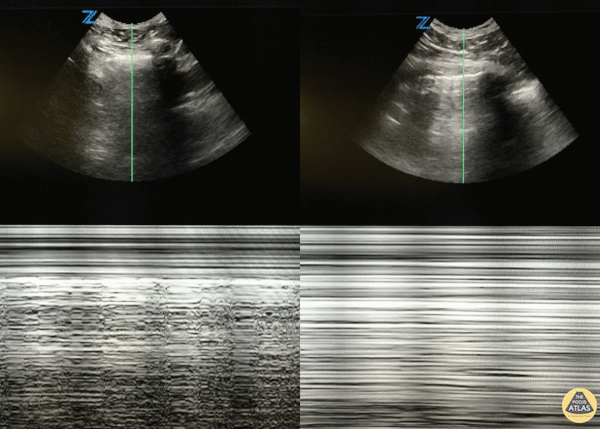
46 y/o M with 20 pack year smoking history with sudden onset right sided chest pain that woke him from sleep. Decreased breath sounds on right side. POCUS with decreased lung slide (right of image) with normal lung slide in left lung (left side of image).
Lung slide can often be appreciated by watching the pleural surfaces move along each other but if you're uncertain, putting the US in m-mode and looking for the classic "seashore sign" (left image) versus the "barcode sign" (right image) can help you figure it out.
Dr. Eric Roseman - Resident Physician, Kings County Emergency/Internal Medicine
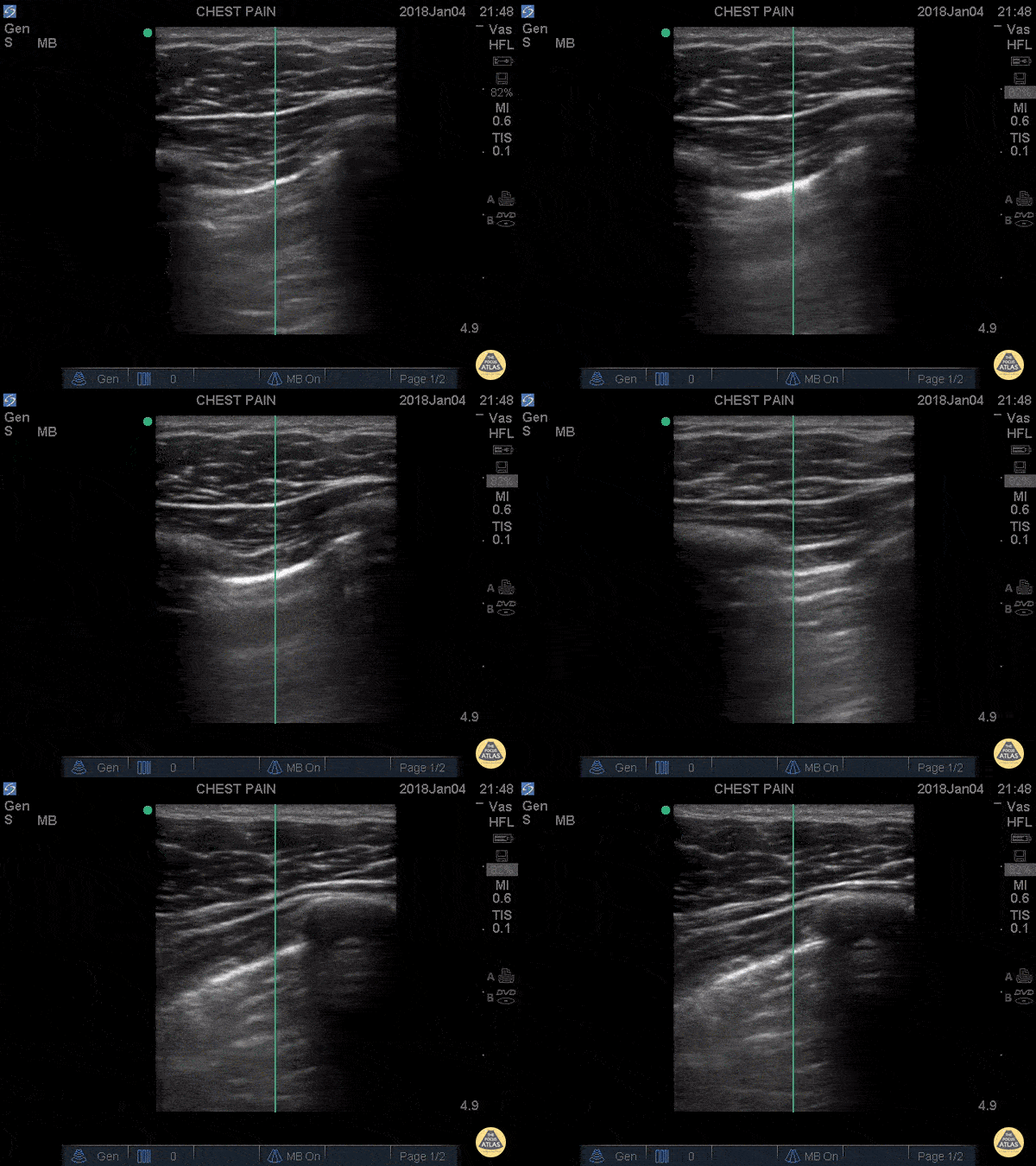
Trauma code to the waiting room... 20 y/o male stabbed to the left chest in the midaxillary line. Patient thrown in a wheelchair and pushed to the resuscitation room and POCUS performed immediately revealing this image: the lung sliding disappearing revealing an area without lung slide. Moved up one rib space to the apex, no lung slide at all.
This junction of slide/no slide is the lung point and its pathognomonic for pneumothorax. It represents the exact point where air begins to separate the parietal from visceral pleura, aka the junction of where we normally see the "ants marching" or the "shimmering" aka the lung slide. This is highly specific.
Don't be fooled when you see the sliding with decreased sliding around it. You're looking at a pneumothorax.
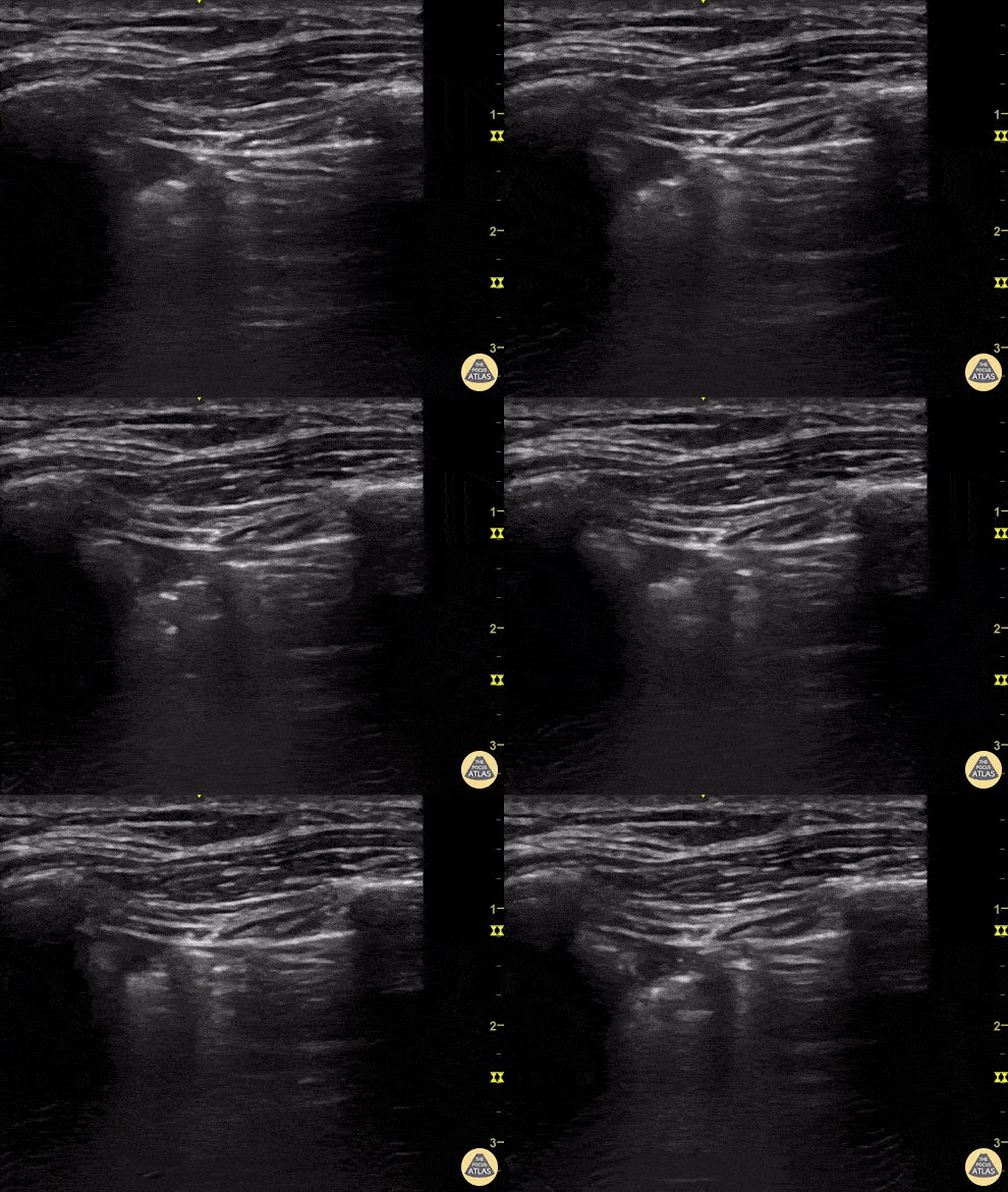
A young man presents to the emergency department with multiple thoracic stab woulds. POCUS quickly identifies absent lung sliding as well as a lung point; findings highly sensitive and specific for our diagnosis of pneumothorax.
Renato Tambelli, @R_Tambelli
Emergency Physician Hospital das Clínicas de Marília
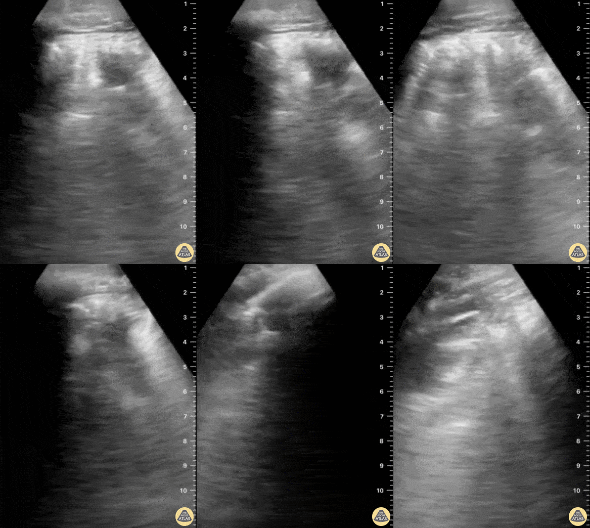
In this patient with history of blunt chest trauma, lung ultrasound reveals a focal area of B-lines as well as a hypoechoic wedge shaped subpleural consolidation.
These findings are consistent with pulmonary contusion and can be identified early in the course of a patient’s care. This contrasts with hours-to-short-days that it often takes to fully appreciate the evolution of pulmonary contusion on chest X-ray.
Aaron Inouye, PA-C, North Canyon Medical Center
@PAintheED
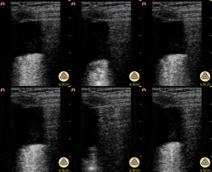
This clip, demonstrating a hydropoint, was taken in a 74 year old M with chest trauma after a fall from 3 meters. A hydropoint shows the air/fluid interface which is suggestive for hemato/hydro/pyo-pneumothorax. It is another sign for diagnosing a pneumothorax described by Volpicelli et al. Critical Ultrasound Journal. 2013
Dr. Van Roosmalen
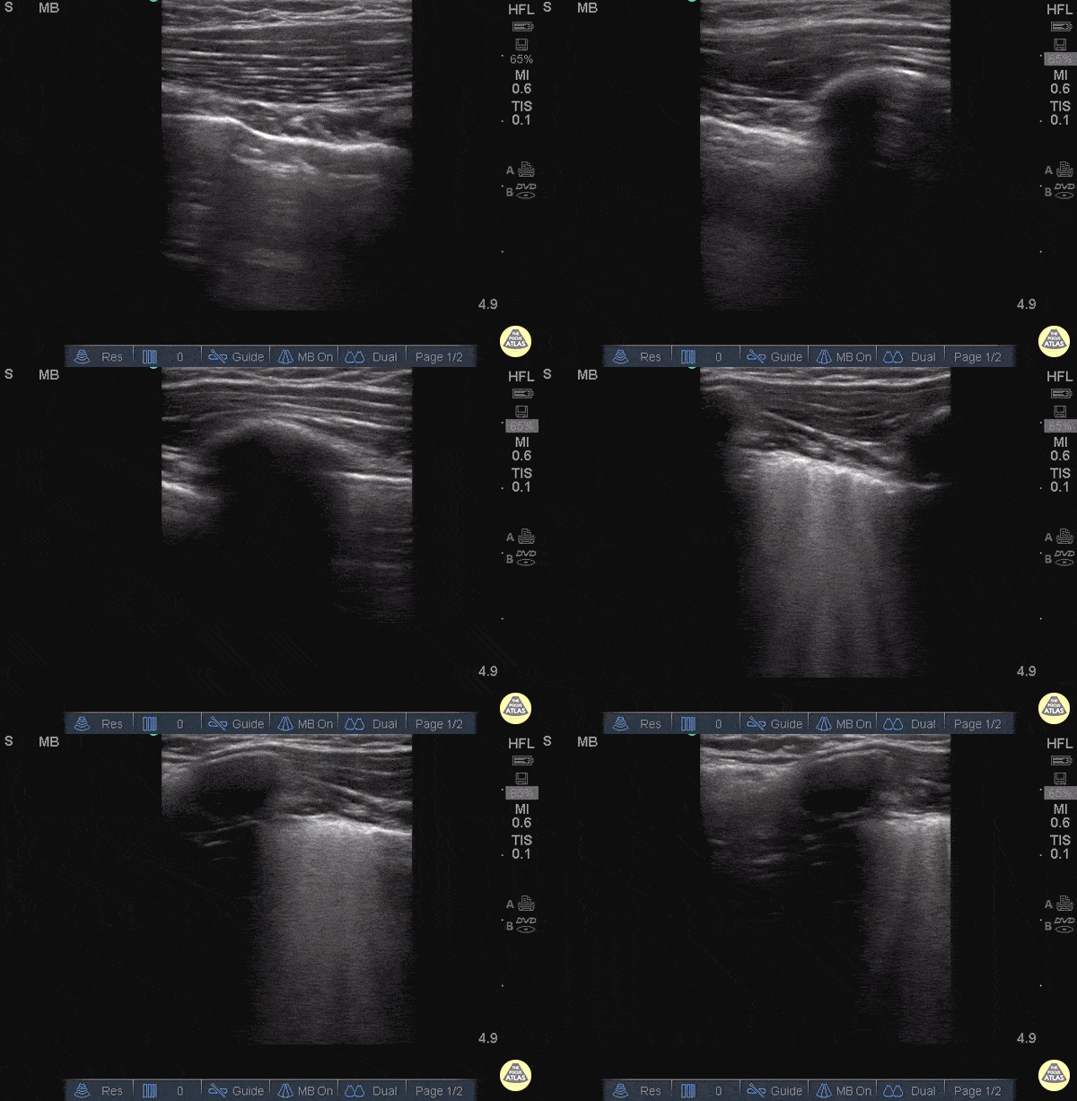
25 y/o female in and MVA with hypotension, hypoxia.
Normal lung with A lines can briefly be seen until the sonographer moves the probe superiorly to reveal and area of B lines adjacent to A line.
In the setting of trauma this is consistent with Pulmonary Contusion.
Images: Dr. Catharine Bon - Kings County Hospital Emergency Medicine
This is an image of a patient's chest wall using a high frequency transducer, with the transducer oriented in a transverse plane between rib spaces. The patient had a pneumothorax and a chest tube was placed. This clip illustrates what happens when the suction is turned 'on'. You will see the pleura slide from right to left as the pneumothorax resolves.
- Jason Tanguay, DO; Ultrasound Leadership Academy Graduate
Decreased lung slide is highly sensitive, it lacks specificity. Lung point however, is a highly specific finding indicating a pneumothorax.
Lung point indicates the transition point between normal pleura with normal lung sliding and where there is air disrupting the pleural space with decreased lung sliding.
In this intercostal space, one can see lung with normal lung slide on the left, and decreased lung slide on the right, and a point where the lung slide changes, which is moving with inspiration. This is the lung point.
Dr. Justin Bowra et al.
55 y/o female complains of left sided chest pain with cough, SOB, and back pain. History of falls from 8-foot ladder (8 weeks ago) and from standing (2 weeks ago).
With patient in supine, US of LUQ lung in coronal view demonstrated a hypoechoic fluid collection above the left hemi-diaphragm consistent with a L pleural effusion.
It is important to scan above and below the diaphragm to differentiate free fluid in thorax vs fluid in subphrenic space. US is more sensitive than plain radiographs for detecting pleural effusion and can detect smaller amounts of fluid.
Crozer Chester Medical Center.
Raghav Sahni, Dr. Melissa Yu, Dr. Brenton Elliot, Dr. Max Cooper.
A male presented to the ED following a gunshot wound to the left chest. The perisplenic window of the FAST exam revealed a left hemothorax with a large subpleural area of clotted blood.
Image courtesy of Robert Jones DO, FACEP @RJonesSonoEM
Director, Emergency Ultrasound; MetroHealth Medical Center; Professor, Case Western Reserve Medical School, Cleveland, OH
View his original post here
Fractures and Musculoskeletal Trauma
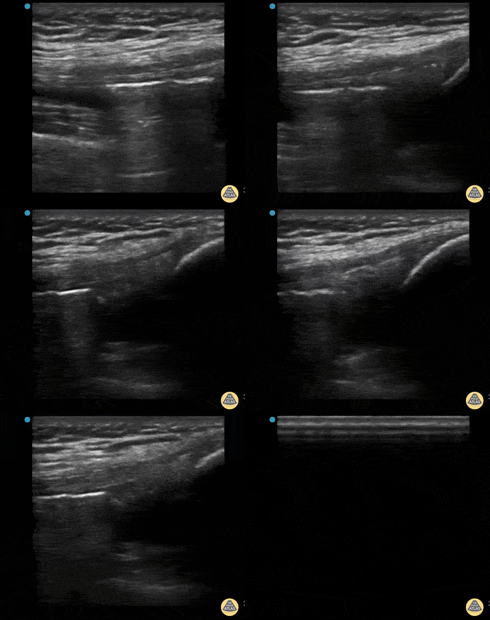
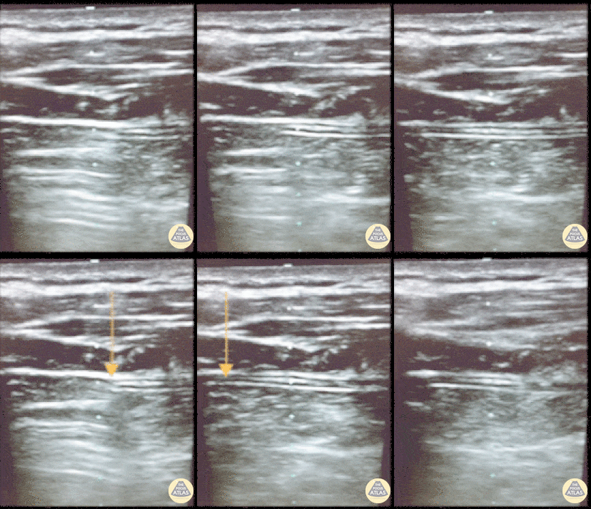
This is an ultrasound clip from a patient who presented to the ED after sustaining a laceration near the knee. There was concern for violation of the knee joint so ultrasound was used to evaluate.
The probe is held in an sagittal orientation, just proximal to the patella, overlying the quadriceps tendon. A hyperechoic line with shadowing (similar in appearance to an A-line seen on lung US) can be seen deep to the quadriceps tendon confirming intra-articular air and therefore violation of the knee joint from the laceration. A small joint effusion can also be appreciated.
Clip courtesy of Dr. Daniel Mantuani and Highland Ultrasound
Twitter: @HGHED
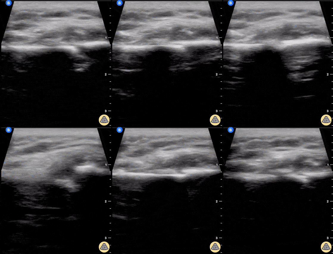
60 year old female with a subacute left clavicular fracture (occurred 2 weeks ago) presented with worsening pain at fracture site of onset while working with occupational therapy. Seen here is the left clavicle (hyperechoic structure) with noted fracture and mild heterogeneous (concern for bloody accumulation) edema around fracture site as observed in long axis view.
Kwasi Ampomah, DO, Eben Alexander IV, DO, Tariq Niazi, MD
EVMS PM&R
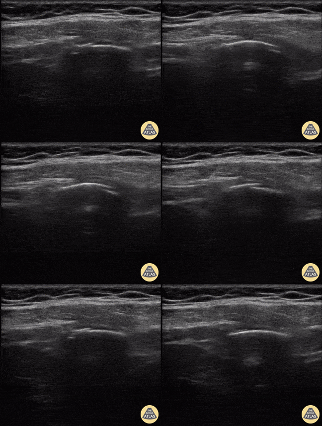
A middle aged man presented 1 week after sustaining a fall with direct injury to his left chest. He reported pain with inspiration and coughing; he localized pain to one specific area of his chest wall. Seen here is the image obtained when the linear probe was placed in the longitudinal plane to his area of point-tenderness. Notice the disruption of the hyperechoic cortex of the rib. Findings were confirmed in the transverse plan. The patient went on to have an anterior serratus nerve block for pain control related to his rib fracture.
Mandy Peach, MD @mandy_peach
Saint John Regional Hospital. NB, Canada
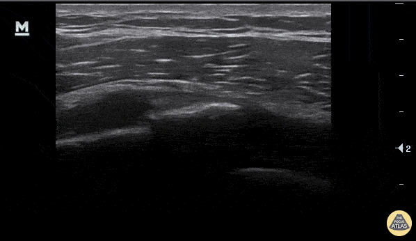
A rib fracture is seen here as disruption in the hyperechoic line or bony cortex. Also note the associated hypoechoic hematoma formation.
Aaron Inouye, PA-C, North Canyon Medical Center
@PAintheED
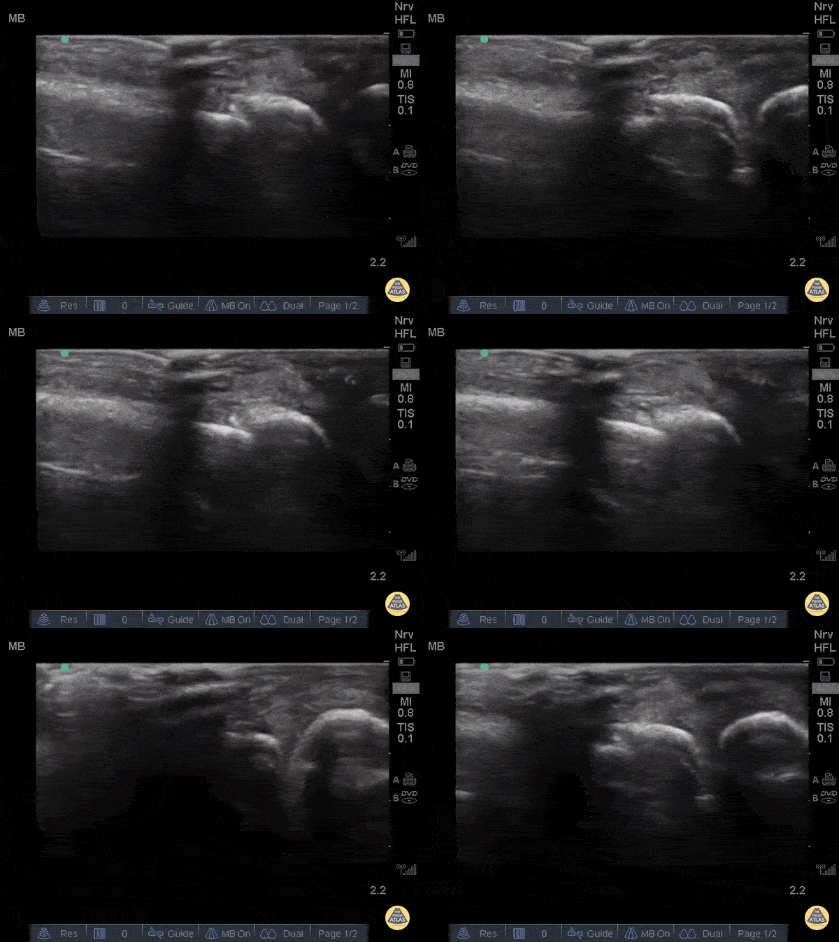
Fractures can easily be diagnosed with POCUS especially in resource limited settings. Just remember... this could be painful so use A LOT of gel and try not to press hard or at all. Gently move the probe along the axis of the bones where you suspect a fracture.
The deepest and most hyperechoic horizontal line is the cortex and discontinuity in the lines represent fracture. Angulation and displacement can be measured. Two planes should be measured.
Sukh Singh, MD, Caption: Matthew Riscinti, MD
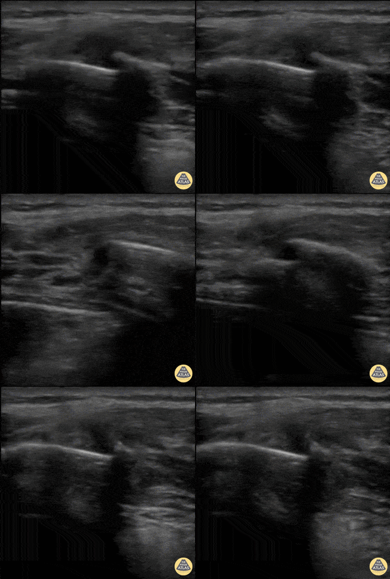
40 y/o M with polysubstance abuse, left-sided rib pain after a traumatic blow. Chest xray was equivocal. The patient was asked to "point to where it hurt", and the linear transducer revealed a displaced rib fracture. He complained of significant pain even after the resident gave two Percocet and was unwilling to leave the ED.
An intercostal nerve block, and that relieved the patient's pain and he went home.
Dr. Stephen Alerhand, Mt Sinai Hospital NYC
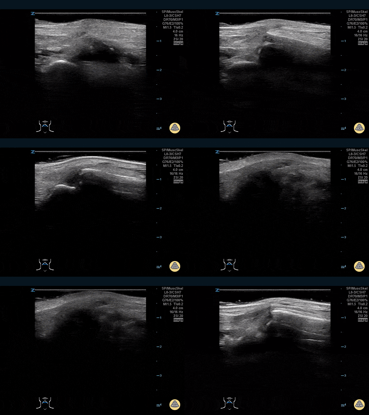
30 year-old male ED resident who injured his thumb at some point while playing football versus the attendings in the annual flag football game. He figured the thumb had merely been sprained, and he kept playing in the game (and scoring touchdowns) while the residents dominated.
Two days later the swelling/ecchymoses seemed to worsen, he used the linear transducer in a water bath to diagnose a fracture of the base of the 1st metacarpal. An x-ray confirmed the diagnosis, and he underwent percutaneous pinning in the operation room the following week.
Dr. Stephen Alerhand, Mt Sinai Hospital, NYC
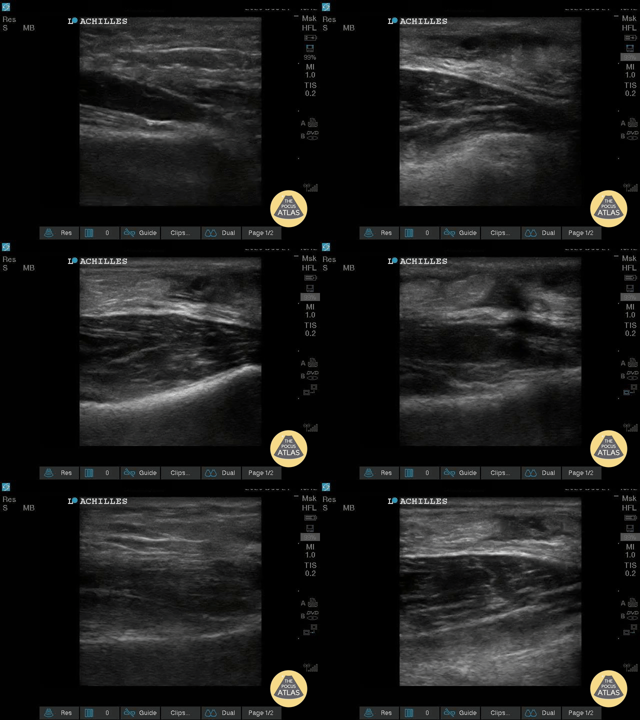
Healthy male in late-20s trying to do a backflip off a diving board. Still was able to plantarflex (very weak and with significant pain).
Submitted by Dr. Elias Jaffa
Pediatric Trauma
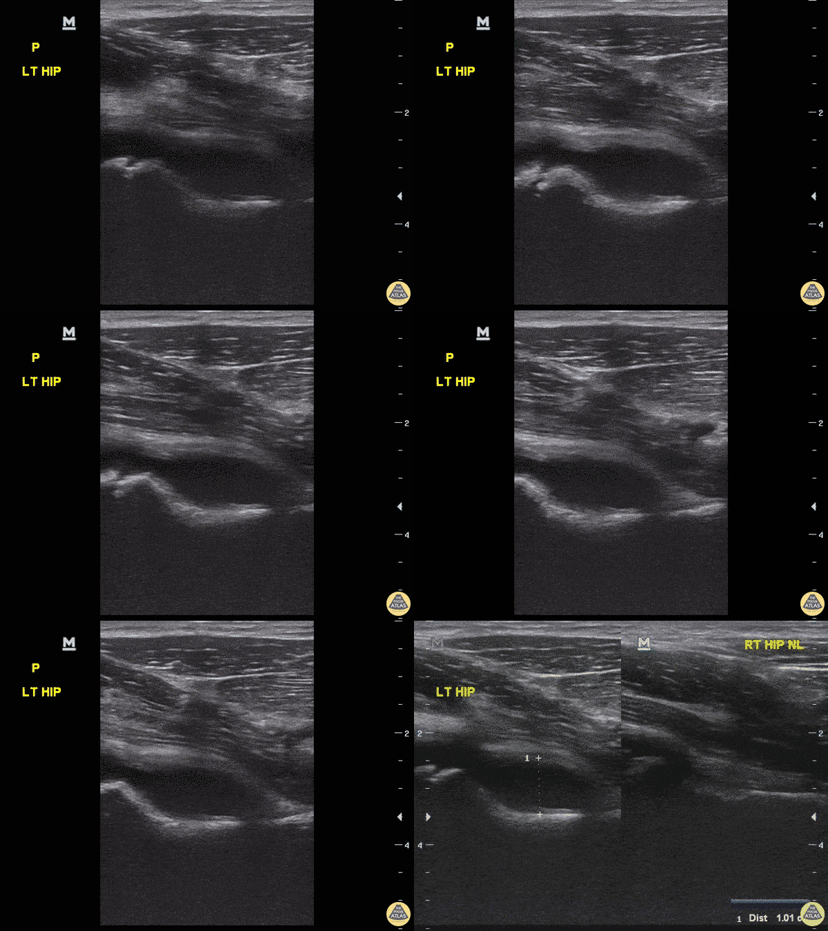
8-year-old female with fever and upper left thigh pain starting last night. Refusing to bear weight and will not flex hip, discomfort with rotating hip.
POCUS performed, revealed effusion. Still image comparing sides confirms effusion. Etiology of effusion remained uncertain.
Dr. Sathya Subramaniam, Pediatric EM Fellow - Kings County/SUNY Downstate
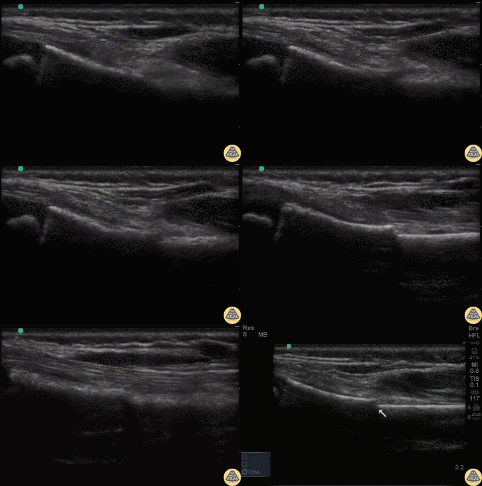
7 year old fallen off monkey bars. Tender over right distal radius with mild swelling.
POCUS reveals a discontinuity in the hyperechoic cortex of the child's distal radius with minimal displacement. This is suggestive of a buckle fracture or minimally displaced distal radius fracture.
Dr. Sathya Subramaniam, Pediatric EM Fellow - Kings County/SUNY Downstate
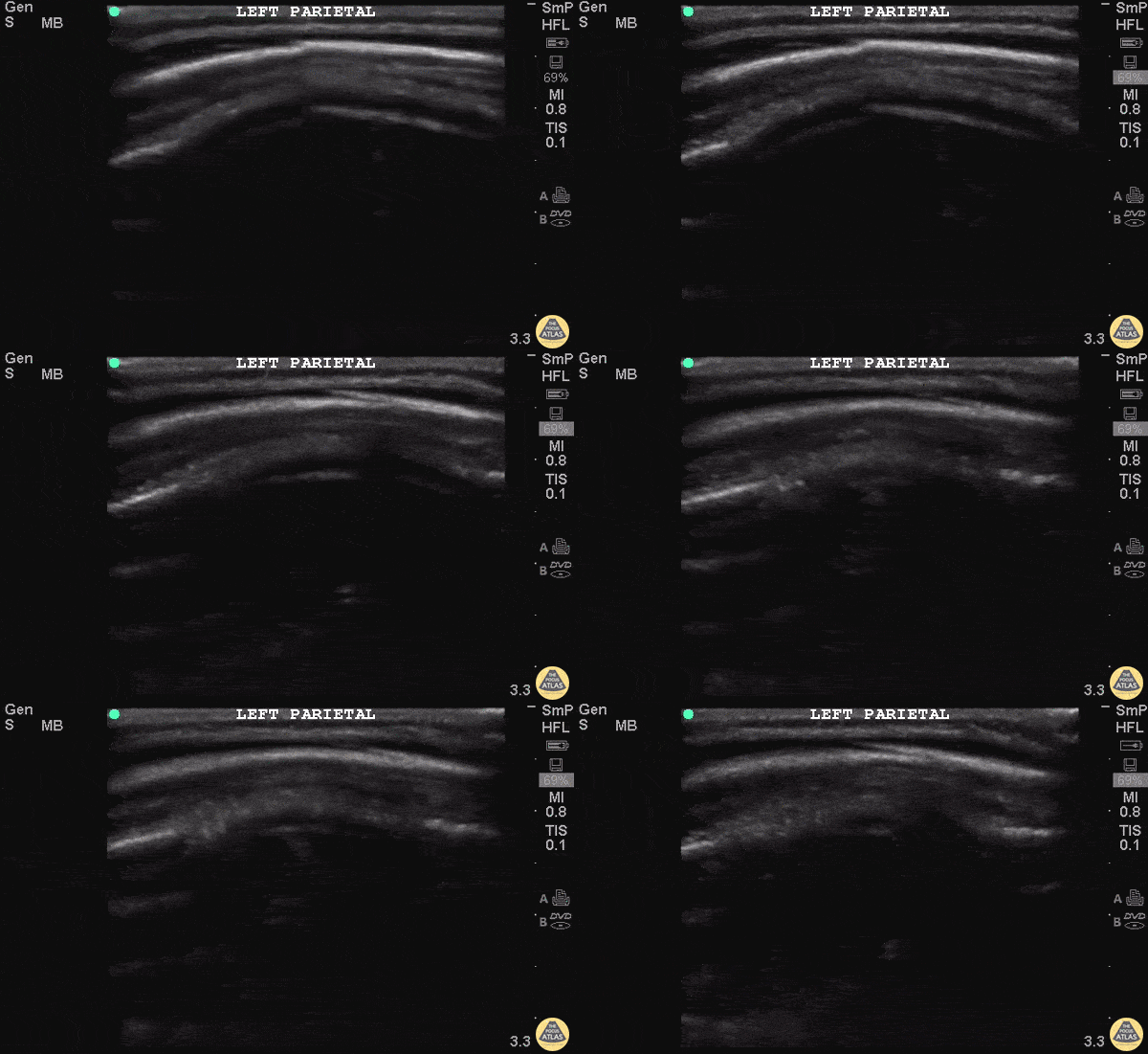
10 month old F presents one day after unwitnessed height level fall. Left parietal hematoma without step-off found on physical exam. Otherwise well appearing with normal vital signs. POCUS found a defect in cortex in area of the hematoma. CT head confirmed non-displaced skull fracture of the left parietal bone.
Patient was observed and did not require neurosurgical intervention. More research is needed into the value of POCUS for pediatrics skull fractures and how it can fit into our PECARN decision rules.
Dr Iain Jeffery - Brooklyn Hospital Emergency Medicine
Dr. Tian Liang and Dr Jeffery Rallo - Kings County Department of Pediatrics Emergency Medicine
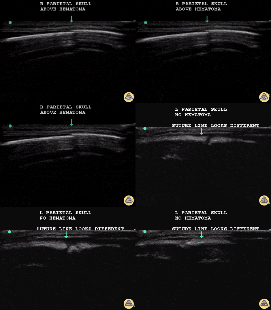
7 m.o fallen from a 4 foot high crib, unwitnessed. On exam small hematoma over right parietal skull, appears tender. POCUS completed to assess for skull fracture.
POCUS reveals a discontinuity in the hyperechoic cortex of the infant skull that is underneath the hematoma. This discontinuity is different from the image of a suture line within the same patient's skull.
Dr. Sathya Subramaniam, Pediatric EM Fellow - Kings County/SUNY Downstate
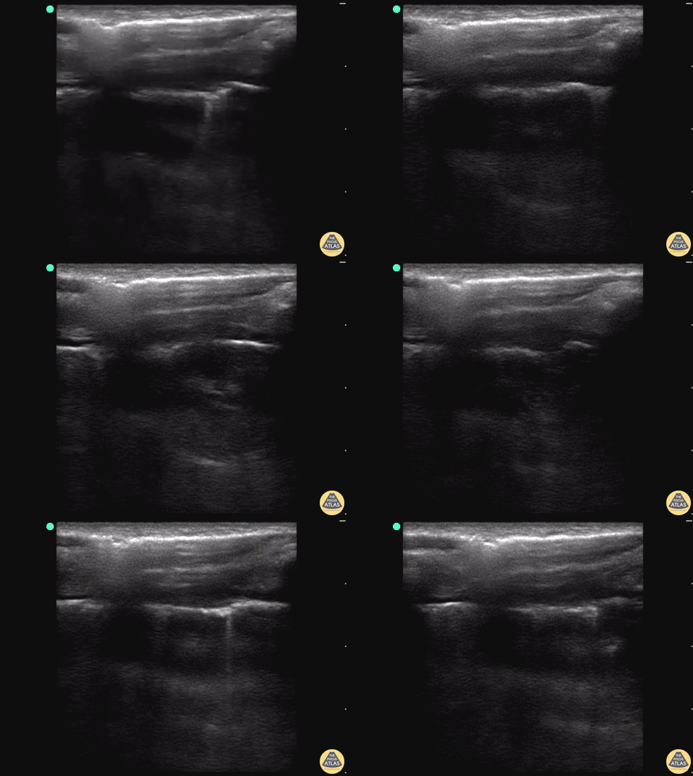
18 y/o M stabbed in the back presents to the trauma bay with left-sided chest pain and shortness of breath. E-FAST revealed decrease lung slide and a clear lung point.
While decreased lung slide is highly sensitive, it lacks specificity. Lung point indicates the transition point between normal pleura with normal lung sliding (on the left side of the image) and where there is air disrupting the pleural space with decreased lung sliding (on the right side of the image). Lung point is a highly specific finding indicating a pneumothorax.
Dr. Sathya Subramaniam, Pediatric EM Fellow - Kings County/SUNY Downstate
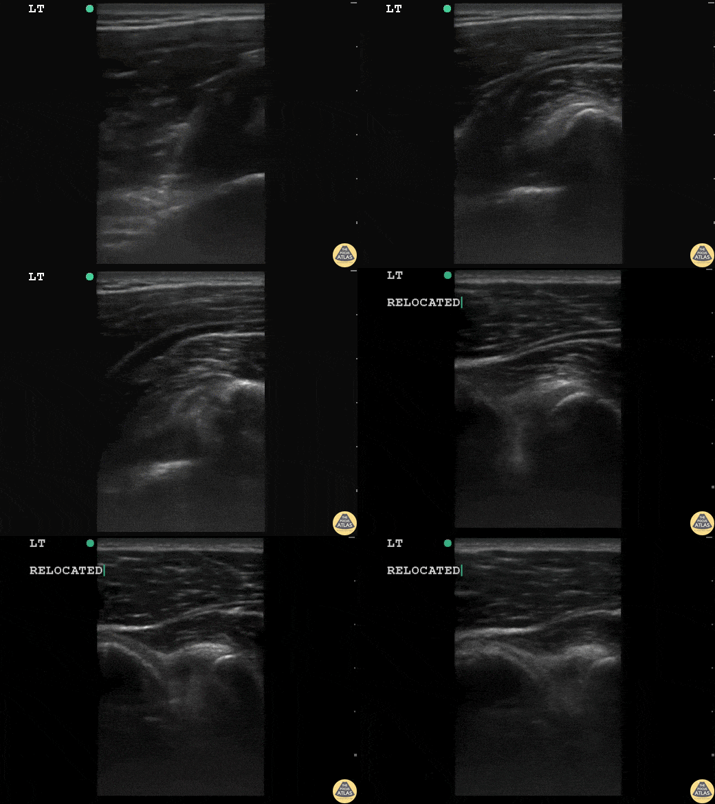
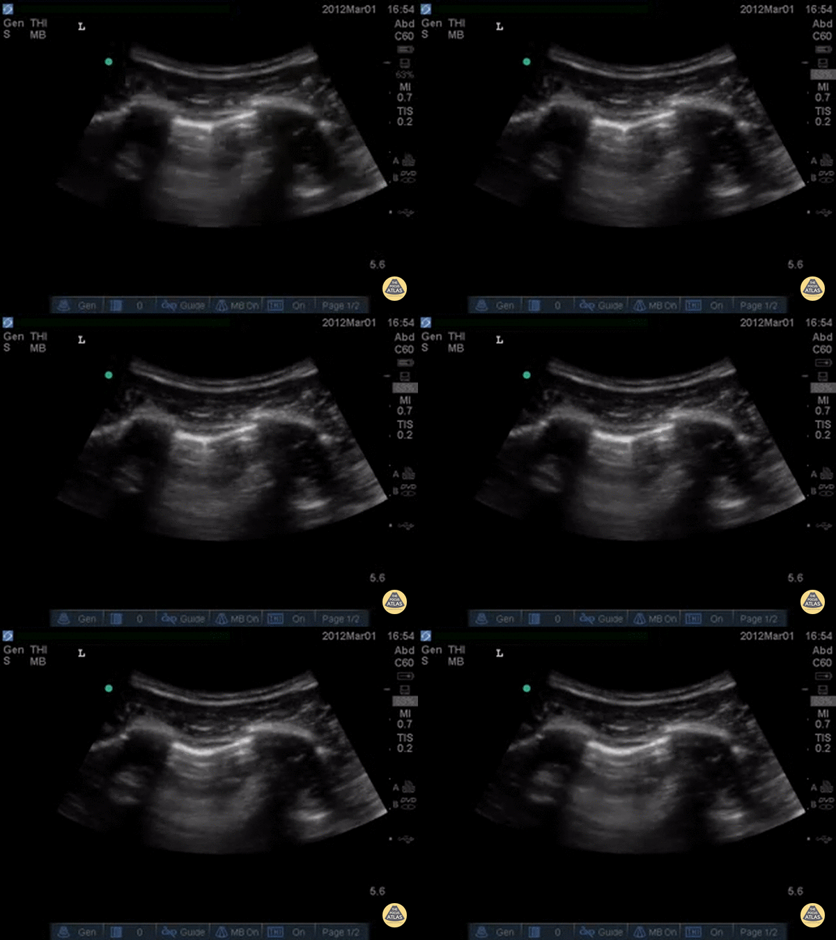
17 y/o basketball player with acute onset left shoulder pain after throwing a basketball across the court. "Feels like my arm is out of the socket." The patient was relocated with simple traction in less then 2 minutes. No x-rays were required.
The head of humerus is dislocated posterior to the glenoid. After relocation it is flush with glenoid as seen. You can appreciate the musculature and rotator cuff throughout both images.
Dr. Sathya Subramaniam, Pediatric EM Fellow - Kings County/SUNY Downstate
Tags
- adrenal
- adrenal hematoma
- adrenal hemorrhage
- air bubble
- ants marching
- artifact
- ascites
- Bladder
- blood
- blunt trauma
- cavity
- clot
- clotted
- comet tail
- Diaphragm
- extravasation
- fall
- fast
- FAST
- fat
- fb
- flank pain
- fluid
- foreign body
- free air
- gastric rupture
- gsw
- gunshot
- gunshot wound
- hematoma
- hemoperotineum
- hemorrhage
- hepatic
- hepatic cyst
- hepatic laceration
- heroperitoneum
- hydatid cyst
- hypotension
- injuries
- injury
- kidney
- laceration
- liver
- lung curtain
- LUQ pain
- metallic fb
- metallic foreign body
- Morrison's
- MVC
- Negative
- negative fast
- normal anatomy
- pain
- Pathology
- Pelvis
- perihepatic
- perinephric
- perisplenic
- peritoneum
- pleural effusion
- pneumoperitoneum
- POCUS
- positive FAST
- positive fast
- quadrant
- retroperitoneal hematoma
- retroperotineal
- reverberation
- right
- rupture
- ruq
- RUQ
- RUQ FAST
- Spleen
- spleen
- splenic
- splenic hematoma
- splenic laceration
- splenic rupture
- Trauma
- Trauma Pathology
- traumatic
- upper