
Choosing Wisely: Ultrasound For Suspected Nephrolithiasis

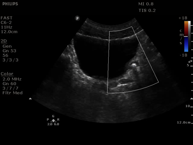

This is a 48 year old man that presented with first episode of severe left lower quadrant abdominal pain and vomiting. The differential diagnosis included diverticulitis, aortic aneurysm and nephrolithiasis. Point of care ultrasound (POCUS) was performed obtaining images of left and right kidneys, bladder and aorta. The images show left mild to moderate hydronephrosis and a 7 mm stone seen in 2 planes (with twinkle artifact). His aorta had normal measurements.
Renal POCUS is easy, safe and makes a big difference in this common emergency presentation. The last patient I saw with uncomplicated renal colic told me he’s had 22 CT (KUB) scans since his first kidney stone, lets not be part of this.
Maria Perez, Emergency Registrar; St Vincent’s Hospital; Melbourne - Australia
Learn More
Article: Ultrasonography versus computed tomography for suspected nephrolithiasis

Article: Sonographic diagnosis of symptomatic ureteral calculi: usefulness of the twinkling artifact

Video: Renal Ultrasound Part 1 Part 2
ACEP Recommendation: Choosing Wisely
