

Echo
Echo
One Minute Image Review
Normal Views

In this video, Dr. Elias Jaffa discusses how to properly obtain the subxiphoid view along with the pertinent visualized sonoanatomy.

In this video, Dr. Elias Jaffa discusses how to differentiate between the aorta and the IVC on ultrasound.
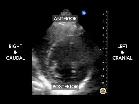
In this video, Elias Jaffa discusses imaging of the normal parasternal short axis view including probe orientation and sonoanatomy.
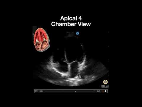
In this video, we discuss a normal apical 4 chamber view including features that suggest proper imaging as well as the utility of this view in point of care echocardiographic evaluation.
Pathology
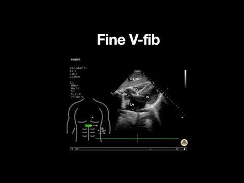
In this video, we discuss a cardiac arrest case demonstrating fine ventricular fibrillation initially thought to be asystole based on rhythm evaluation!
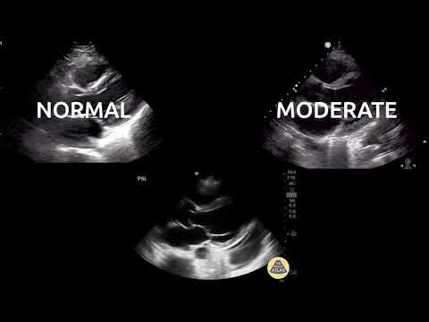
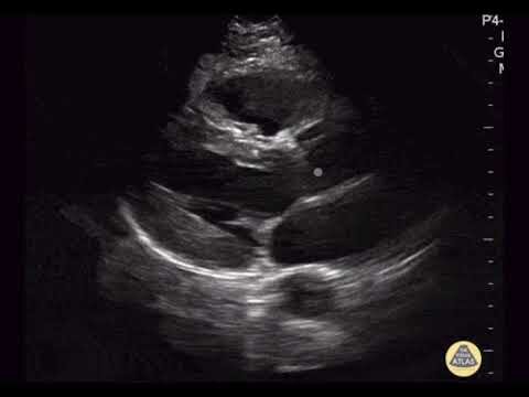
In this video Dr. Elias Jaffa discusses specific characteristics of the left ventricle that can be utilized when evaluating left ventricular function on the parasternal long axis view (PSLA).
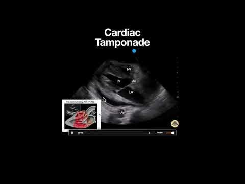
In this video we discuss findings of a pericardial effusion with evidence of cardiac tamponade in a parasternal long axis view. Specifically we discuss the finding of right ventricular diastolic collapse.
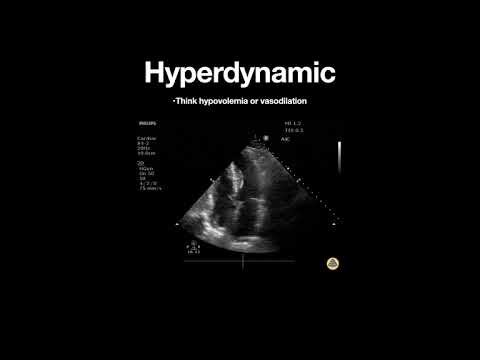
In this video we discuss findings in a hyperdynamic left ventricle and its clinical implications.
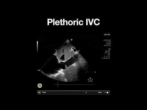
In this video, we discuss a normal apical 4 chamber view including features that suggest proper imaging as well as the utility of this view in point of care echocardiographic evaluation.
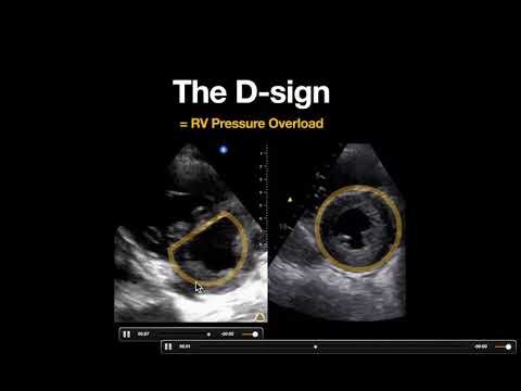
In this clip we discuss the important ultrasound finding known the D-Sign. When the D sign is present throughout systole and diastole, this indicated RV pressure overload which may be from etiologies including pulmonary embolism or pulmonary hypertension.
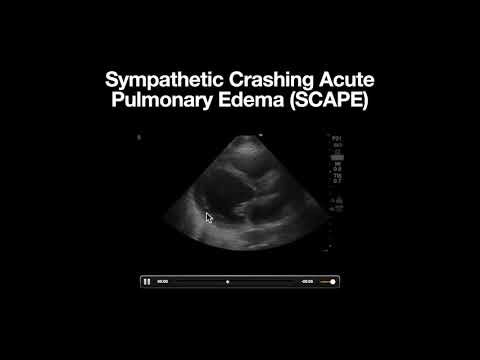
In this video we discuss ultrasound findings seen in a patient with SCAPE. In general these patients are very sick from acute redistribution of fluid into their lungs and the treatment should focus on NIPPV and high dose nitroglycerine.
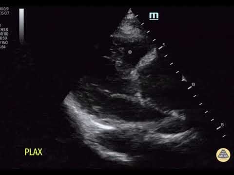
In this image we discuss an enlarged aortic root seen on parasternal long axis in the setting of acute aortic dissection. The aortic root is normally < 4cm and any dilation in the setting of acute chest symptoms should raise suspicion for Type A aortic dissection and further imaging and specialist consultation should be considered.
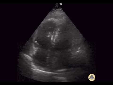
In this clip we discuss several signs seen in pulmonary embolism with right ventricular strain including: RV dilation, McConnell’s sign and thrombus (clot)-in-transit.