
Cholecystitis
Hepatobiliary - Cholecystitis
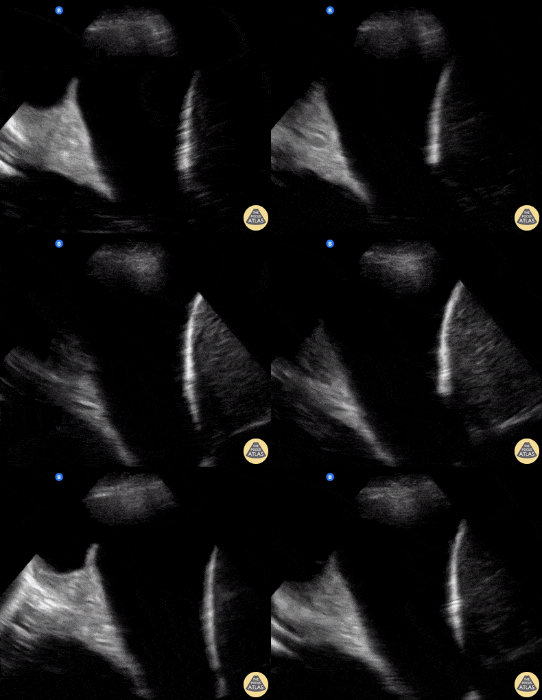
![Thick Walled Gallbladder in Dengue Fever (Short Axis) [2/2]](https://images.squarespace-cdn.com/content/v1/58118909e3df282037abfad7/1728883282342-87F9ZQFLQ314C6WAFAAC/image-asset.gif)
A 28 year old female comes to the ER with fever, myalgia, hypotension, and abdominal pain.
This case illustrates one of the ultrasound findings in Dengue Fever in the Leakage Syndrome Stage. We can see a large, thick-walled gallbladder and the presence of pericholecystic fluid. This clip demonstrates the gallbladder in short-axis view.
Contributor: Renato Tambelli (@R_Tambelli @JediPocus)
![Thick Walled Gallbladder in Dengue Fever (Long Axis) [1/2]](https://images.squarespace-cdn.com/content/v1/58118909e3df282037abfad7/1728882762911-WS6YZE9IZ5WVP4XD8BX1/image-asset.gif)
A 28 year old female comes to the ER with fever, myalgia, hypotension, and abdominal pain.
This case illustrates one of the ultrasound findings in Dengue Fever in the Leakage Syndrome Stage. We can see a large, thick-walled gallbladder and the presence of pericholecystic fluid. This clip demonstrates the gallbladder in long-axis view.
Contributor: Renato Tambelli (@R_Tambelli @JediPocus)
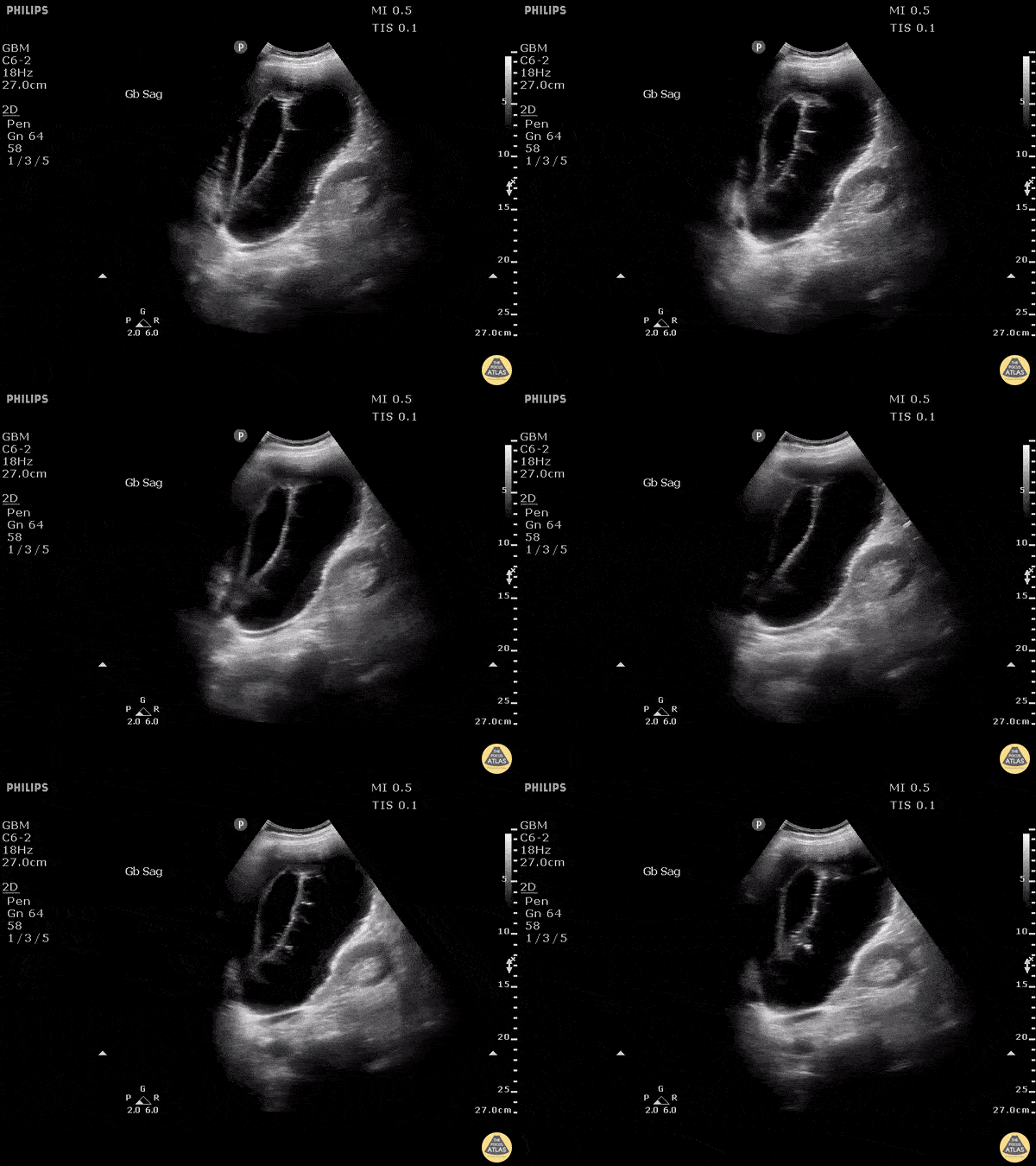
Stone trapped in cystic duct resulted in gallbladder distention as well as sloughing of intraluminal membranes.
Image courtesy of Robert Jones DO, FACEP @RJonesSonoEM
Director, Emergency Ultrasound; MetroHealth Medical Center; Professor, Case Western Reserve Medical School, Cleveland, OH
View his original post here
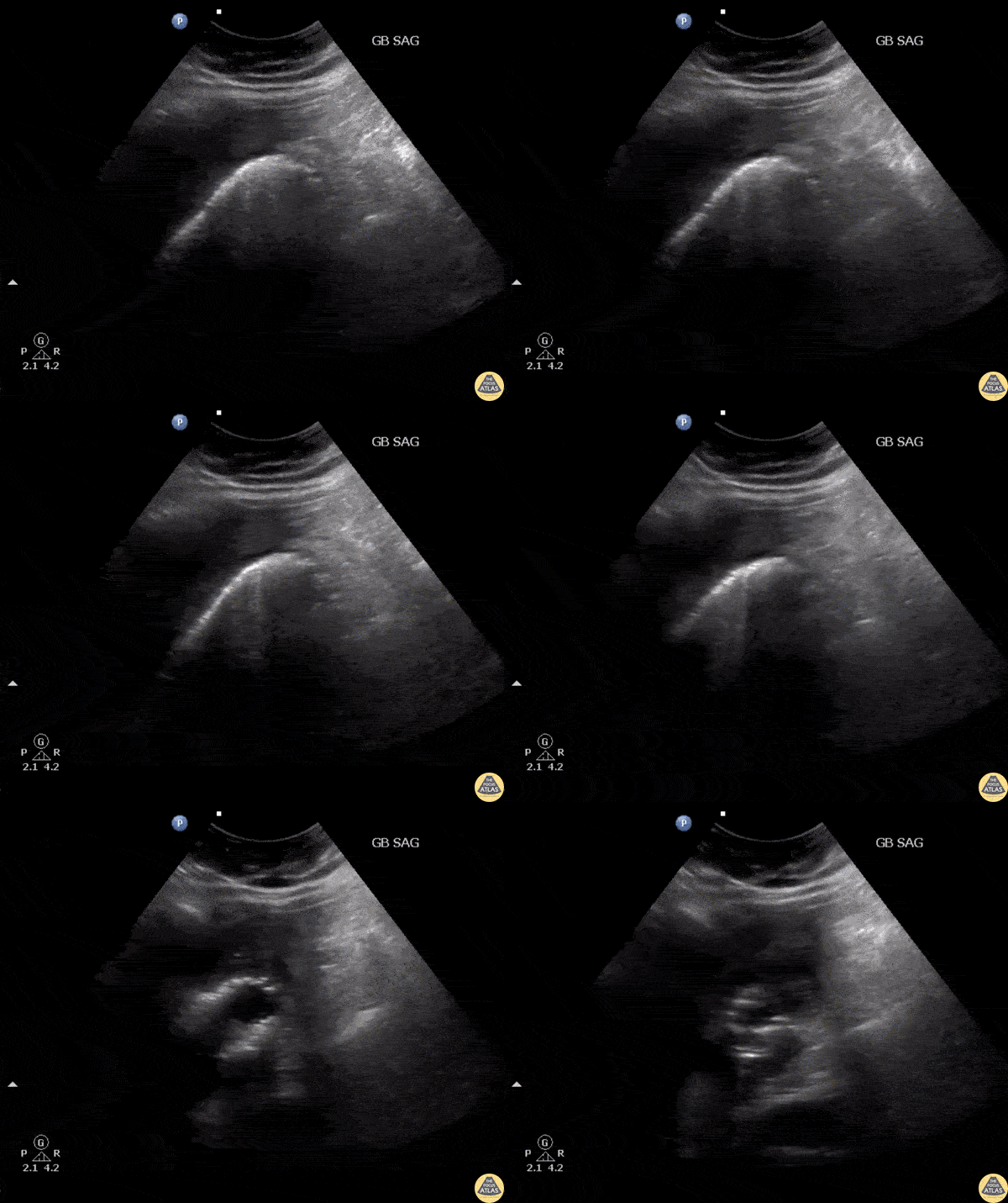
RUQ US of a patient revealed air within the gallbladder consistent with emphysematous cholecystitis, which can easily be confused with bowel gas.
Image courtesy of Robert Jones DO, FACEP @RJonesSonoEM
Director, Emergency Ultrasound; MetroHealth Medical Center; Professor, Case Western Reserve Medical School, Cleveland, OH
View his original post here
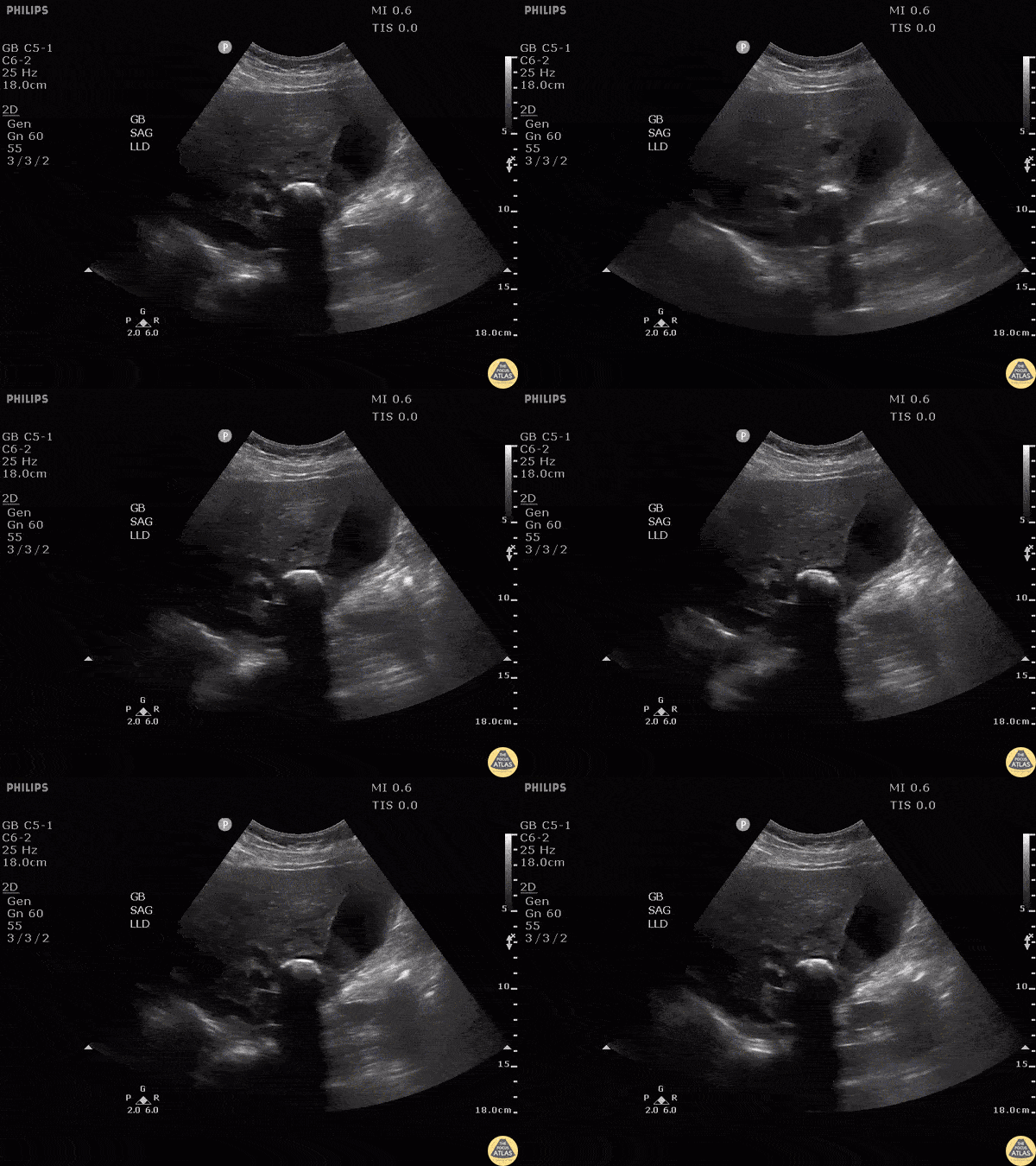
Note the hyperechoic structure with posterior acoustic shadowing indicative of a gallstone. Patient was moved and the stone remained lodged in the neck. Additionally, gallbladder sludge can be seen. The patient was found to have elevated LFTs. MRCP performed demonstrating external compression of biliary tree this large stone in GB neck consistent with Mirizzi’s syndrome.
Image courtesy of Robert Jones DO, FACEP @RJonesSonoEM
Director, Emergency Ultrasound; MetroHealth Medical Center; Professor, Case Western Reserve Medical School, Cleveland, OH
View his original post here
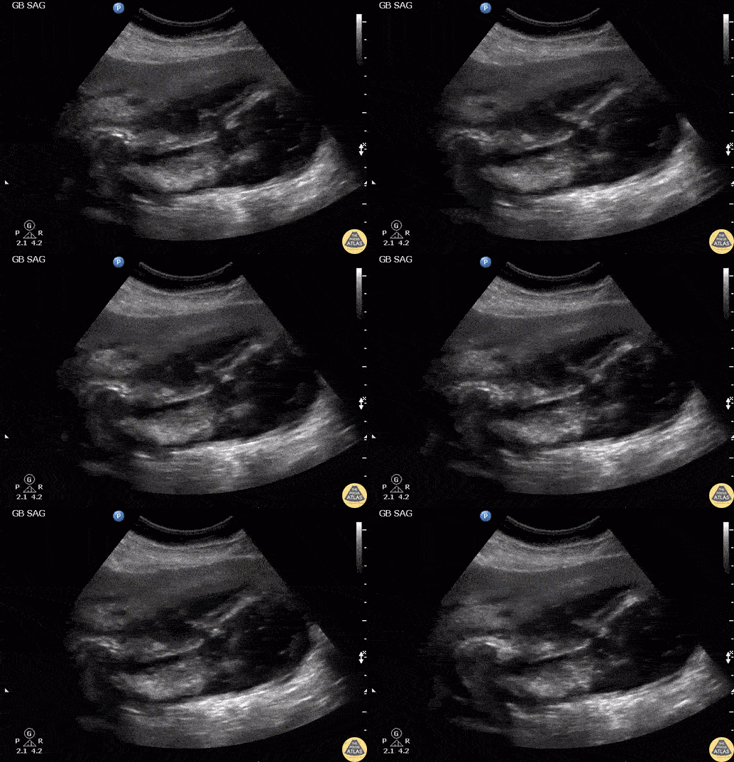
Patient presented with right upper quadrant abdominal pain and fever. RUQ ultrasound showed an impacted stone in the gallbladder neck with gallbladder wall perforation. Note the flow through the perforation and the associated pericholecystic abscess. Labelled image can be found on the original post with the link below.
Image courtesy of Robert Jones DO, FACEP @RJonesSonoEM
Director, Emergency Ultrasound; MetroHealth Medical Center; Professor, Case Western Reserve Medical School, Cleveland, OH
View his original post here
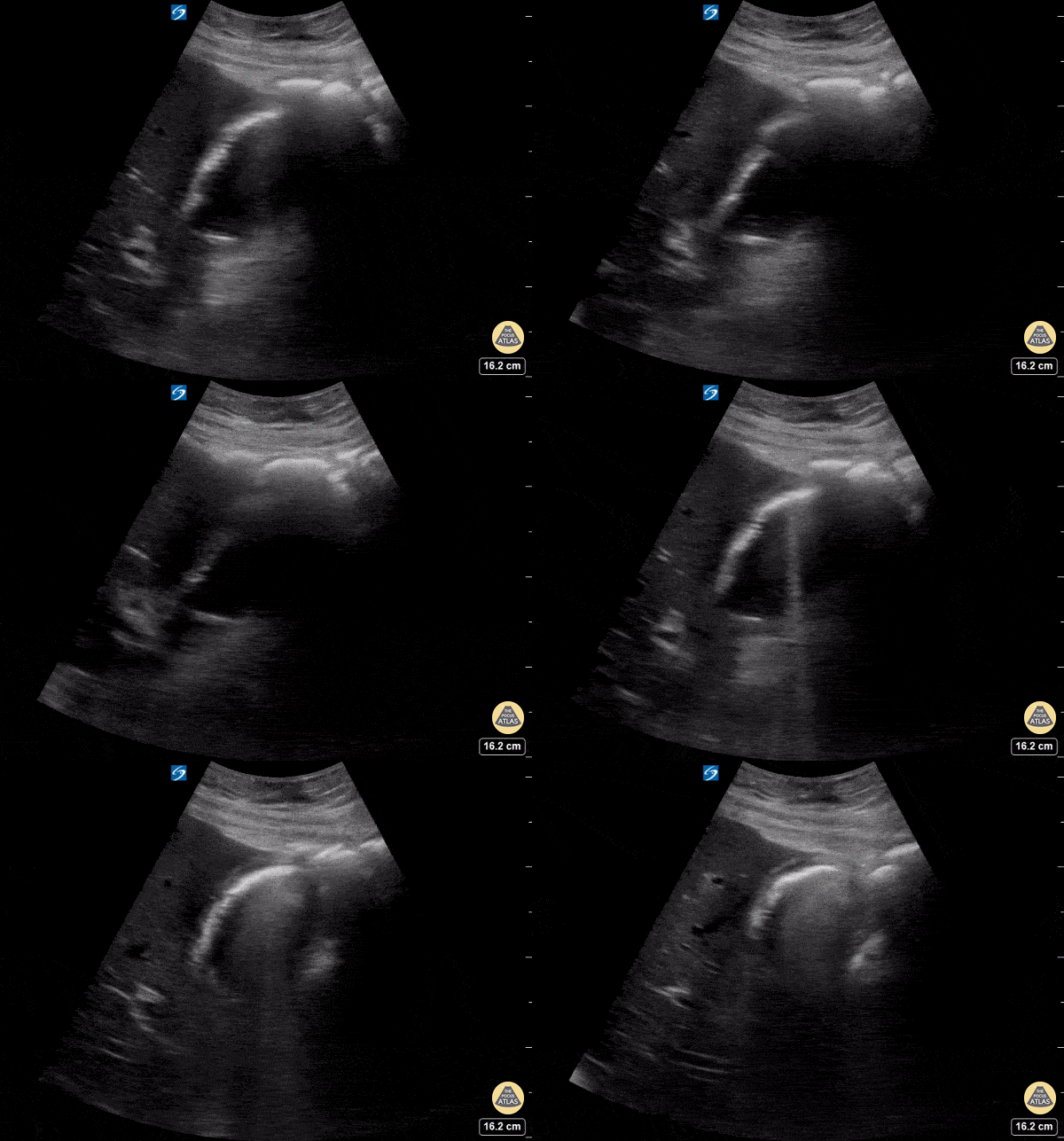
A patient presented with altered mental status and anorexia; clinically he had findings consistent with sepsis. POCUS revealed one angry gallbladder! You can appreciate gallbladder wall thickening (measuring 6mm), trace pericholecystic fluid, and shadowing from gallbladder sludge.
Garrett Ghent, Resuscitationst and diagnostician; Norfolk, VA
@garrettghentMD
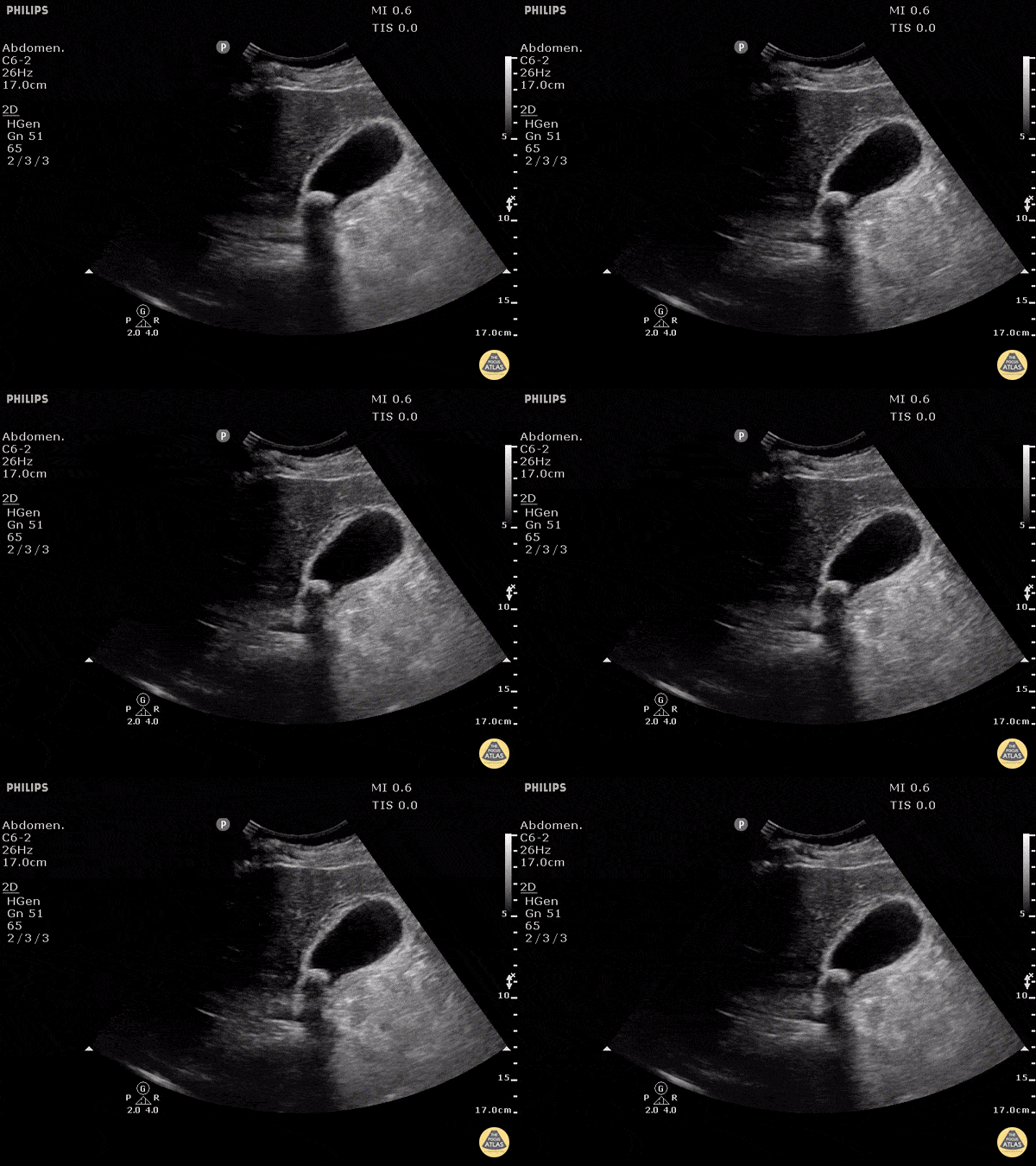
This clearly demonstrates a stone in the gallbladder neck as a hyperechoic structure with posterior shadowing. The neck is not always clearly visualized on first glance so it is important to scan through the gallbladder in two planes to exclude stones in the neck. Other signs of acute cholecystitis include pericholecystic fluid, gallbladder wall thickening (>3mm) common bile duct dilatation, and positive sonographic Murphy's sign.
Justin Bowra MBBS, FACEM, CCPU Emergency Physician, RNSH et al. (Mo)
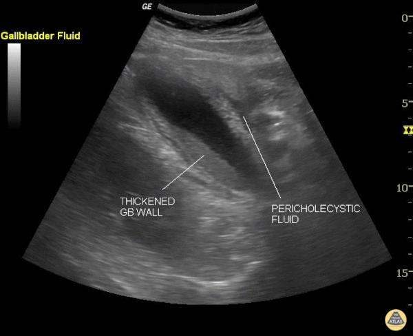
This still image shows two clear signs of acute cholecystitis. The gallbladder wall is thickened and there is pericholecystic fluid. Of note, when measuring the gallbladder wall it's better to measure the anterior wall as the posterior wall can appear enlarged due to artifact. The cut off for wall thickening is 3mm. The anterior wall is thickened in this case but is not labeled. Other signs to look for are stones, sludge, common bile duct dilatation, and overall gallbladder enlargement.
Of note, a positive sonographic Murphy's carries a sensitivity of 63%, specificity of 93.6% and positive predictive value of 72.5%.
Justin Bowra MBBS, FACEM, CCPU Emergency Physician, RNSH et al. (Sharon)
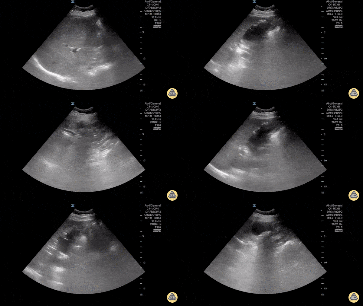
Emphysematous cholecystitis is the result of gas formation within the wall or lumen of the gallbladder as a result of infection by a gas forming organism. It is a surgical emergency. The air in the gallbladder appears highly echogenic with mild posterior shadowing, as air next to tissue is highly reflective.
Sukh Singh, MD
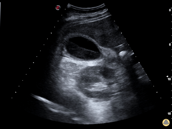
The image above is from a patient presenting to the emergency department with recurrent abdominal pain. The image demonstrates a large amount of biliary sludge within a normal sized gallbladder. Biliary colic from intermittent obstruction was suspected. While the gallbladder wall is noted to be mildly thick here, cholecystitis was ruled out based on other clinical factors.
Marco Garrone, MD - Torino, Italy
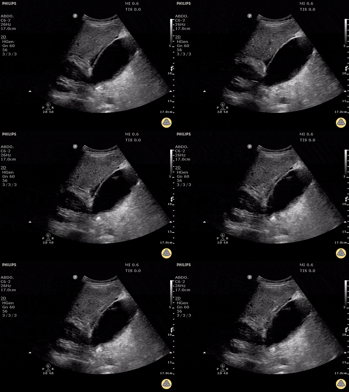
Free fluid can be appreciated around this gallbladder. This is called pericholecystic fluid. However there are no stones, nor gallbladder wall thickening, and the sonographic Murphy's is negative. This free fluid is unlikely to be caused by cholecystitis. In this case, the free fluid came from a different source.
Justin Bowra MBBS, FACEM, CCPU Emergency Physician, RNSH et al.