
Cholelithiasis
Hepatobiliary - Cholelithiasis
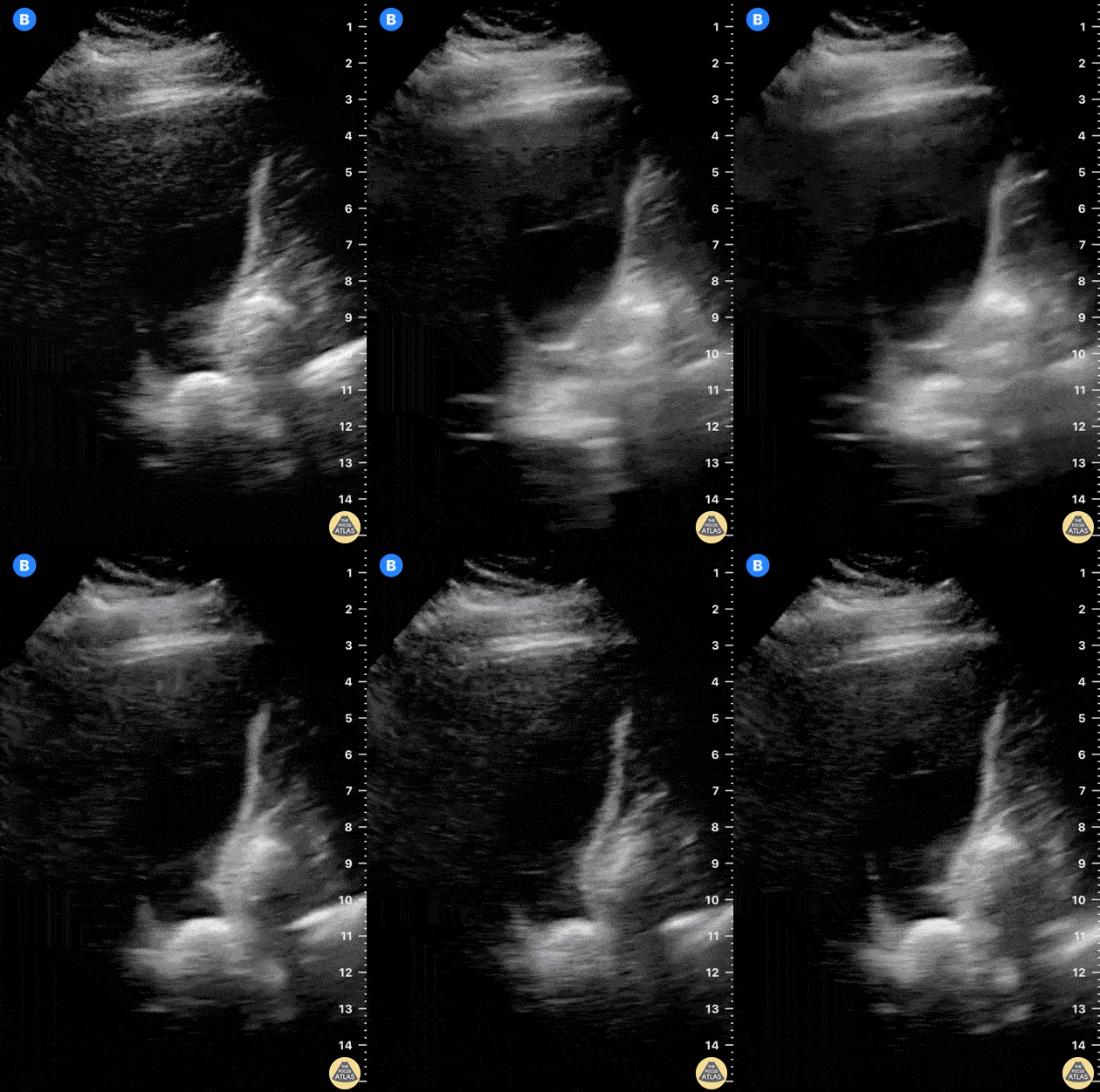
It’s fundamental to study the gallbladder in its whole extension. In this image, there is an impacted stone at the neck.
Dr. Felipe Urriola P.
www.thepocus.com
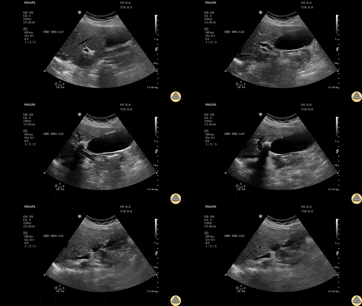
Longitudinal view of the gallbladder revealing two unique findings. A hyperechoic structure with posterior acoustic shadowing indicative of a stone is located in the neck. Two smaller hyperechoic structures can be seen attached to the wall of the GB body, likely adenomyomatosis.
Image courtesy of Robert Jones DO, FACEP @RJonesSonoEM
Director, Emergency Ultrasound; MetroHealth Medical Center; Professor, Case Western Reserve Medical School, Cleveland, OH
View his original post here
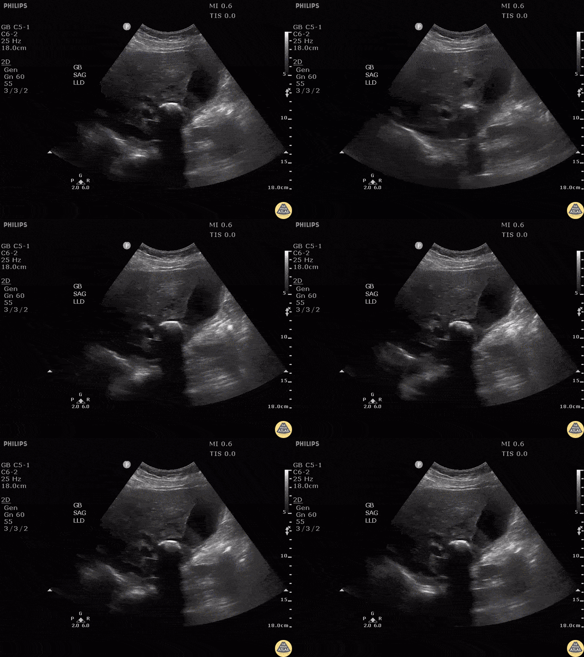
Note the hyperechoic structure with posterior acoustic shadowing indicative of a gallstone. Patient was moved and the stone remained lodged in the neck. Additionally, gallbladder sludge can be seen. The patient was found to have elevated LFTs. MRCP performed demonstrating external compression of biliary tree this large stone in GB neck consistent with Mirizzi’s syndrome.
Image courtesy of Robert Jones DO, FACEP @RJonesSonoEM
Director, Emergency Ultrasound; MetroHealth Medical Center; Professor, Case Western Reserve Medical School, Cleveland, OH
View his original post here
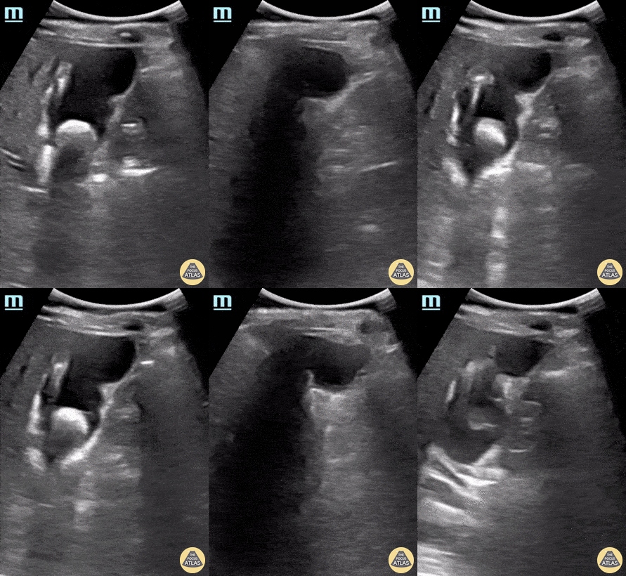
An elderly female presented to the emergency department reporting abdominal pain. POCUS seen here revealed the presence of a gallstone within her gallbladder with shadowing; and a notable absence of findings consistent with cholecystitis including gallbladder sludge, wall thickening, and/or ductal dilatation. This enabled appropriate triage of this patient to outpatient follow-up rather than considering immediate surgical intervention.
Rupinder Sekhon, MD & Peter Biggane, MD
Central Michigan University, Emergency Medicine
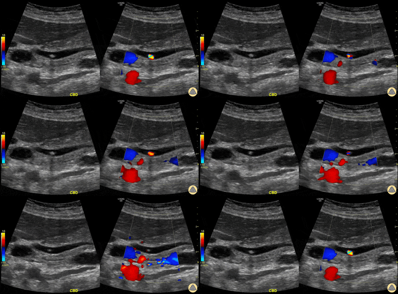
A patient presented with RUQ abdominal pain.
Ultrasound showed cholelithiasis and choledocholithiasis. Left image shows a portion of the common bile duct containing a small hyperechoic shadowing mobile stone. Right image shows the same with color Doppler added demonstrating twinkling artifact.
Learning point: Twinkling artifact is most often associated with urinary stones but also can be caused by gallstones (depending on composition), bowel gas, foley balloons, metallic objects, and many others. Twinkling artifact may help identify small stones that do not cause prominent acoustic shadowing.
Michael Cover, MD
@michaelc0ver
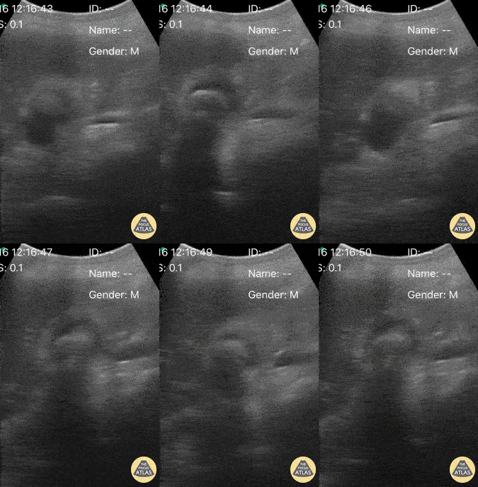
A 33-year-old female presented to the ED reporting acute onset abdominal pain immediately after eating a meal. She was well-appearing and afebrile; physical exam notable for reproducible tenderness to palpation of her upper abdomen. POCUS long axis view obtained to right of epigastrium notable for hyperechoic material within the lumen of the gallbladder (anechoic) producing posterior shadowing. This confirmed our clinical suspicion of biliary colic, and the patient was able to be discharged with scheduled outpatient elective surgery.
Victor Bang, Medical Student Brazil
@vmjbang
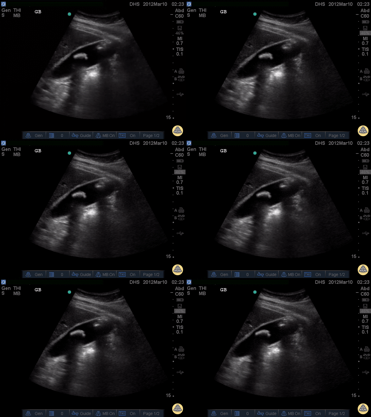
Two large, hyperechoic stones can be appreciated in this gallbladder. These are likely two stones because of the degree of echogenicity and posterior shadowing. If you have one or two objects in the gallbladder and you're not sure if they're stones, it can be useful to have the patient lay on their side. If the objects moves when the patient moves, they are stones. If not, they are likely polyps or other masses growing from the gallbladder wall.
Justin Bowra MBBS, FACEM, CCPU Emergency Physician, RNSH et al. (Dr. Darmas)
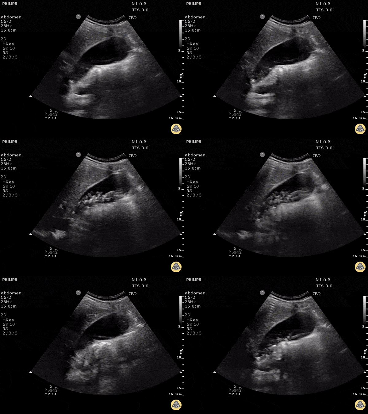
This non-inflamed gallbladder contains many stones. The stones are hyperechoic with posterior shadowing. The gallbladder wall is not thickened, there is no pericholecystic fluid, and no sonographic Murphy's sign, thus cholecystitis is unlikely.
Justin Bowra MBBS, FACEM, CCPU Emergency Physician, RNSH et al. (Dr. Ken Lee)
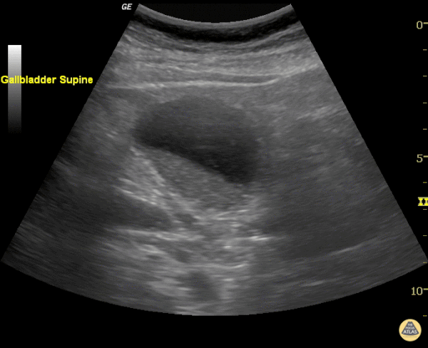
Echogenic material can be seen within this gallbladder but there are no stones. It is not bright white (it is not highly echogenic) and furthermore there isn't any shadowing. When there is no shadowing consider polyps or in this case biliary sludge.
Justin Bowra MBBS, FACEM, CCPU Emergency Physician, RNSH et al. (Sharon)
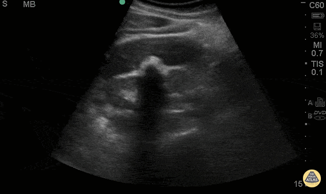
38 y/o F presented with abdominal pain, nausea & vomiting. POCUS shows the WES (Wall-Echo-Shadow) sign. This shows a curvelinear hyperechoic line representing the gallbladder wall, followed by a thin hypoechoic line representing a small amount of bile, then a curvelinear hyperechoic line, followed by acoustic shadowing. This sign is seen when the gallbladder has either a large gallstone or multiple small gallstones in a contracted gallbladder taking up the extent of the lumen. Sometimes this sign is misinterpreted as an air filled loop of adjacent bowel.
Jaramillo, Juliana MD; Shah, Rushabh MD; Maurelus, Kelly MD - Kings County Emergency Medicine
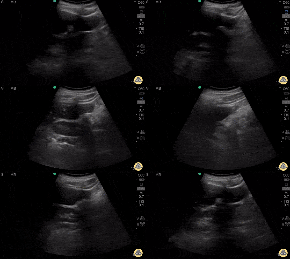
38 y/o F presented with abdominal pain, nausea & vomiting. POCUS shows the WES (Wall-Echo-Shadow) sign. This shows a curvelinear hyperechoic line representing the gallbladder wall, followed by a thin hypoechoic line representing a small amount of bile, then a curvelinear hyperechoic line, followed by acoustic shadowing. This sign is seen when the gallbladder has either a large gallstone or multiple small gallstones in a contracted gallbladder taking up the extent of the lumen. Sometimes this sign is misinterpreted as an air filled loop of adjacent bowel.
Jaramillo, Juliana MD; Shah, Rushabh MD; Maurelus, Kelly MD - Kings County Emergency Medicine