
Common Bile Duct Dilation
Hepatobiliary - Common Bile Duct
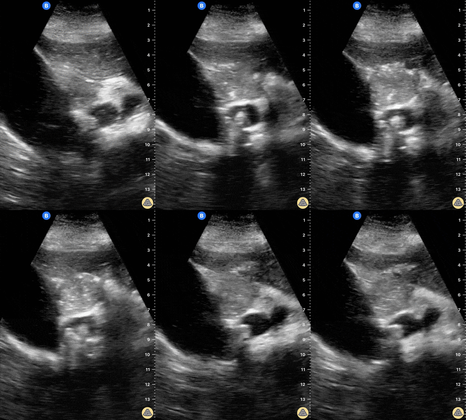
A 65-year-old female with no PMH presented to ED with 6 hour hx RUQ abdominal pain. She reported no associated vomiting or fever.
POCUS seen here reveals the cystic duct emerging from the GB neck to then reach a severely dilated CBD. A hyperechoic round structure with posterior shadowing lies inside the CBD, consistent with a diagnosis of choledocholithiasis.
Dr. Felipe Urriola
Emergency Unit. Puerto Aysen Hospital, Chilean Patagonia.
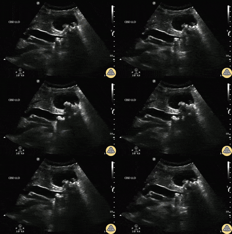
A patient presented to the ED with RUQ pain. Sagittal view of the common bile duct revealed dilation and stones indicative of choledocolithiasis.
Image courtesy of Robert Jones DO, FACEP @RJonesSonoEM
Director, Emergency Ultrasound; MetroHealth Medical Center; Professor, Case Western Reserve Medical School, Cleveland, OH
View his original post here
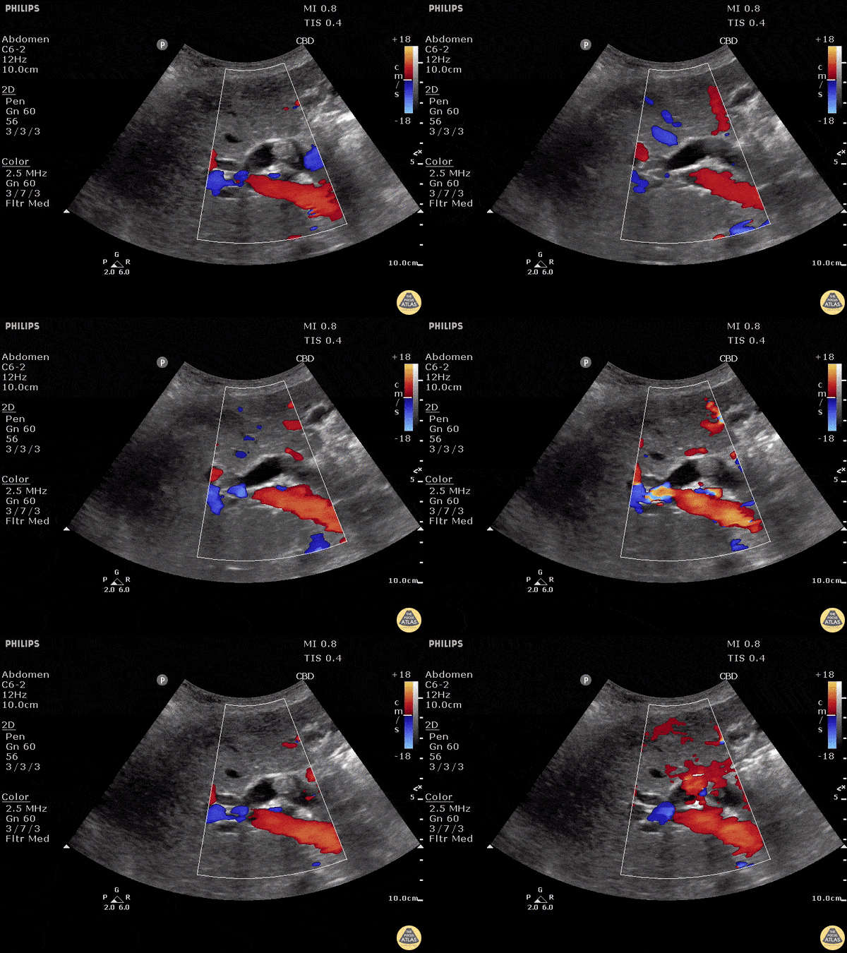
A normal common bile duct should be < 4mm plus 1mm per decade after 40 years of age. A stone can clearly be seen in the area of color flow doppler. The bile duct is obviously dilated here (although not measured).
When searching for the CBD, it can be useful to turn color flow doppler on to ensure you're visualizing a biliary structure and not vessels. This technique was employed here.
POCUS is highly sensitive for acute cholecystitis but as many people already know, it can be hard to find the CBD, which is reflected in the lower sensitivity and specificity for this indication in many studies.
Justin Bowra MBBS, FACEM, CCPU Emergency Physician, RNSH et al. (Dr. Dop Ahilan)
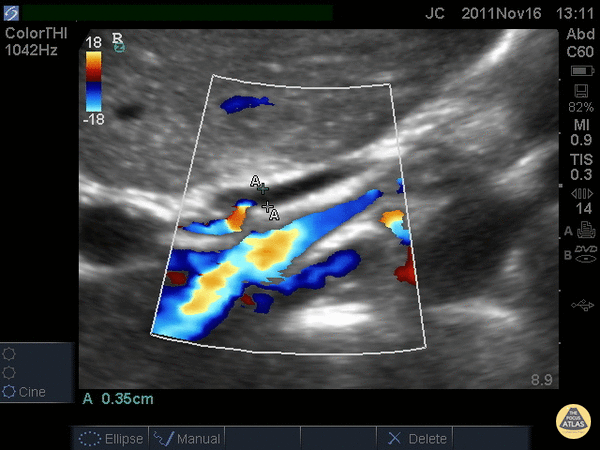
When searching for the common bile duct (CBD), color doppler can be useful to differentiate vascular structures from non-vascular structures. In this case the CBD is being measured anteriorly and does not have any pulsations on color doppler. The portal vein and artery can be seen and with the CBD, make the portal triad.
A normal CBD should be less than 4mm plus 1mm per decade after 40 years old. It should be measured inner wall to inner wall.
Justin Bowra MBBS, FACEM, CCPU Emergency Physician, RNSH et al. (Dr. Chiang)
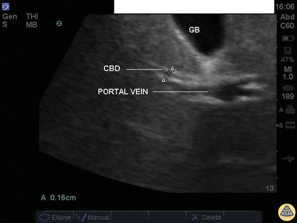
A normal common bile duct should be <4mm plus 1mm per decade after 40 years of age. It should be measured from inner wall to inner wall.
You should attempt to find the portal triad: the CBD, the portal vein, and the hepatic artery. You may need color flow to find all three. Here the CBD and portal vein are labeled (no labeled hepatic artery).
To get better images, especially if you're dealing with a lot of rib shadow, it can be helpful to have your patient take a deep breath and/or roll onto their left side.
Justin Bowra MBBS, FACEM, CCPU Emergency Physician, RNSH et al.
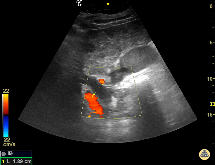
The CBD measures at 1.89cm indicating it is grossly dilated, as a normal common bile duct should be <4mm plus 1mm per decade after 40 years of age. It should be measured from inner wall to inner wall. The color doppler is used to ensure that the dilated structure is indeed a bile duct rather than a vascular abnormality.
Sukh Singh, MD
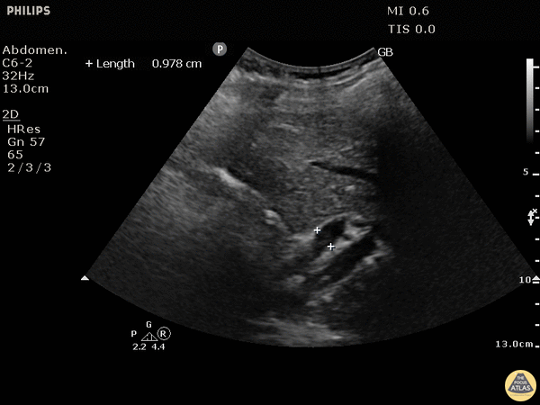
This patient has a dilated common bile duct and obstructive pattern LFTs, highly suggestive of choledocholithiasis or other biliary obstruction. No stone is visualized on this study.
A normal common bile duct should be <4mm plus 1mm per decade after 40 years of age. Here it is 9.78 mm.
POCUS is highly sensitive for acute cholecystitis but as many people already know, it can be hard to find the CBD, which is reflected in the lower sensitivity and specificity for this indication in many studies.
Justin Bowra MBBS, FACEM, CCPU Emergency Physician, RNSH et al. (Dr. Ken Lee)
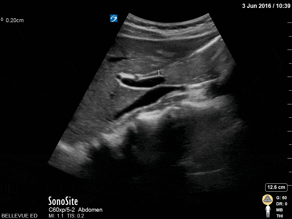
The “olive sandwich” sign: the small round hepatic artery (in cross section) sandwiched between long axis views of the CBD superiorly and portal vein inferiorly. The IVC is seen in long axis deep to the portal vein.
Hannah Kopinksi and Dr. Lindsay Davis - NYU Emergency Medicine