Biliary Evidence
POCUS Evidence Atlas - Biliary, Gallbladder, Cholecystitis. EBM review and resource of POCUS for biliary diagnosis. Sensitivity Specificity positive and negative likelihood ratios. Point of care ultrasound evidence.
The Evidence Atlas: Hepatobiliary
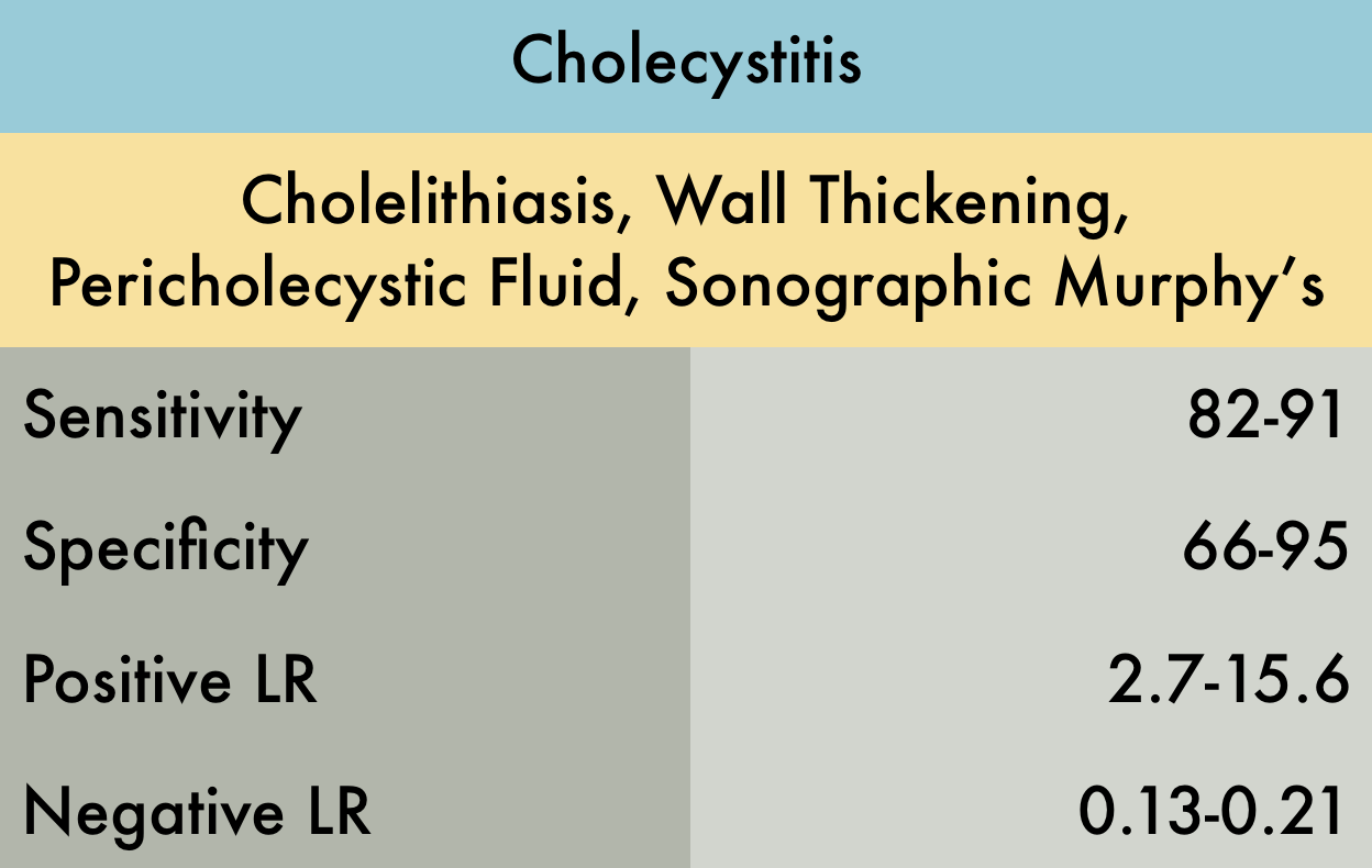
This was a systematic review including 4 prospective studies evaluating the operating characteristics of bedside ultrasound for acute cholecystitis (AC) in adult patients seen in the emergency department with a clinical suspicion for AC or right upper quadrant pain. Sample size of the studies varied from 30 to 193 subjects. Reference standard was surgical pathology. The experience of the sonographers varied between the studies and in one study no documentation of sonographer experience was noted. There was significant heterogeneity across the included studies precluding the authors ability to pool the results hence a range is noted in the operating characteristics table.
PMID: 27862628
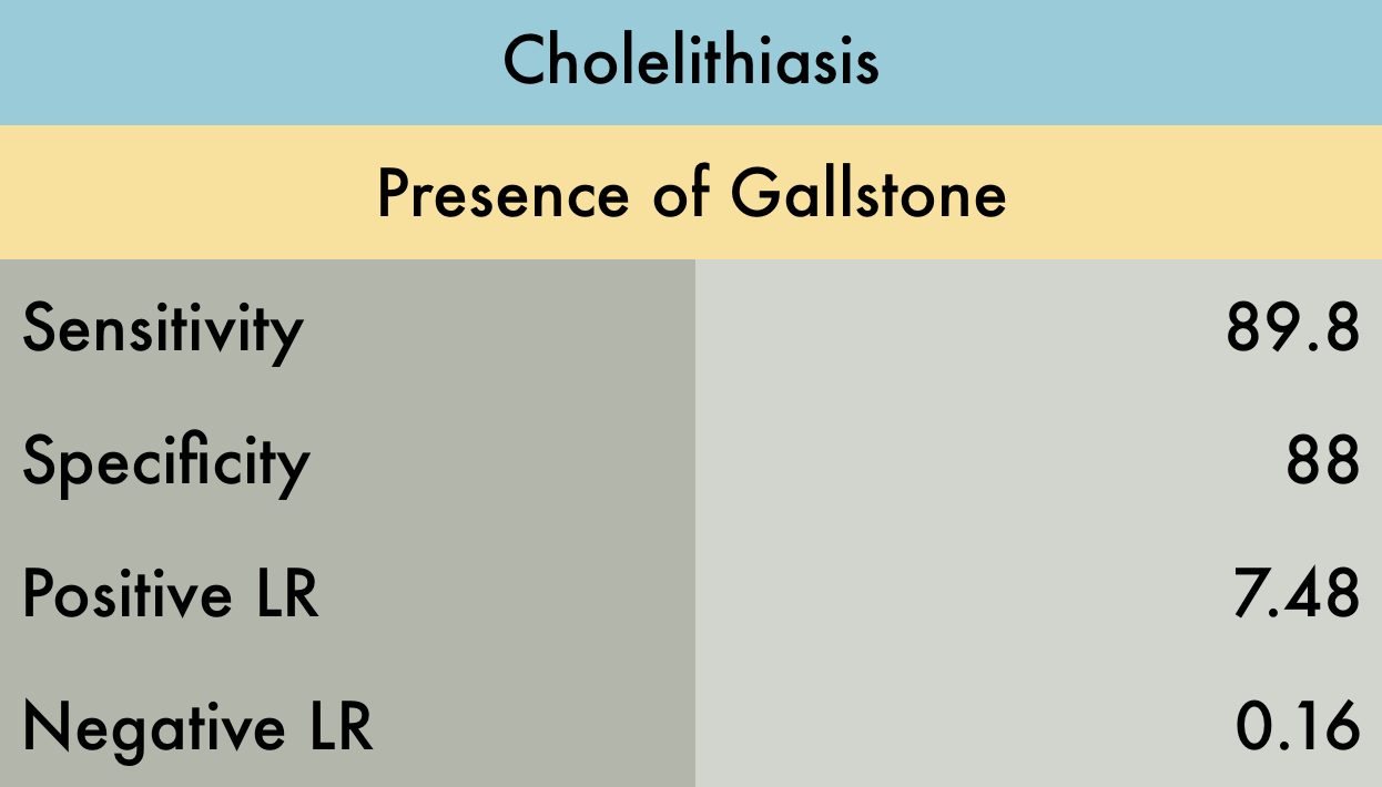
This was a meta-analysis including 8 studies (n=710) evaluating the operating characteristics of emergency ultrasound (EUS) for identifying cholelithiasis in adult patients presenting to the emergency department with symptoms suggestive of biliary colic (RUQ pain, epigastric pain, or right flank pain). Reference standards included radiology performed ultrasound, CT, MRI, and/or surgical pathology. There was quite a variation in the technical ability between operators which may distort the pooled sensitivity and specificity.
PMID: 21401784
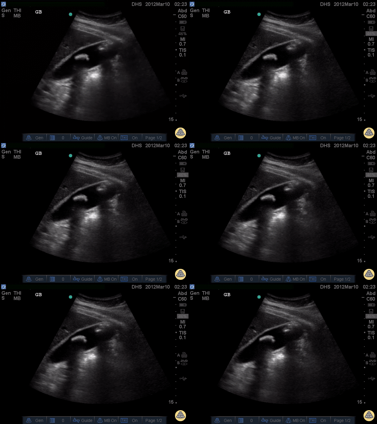
Two large, hyperechoic stones can be appreciated in this gallbladder. These are likely two stones because of the degree of echogenicity and posterior shadowing. If you have one or two objects in the gallbladder and you're not sure if they're stones, it can be useful to have the patient lay on their side. If the objects moves when the patient moves, they are stones. If not, they are likely polyps or other masses growing from the gallbladder wall.
Justin Bowra MBBS, FACEM, CCPU Emergency Physician, RNSH et al. (Dr. Darmas)
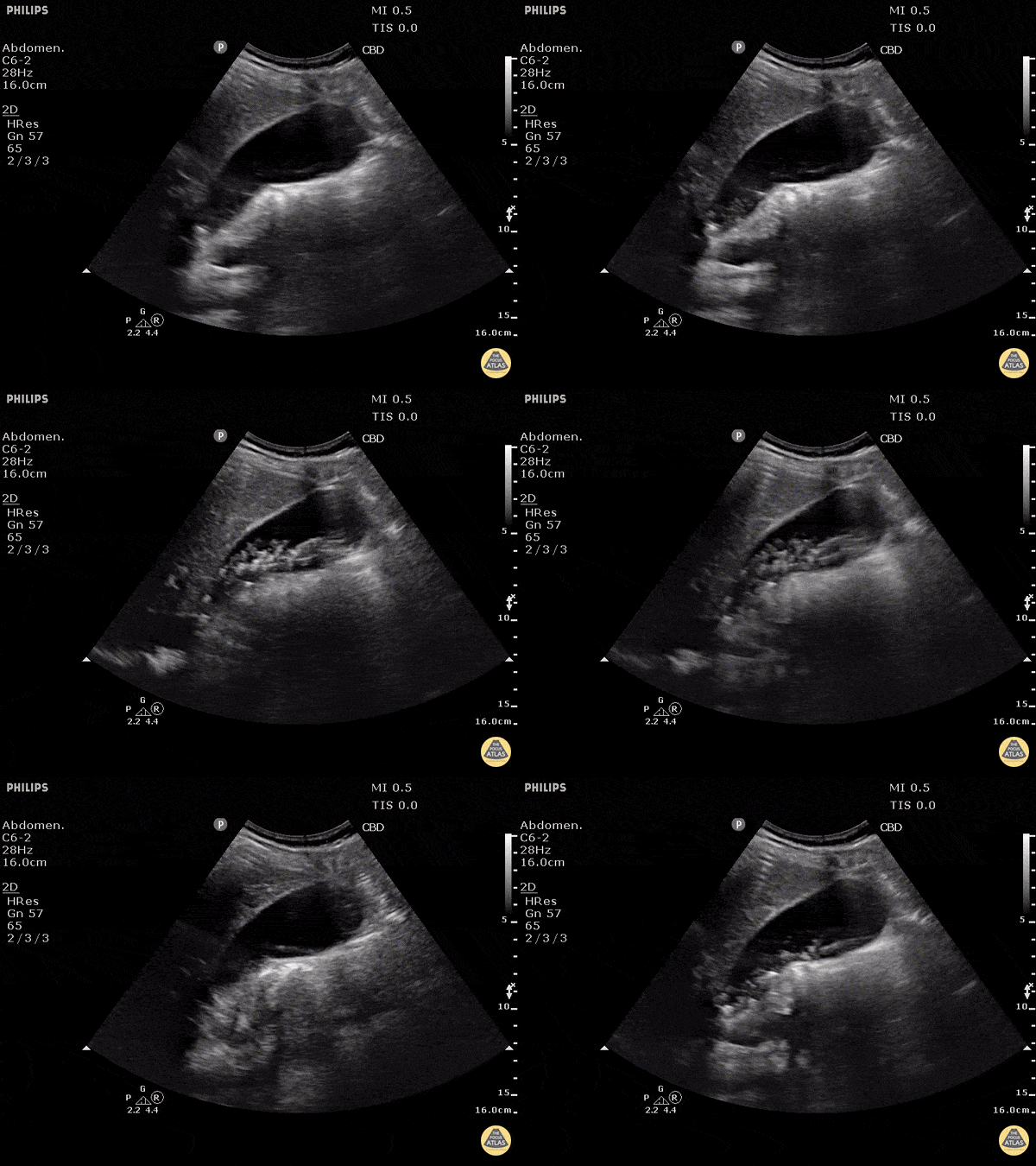
This non-inflamed gallbladder contains many stones. The stones are hyperechoic with posterior shadowing. The gallbladder wall is not thickened, there is no pericholecystic fluid, and no sonographic Murphy's sign, thus cholecystitis is unlikely.
Justin Bowra MBBS, FACEM, CCPU Emergency Physician, RNSH et al. (Dr. Ken Lee)
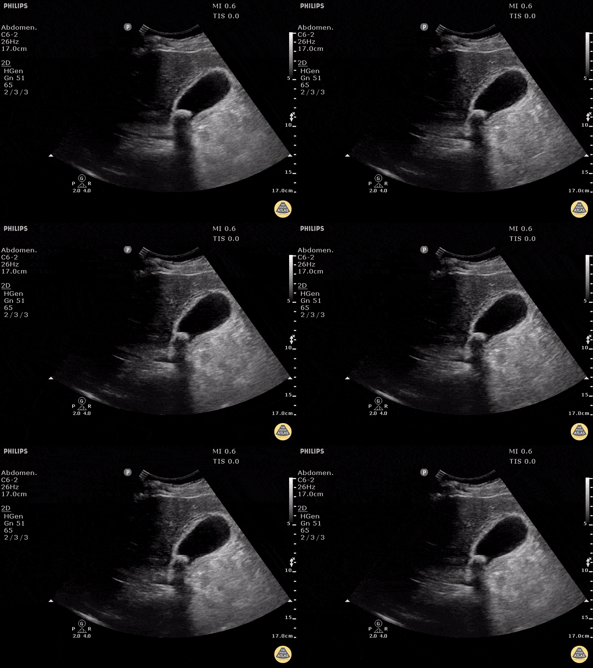
This clearly demonstrates a stone in the gallbladder neck as a hyperechoic structure with posterior shadowing. The neck is not always clearly visualized on first glance so it is important to scan through the gallbladder in two planes to exclude stones in the neck. Other signs of acute cholecystitis include pericholecystic fluid, gallbladder wall thickening (>3mm) common bile duct dilatation, and positive sonographic Murphy's sign.
Justin Bowra MBBS, FACEM, CCPU Emergency Physician, RNSH et al. (Mo)
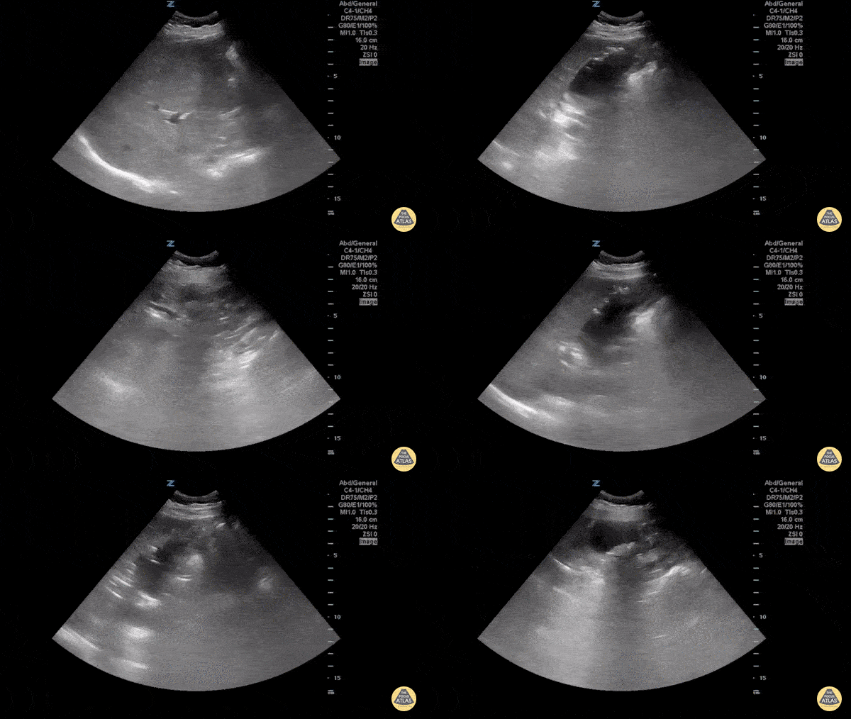
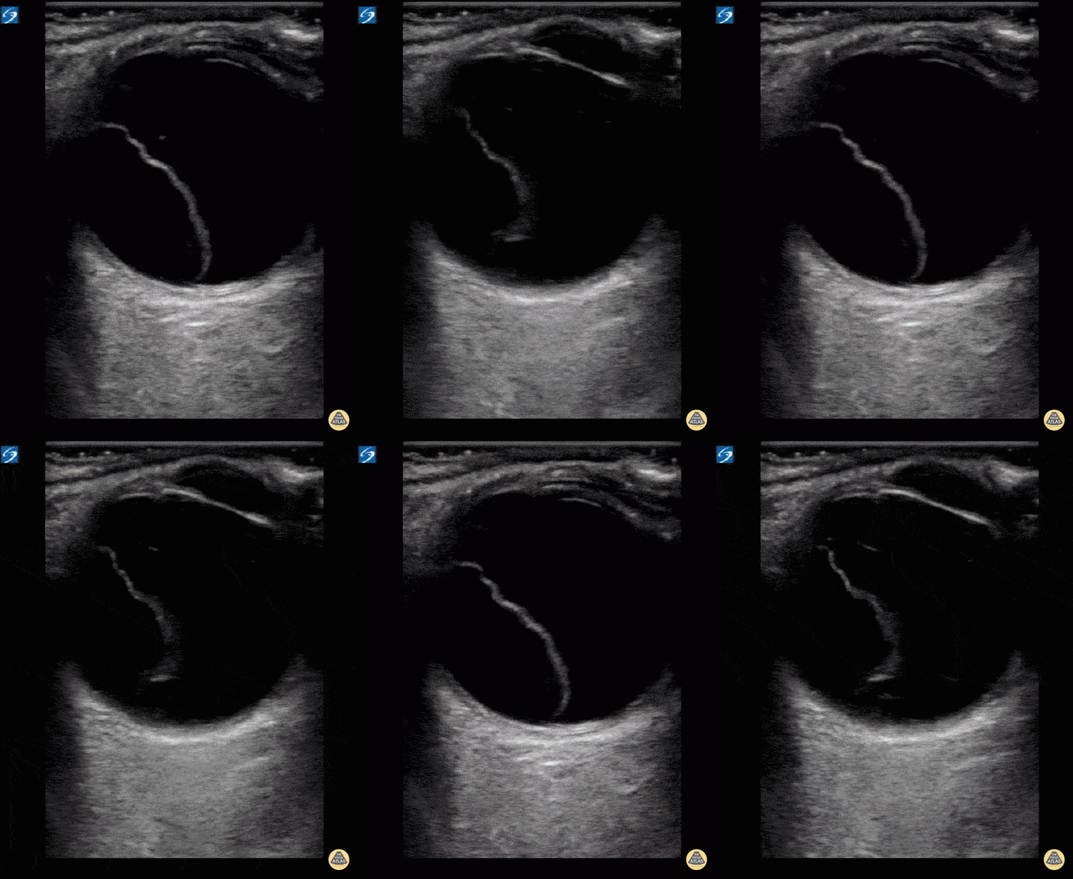
Emphysematous cholecystitis is the result of gas formation within the wall or lumen of the gallbladder as a result of infection by a gas forming organism. It is a surgical emergency. The air in the gallbladder appears highly echogenic with mild posterior shadowing, as air next to tissue is highly reflective.
Sukh Singh, MD
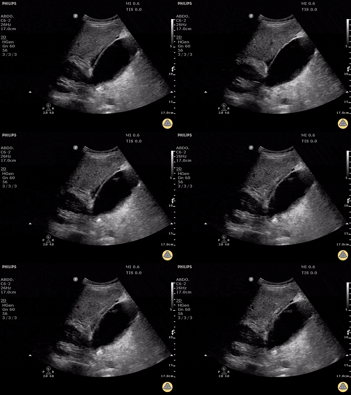
Free fluid can be appreciated around this gallbladder. This is called pericholecystic fluid. However there are no stones, nor gallbladder wall thickening, and the sonographic Murphy's is negative. This free fluid is unlikely to be caused by cholecystitis. In this case, the free fluid came from a different source.
Justin Bowra MBBS, FACEM, CCPU Emergency Physician, RNSH et al.