Aorta Evidence
POCUS Evidence Atlas - Aorta. EBM review resource of POCUS for aorta diagnosis. Sensitivity Specificity positive and negative likelihood ratios.
The Evidence Atlas: Aorta
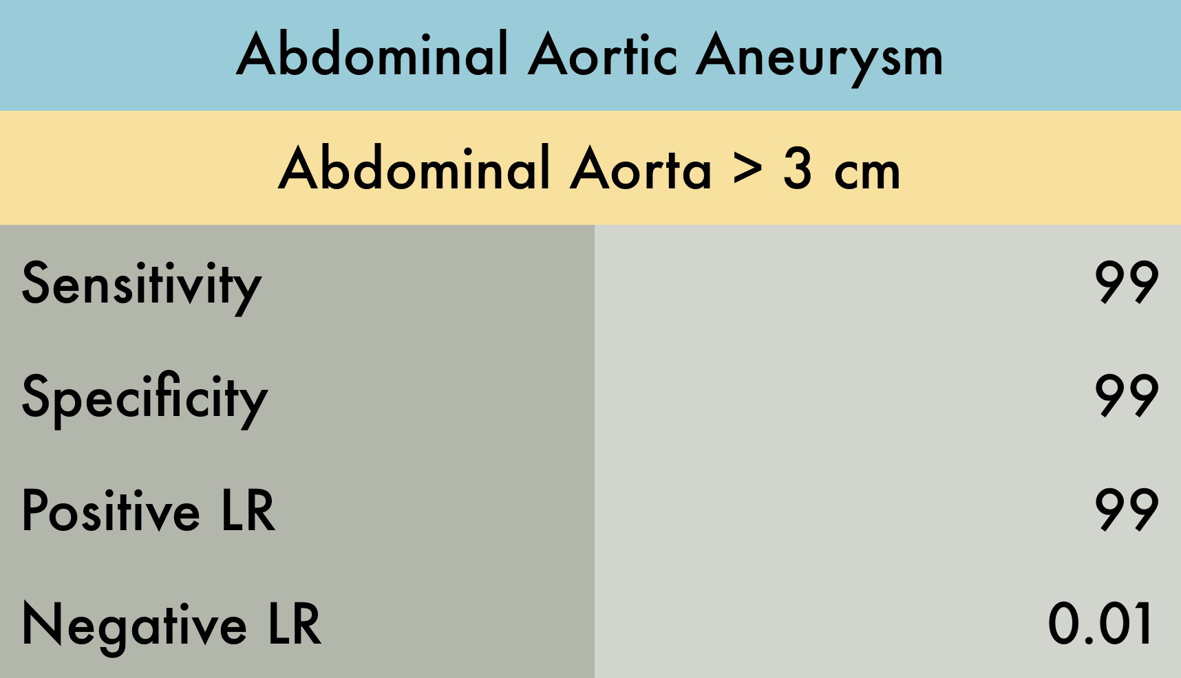
This was a systematic review evaluating the operating characteristics of emergency department (ED) performed ultrasonography for abdominal aortic aneurysm (AAA). A total of 7 studies (n = 655) were included in the analysis, all of which were prospective studies which enrolled adult patients with symptoms/signs suggestive of AAA. AAA was defined as > 3 cm dilation of the aorta. The reference standard was varied among studies including CT, MRI, aortography, radiology performed ultrasound, exploratory laparotomy, or autopsy results. The operator training and experience, as well as the number of participating emergency physicians in each study, was also quite variable. While the pooled data is displayed in the included table, the individual sensitivity and specificity range for AAA detection among the 7 studies were as follows: sensitivity 97.5-100%, specificity 94.1-100%, LR+ 10.8 - infinite, and LR- 0.00-0.025.
PMID: 23406071
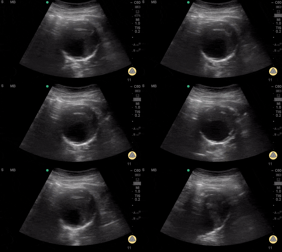
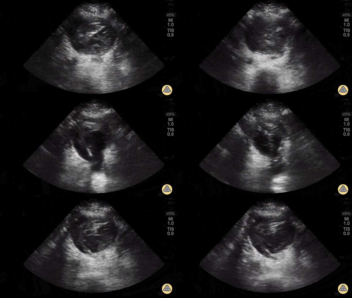
74 y/o F hx stage 4 lung cancer, AAA, presented with chest pain and SOB x 1 day. POCUS shows the aorta is larger than the normal diameter of 3 cm, representing an abdominal aortic aneurysm, and measuring approximately 5.6 cm at its largest point. On cross section you are able to see a large mural thrombus with decreased diameter of blood flow through the center.
Juliana Jaramillo MD - Kings County Emergency Medicine
Approximately 6 cm abdominal aortic aneurysm with intramural thrombus.
Frances Russell, MD, RDMS
Assistant Professor of Emergency Medicine Division Chief, Ultrasound Fellowship Director, Ultrasound
74 y/o F hx stage 4 lung cancer, AAA, presented with chest pain and SOB x 1 day. POCUS shows the aorta is larger than the normal diameter of 3 cm, representing an abdominal aortic aneurysm, and measuring approximately 5.6 cm at its largest point. On cross section you are able to see a large mural thrombus with decreased diameter of blood flow through the center.
Juliana Jaramillo MD - Kings County Emergency Medicine
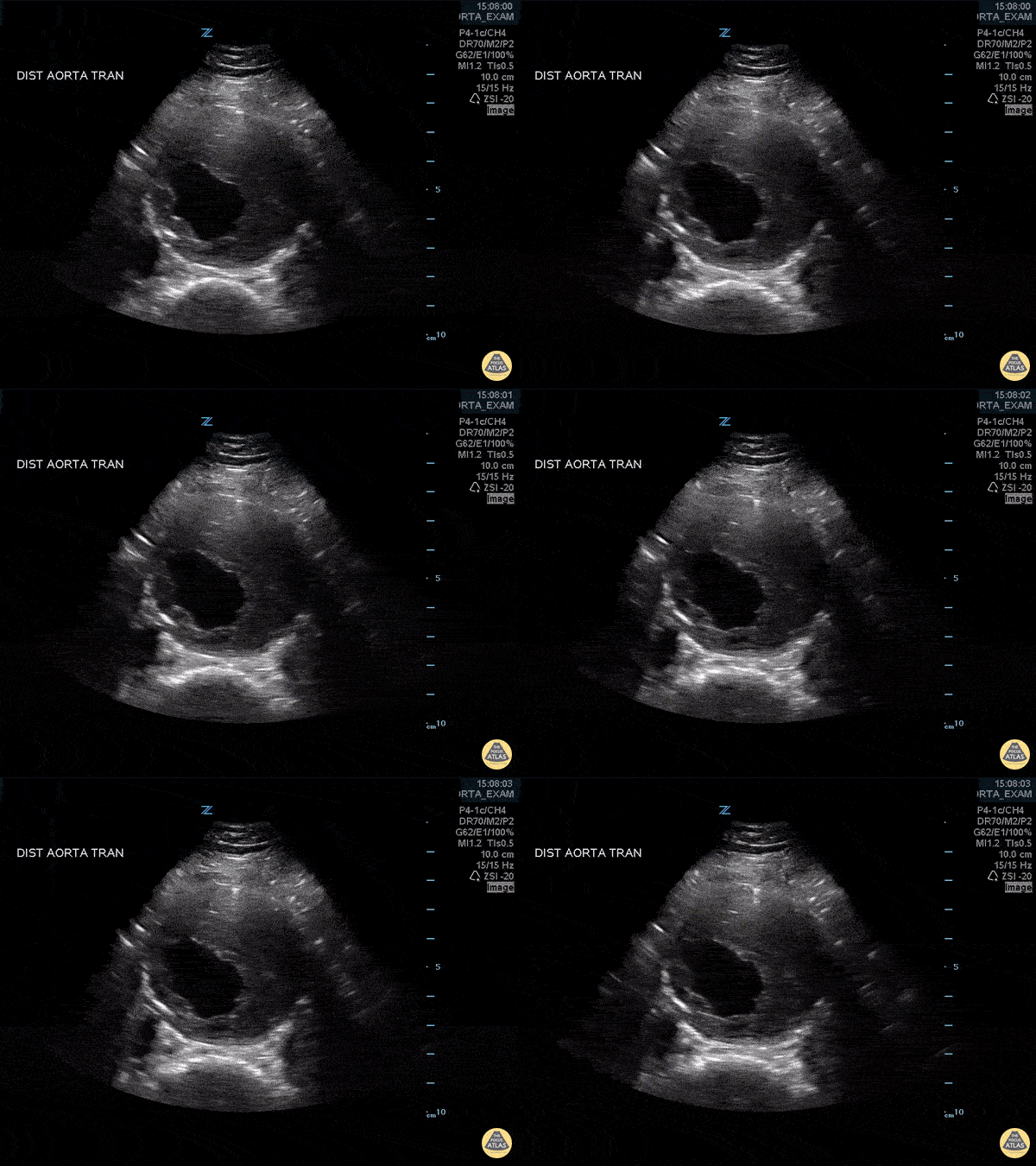
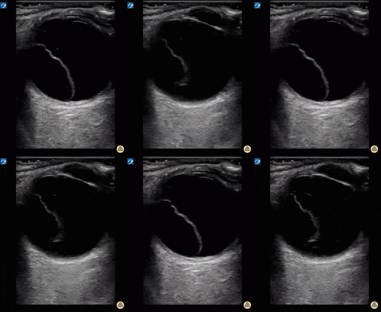
You may think this a dissection but it's actually an aortic aneurysm filled with thrombus, "with holes" making it a very "happy flappy." CT Confirmed.
Dr. Vincent Rietveld - Amsterdam, The Netherlands
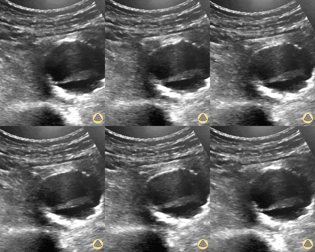
63yoM witnessed collapsed. Arrived VSA with PEA rhythm. A fast look abdominal aorta POCUS showed a large abdominal aortic aneurysm with internal thrombus and rupture through the posterior wall. Note the AAA is measured from echogenic outer wall to outer wall, not the hypoechoic internal lumen that is seen pulsating. The patient was resuscitated aggressively with blood and initially survived operative repair. Unfortunately he died in the ICU several days later from multiorgan failure.
Dr. Joey Newbigging