Renal Evidence
POCUS Evidence Atlas - Biliary, Gallbladder, Cholecystitis. EBM review and resource of POCUS for biliary diagnosis. Sensitivity Specificity positive and negative likelihood ratios. Point of care ultrasound evidence.
The Evidence Atlas: Renal
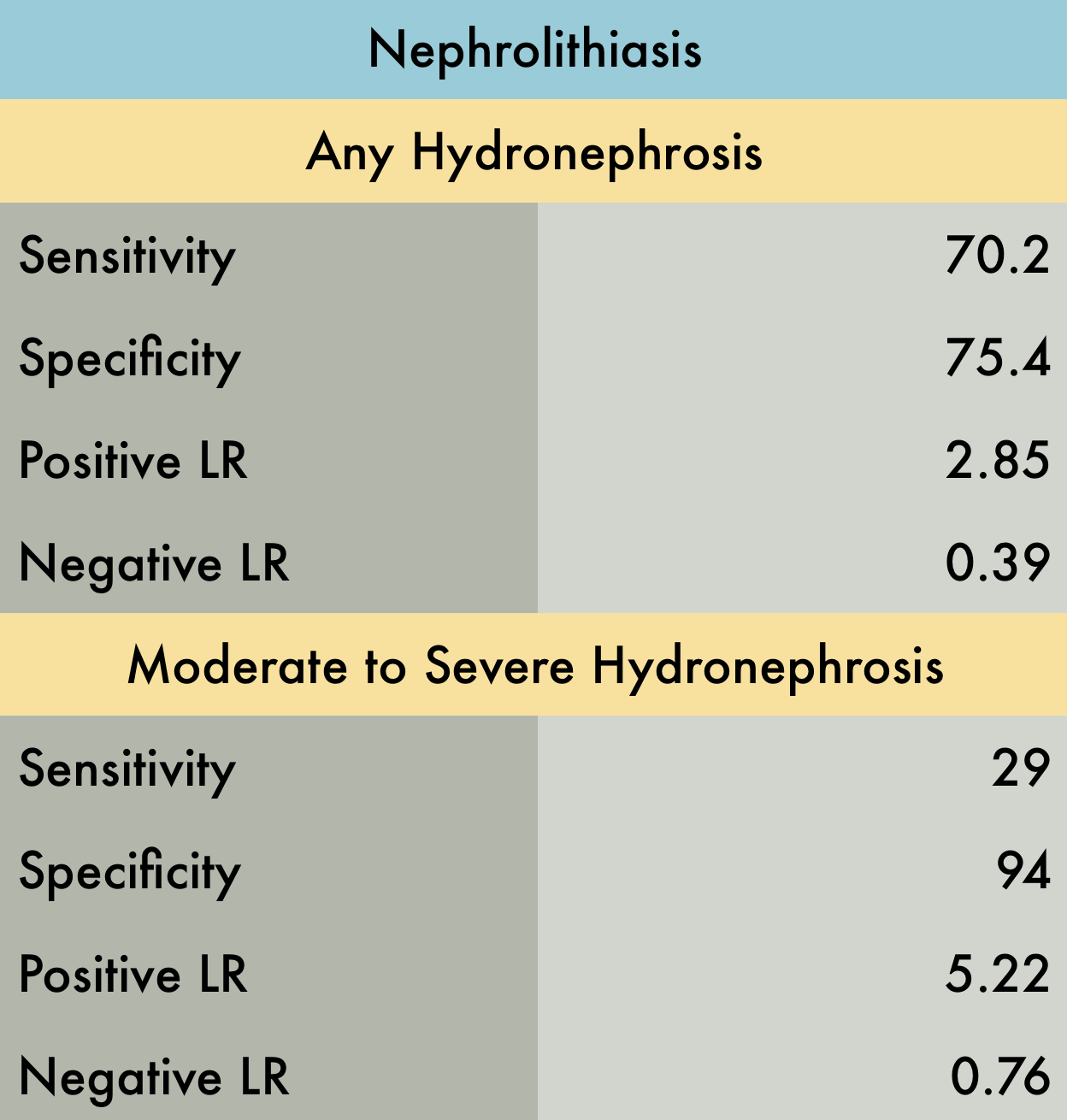
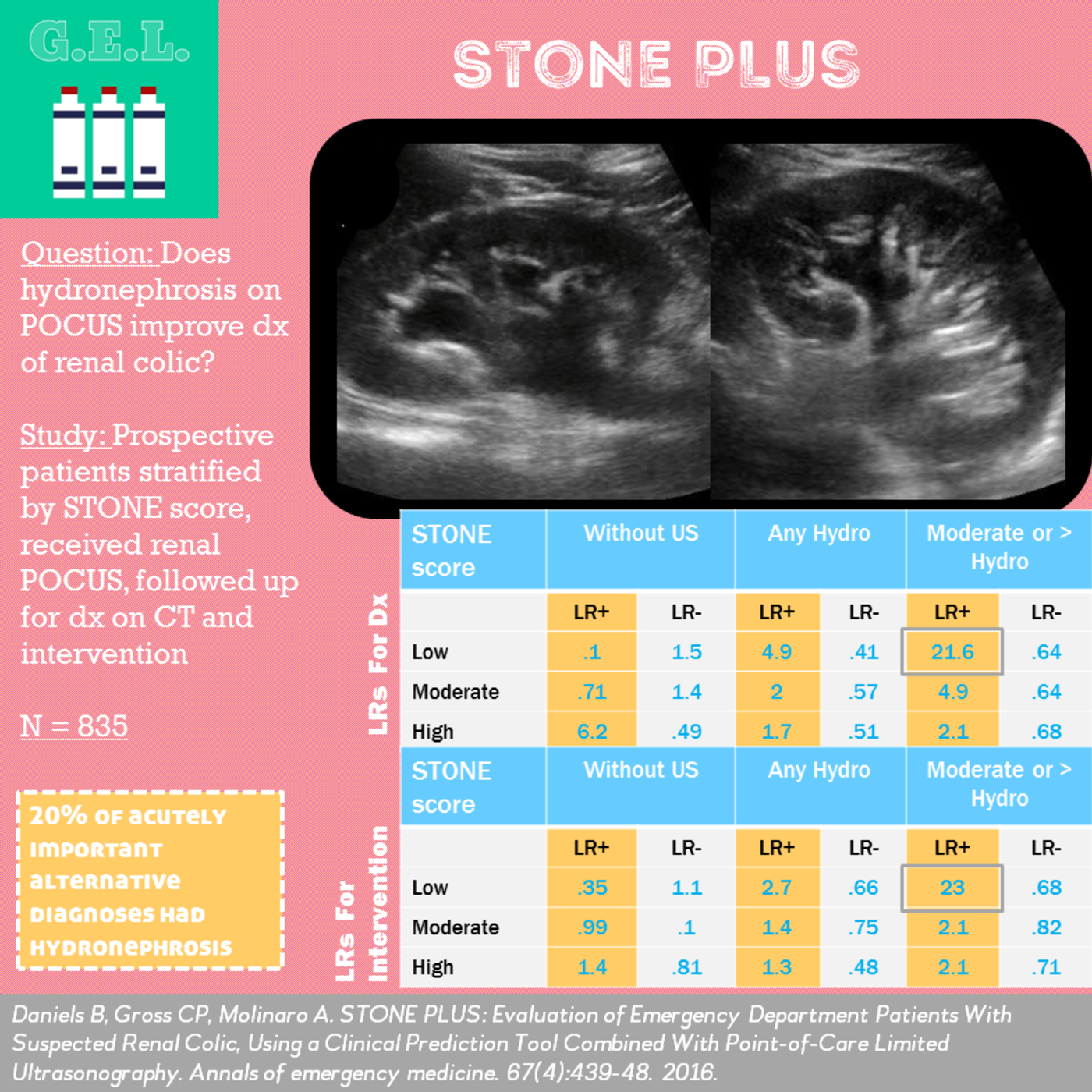
This was a meta-analysis including 5 studies (n=1773) evaluating the accuracy of POCUS to diagnose nephrolithiasis in adult patients presenting to the emergency department with symptoms suggestive of renal colic (flank pain, dysuria, abdominal pain radiating to groin). Reference standards included CT, direct stone visualization, or surgical findings. Specificity improved significantly (~94.4%) for moderate to severe hydronephrosis (i.e. exclusion of mild hydronephrosis). This systematic review has some flaws (most importantly not having a single uniform gold standard) but appears to be the best available evidence.
PMID: 29427476
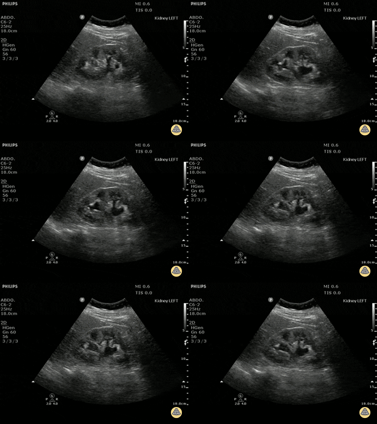
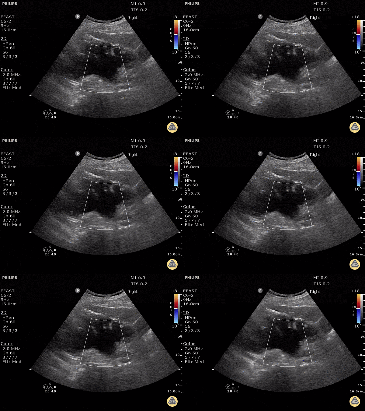
Mild - grade two hydronephrosis with dilation of the renal pelvis and dilation of the some calyces. Grade one would not include dilated calyces.
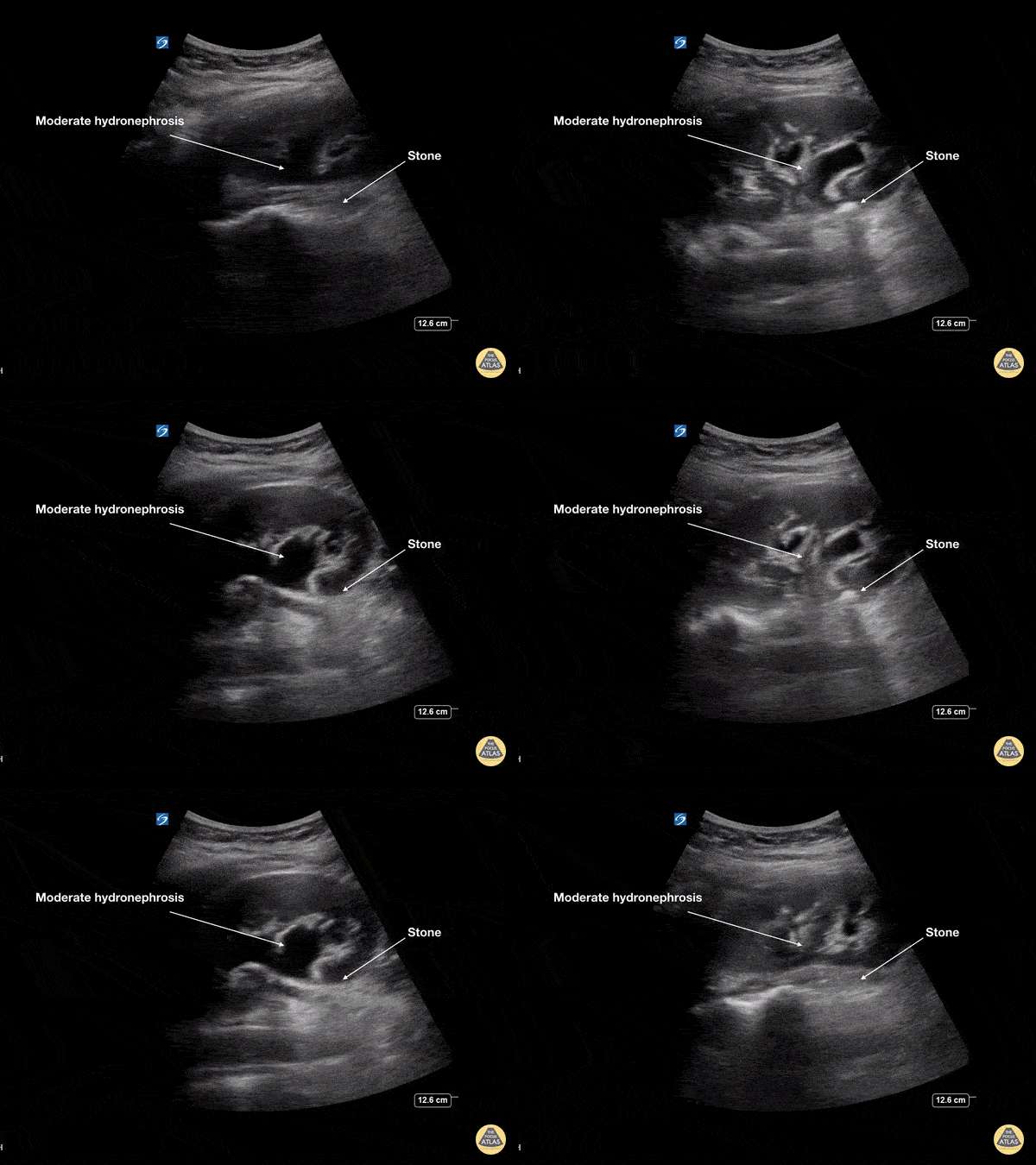
This young female presented with colicky left flank pain worsening over the previous 24 hours. POCUS showed moderate hydronephrosis with rounding of the calyces of the left kidney collecting system. The stone is seen at the ureteropelvic junction (UPJ) as a hyperechoic structure with posterior acoustic shadowing measuring 7mm on CT. At ureteroscopy, the stone was retrieved, fragmented and a double J stent was placed
Dr Cian McDermott, Mater University Hospital, Dublin, Ireland
Moderate (grade three) hydronephrosis can be appreciated here with dilation of both the renal pelvis and calcyces. The renal cortex is also thinned. There is not gross atrophy.
Dr. Justin Bowra et al. (Dr. Yogi)
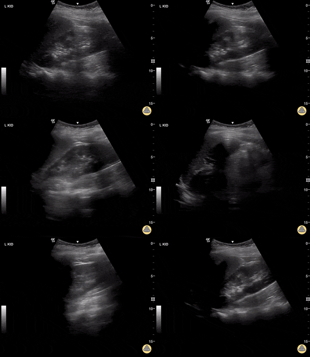
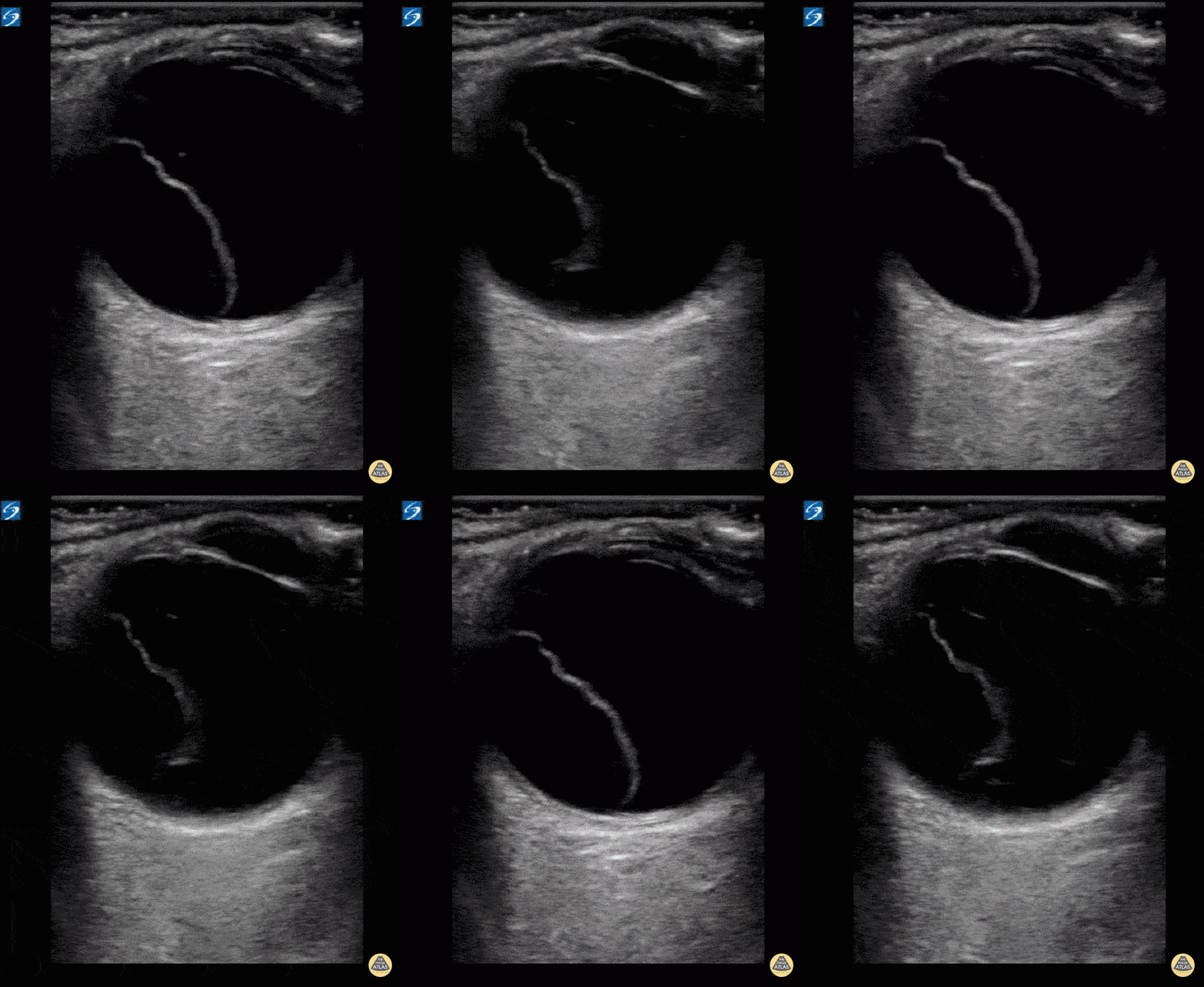
In this patient with severe hydronephrosis, there is gross dilation of both the renal pelvis and calyces, which are ballooned. The cortex of the kidney has atrophied and is very thin. This is severe, or grade 4.
Dr. Justin Bowra et al. (Dr. Browne and Dr. Knights)