OB/GYN Evidence
POCUS Evidence Atlas - OB GYN ectopic pregnancies and pregnancy. EBM review and resource of POCUS for ectopic pregnancy positive fast exam diagnosis. Sensitivity Specificity positive and negative likelihood ratios.
The Evidence Atlas: OB/GYN
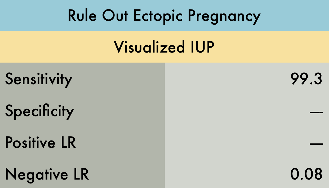
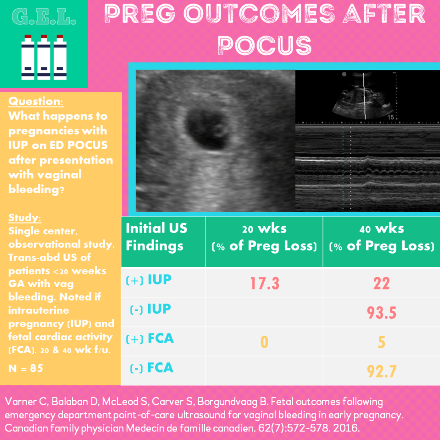
This was a meta-analysis including 10 studies (n=2057) examining the operating characteristics of bedside emergency provider (EP) performed ultrasound to rule out ectopic pregnancy. All studies used the visualization of an IUP (intrauterine pregnancy) on ultrasound as the rule out criteria. The ultrasound examinations performed by EPs included transabdominal, transvaginal or both. Reference standards included formal radiology US, gynecology US, over-read of EP performed US by radiology, or clinical record review. They did not report specificity and positive LR's because of significant heterogeneity in these results when data was pooled. This study supports the utility of EP performed ultrasound as a strong rule out test for ectopic pregnancy.
PMID: 20828874
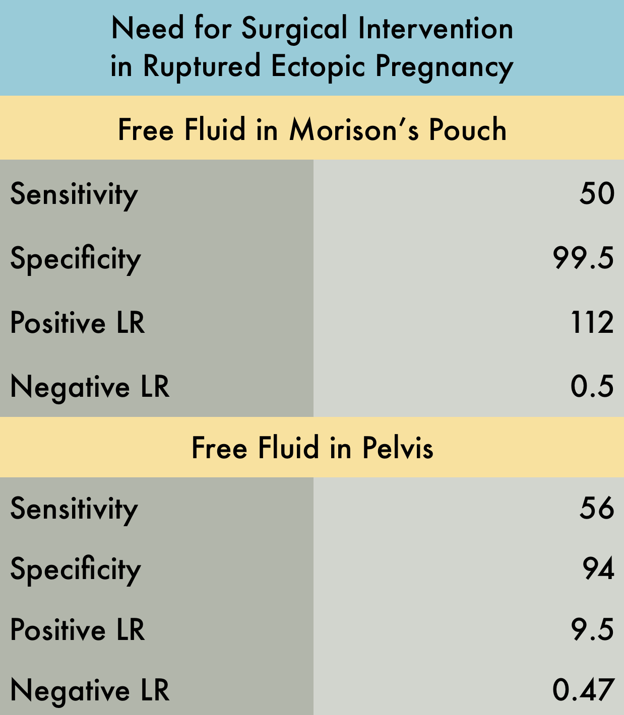
This was a prospective observational study (n=242) of patients presenting to the emergency department with suspected ectopic pregnancy, evaluating if free fluid in the peritoneal cavity identified by bedside ultrasound was predictive of the need for operative intervention. The included patients were suspected to have an ectopic pregnancy based on positive pregnancy test results, in their first trimester, with either abdominal pain or vaginal bleeding. All examinations were performed by emergency providers (EPs) using a transabdominal approach, to determine if free fluid was present in the hepatorenal space (Morison’s Pouch) and/or pelvis. Reference standard was chart review by one of four study investigators who were blinded to the EP performed US results. There was one patient who had free fluid in Morison’s Pouch but had a confirmed IUP (suspected to be a ruptured corpus luteum cyst) and no ruptured ectopic pregnancy. This study supports that free fluid present in Morison’s pouch in patients with suspected ectopic pregnancy strongly predicts the need for operative intervention.
PMID: 17554008
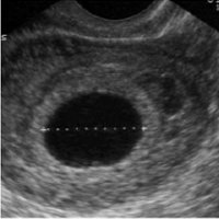
Seen at 4-6 weeks:
- Not diagnostic for IUP
- Ectopic may have pseudogestational sac, though this is rare
- It might be early pregnancy but they will need close follow up
Seen at 5-7 weeks:
- First evidence of IUP
- Appears as ring like structure within the gestational sac
Seen at 6-8 weeks:
- Tissue with similar echogenicity as uterus seen within the gestational sac
- If you see a fetal pole, you should see fetal cardiac activity
- Early on, the yolk sac and fetal pole may be present simultaneously
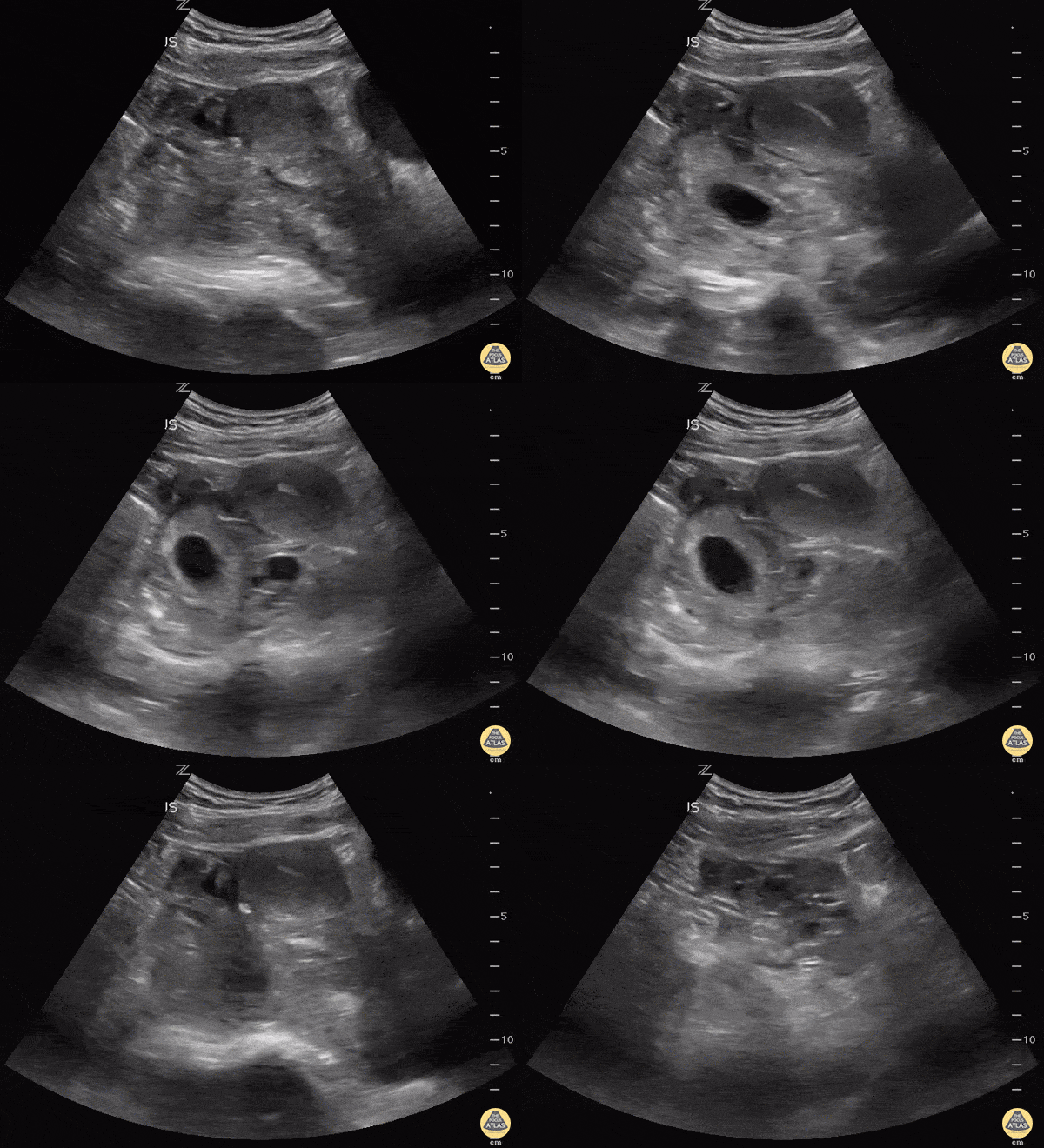
Always make sure the "IUP" is actually in the uterus by observing the uterus superior to the bladder in the sagittal view. This young lady could have been diagnosed easily with IUP and her abdomen pain dismissed as pain with pregnancy instead of a 10 week ectopic.
Matt Rutz, MD
@IUEM_Ultrasound
Indiana University School of Medicine
Department of Emergency Medicine
Division of Ultrasound
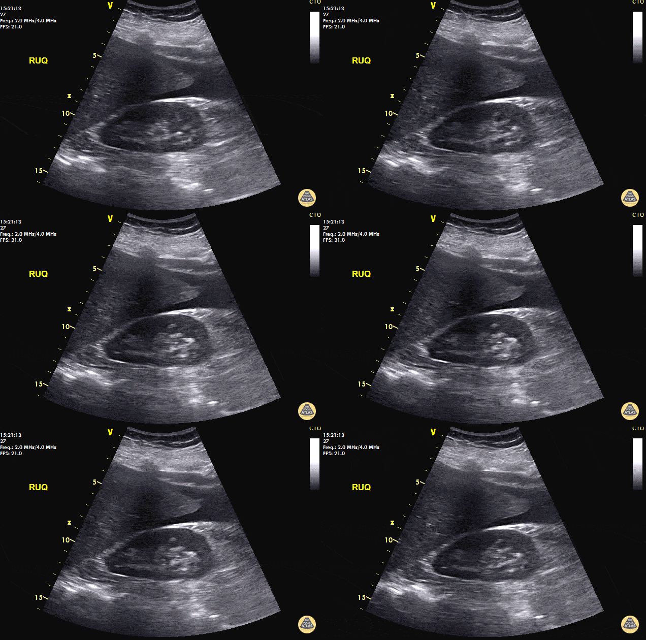
40 y/o F with abdominal pain, syncopal, +HCG. POCUS reveals free fluid in RUQ at inferior pole of the kidney and caudal tip of liver. Patient taken immediately to the OR and 800mL of blood evacuated. A left fallopian ectopic pregnancy was found.
Always look for free fluid at the inferior pole of the kidney. POCUS saves lives.
Dr. Cian McDermott - Dublin, Ireland