Bowel Evidence
POCUS Evidence Atlas - Bowel, SBO, Appendicitis. EBM review and resource of POCUS for bowel diagnosis. Sensitivity Specificity positive and negative likelihood ratios. Point of care ultrasound evidence.
The Evidence Atlas: Bowel
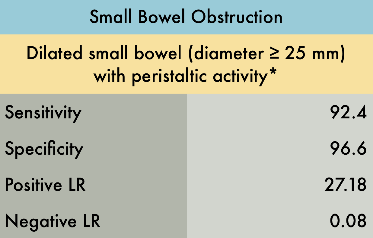
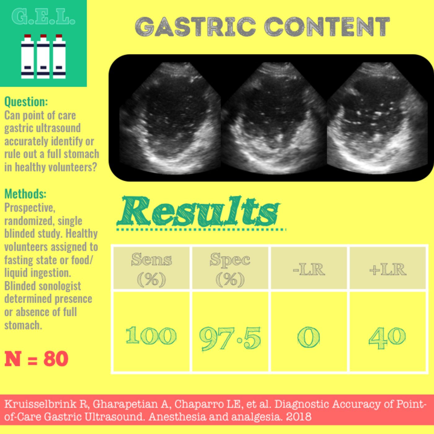
This was a meta-analysis including 11 studies (n=1178) to evaluate the test characteristics of US in diagnosis of small bowel obstruction (SBO). There was mild to moderate heterogeneity in diagnostic criteria, study location, sonographer experience, and reference standard. Specifically, most studies used 2.5 cm as the cutoff to diagnose SBO while one study used the cutoff of 3.0 cm, and several other studies only noted the presence of “dilated bowel loops” as a diagnostic criteria. Of the 11 studies included, only 3 were emergency department studies. Reference standards included surgery, clinical diagnosis, CT, or other advanced imaging. While there were multiple components to the index test and varied reference standard, this does appear to be the best and biggest review on this topic. These operating characteristics suggest ultrasound to be a valuable tool in the diagnosis of SBO, however further studies are needed specifically with regards to the emergency department setting. *Other diagnostic criteria included visualizing collapsed distal loops of bowel with decreased peristalsis
PMID: 28797559
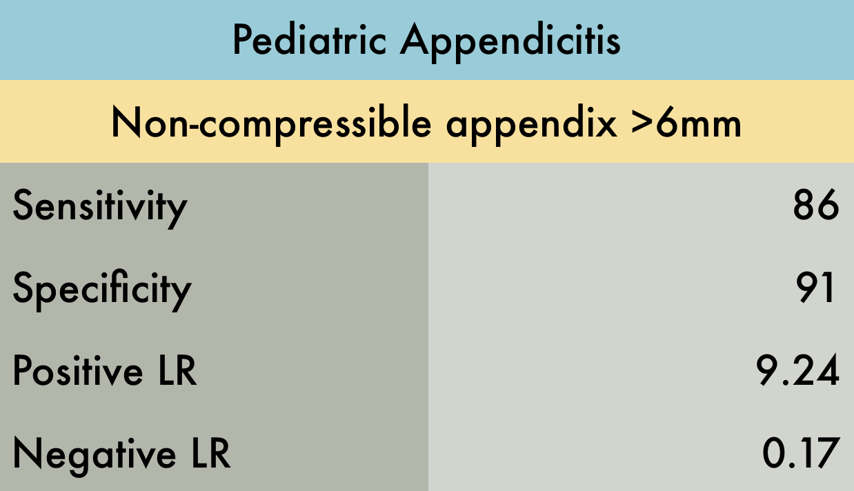
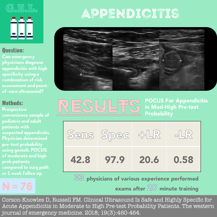
This systematic review and meta-analysis of 4 studies (n=461) evaluated the accuracy of emergency department POCUS performed by EM or PEM physicians for diagnosis of acute appendicitis in children. The main limitation of the study was the high prevalence of equivocal results. However, authors did a sensitivity analysis with and without equivocal cases and results were similar. A mathematical model was used to compare POCUS to CT scan and MRI. The authors concluded that a positive POCUS exam is diagnostic, obviating the need for CT, however if POCUS is equivocal or negative, further imaging with CT or MRI is necessary.
PMID: 28214369
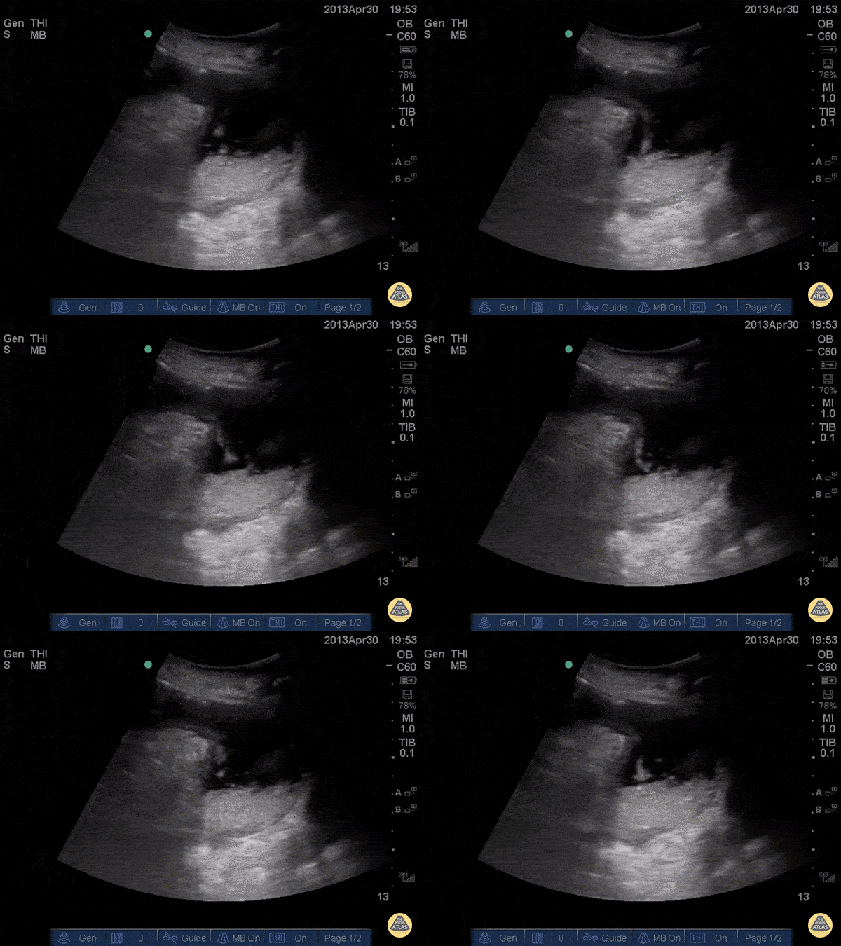
A 60 female presented with 3 days of vomiting, suprapubic distension and left groin pain. POCUS shows distended bowel loops (>2.5cm), 'to-and-fro' motion of hyperechoic bowel contents and extraluminal free fluid. NG was passed for decompression and surgical admission arranged. CT showed small bowel obstruction at the level of distal ileum within a left inguinal hernia
Dr Cian McDermott, Mater University Hospital, Dublin, Ireland
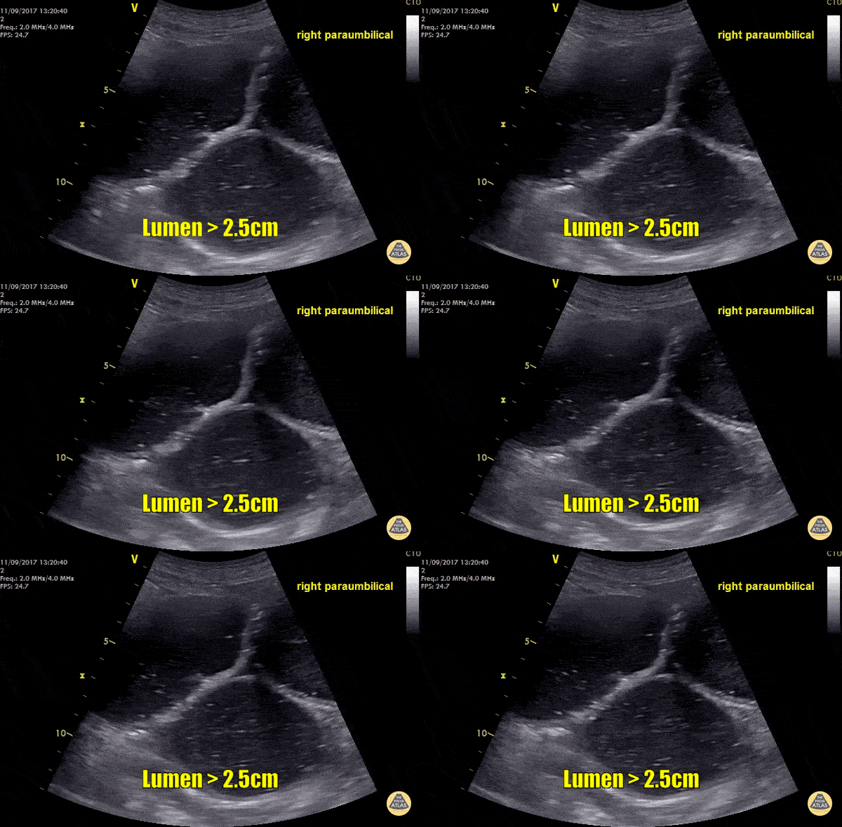
These images were recorded from an elderly lady with abdominal pain and vomiting. Small bowel lumen is dilated more than 2.5cm and there is reduced peristalsis within the lumen - these sonographic signs are consistent with small bowel obstruction (SBO). Other signs of SBO include the presence of hyperechoic material moving back and forth within the bowel lumen and free fluid between the loops of bowel (Tanga sign).
Images by Dr. Rory Whelan
Submitted by Dr. Cian McDermott - Dublin, Ireland
Small bowel obstruction with a steep precipice and tumbling feces. We call this Iguapoo falls.
Andrew Liteplo MD, RDMS - Massachusetts General Hospital
Chief, Division of Ultrasound in Emergency Medicine
Director, Emergency Ultrasound Fellowship
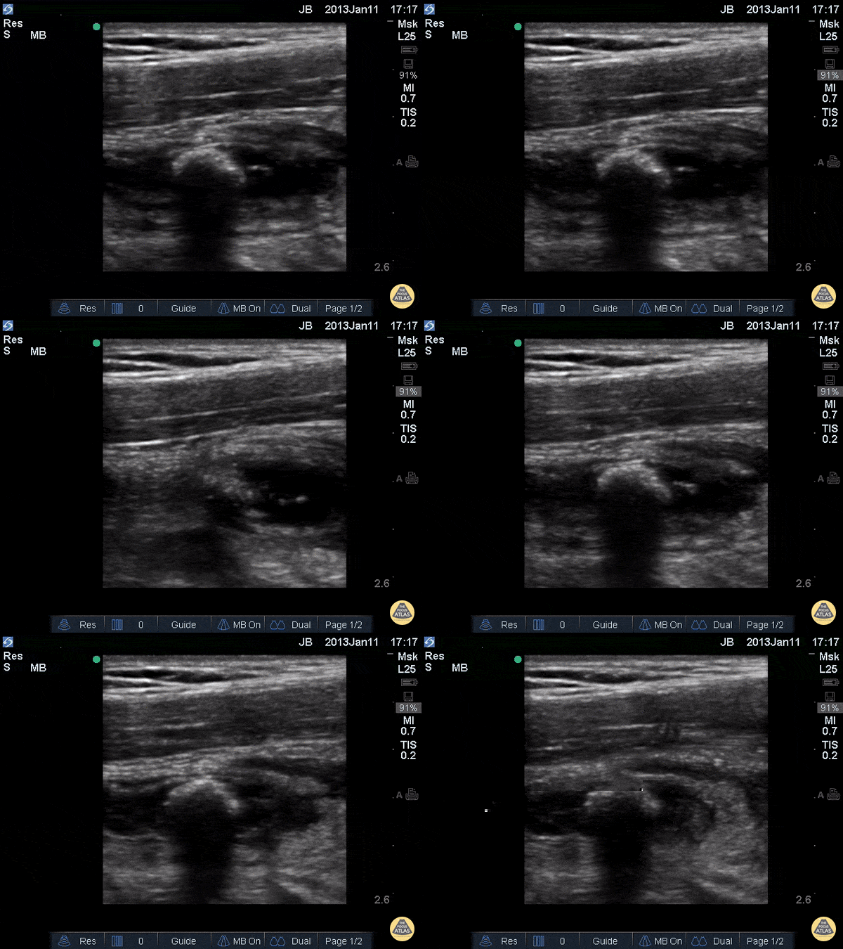
This images demonstrates a longitudinal view of an appendix that appears enlarged with edematous walls, containing a shadowing appendicolith, consistent with acute appendicitis.
Justin Bowra MBBS, FACEM, CCPU Emergency Physician, RNSH et al.
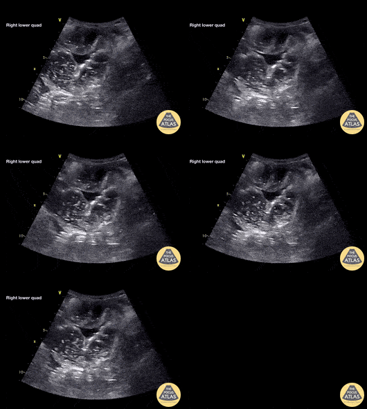
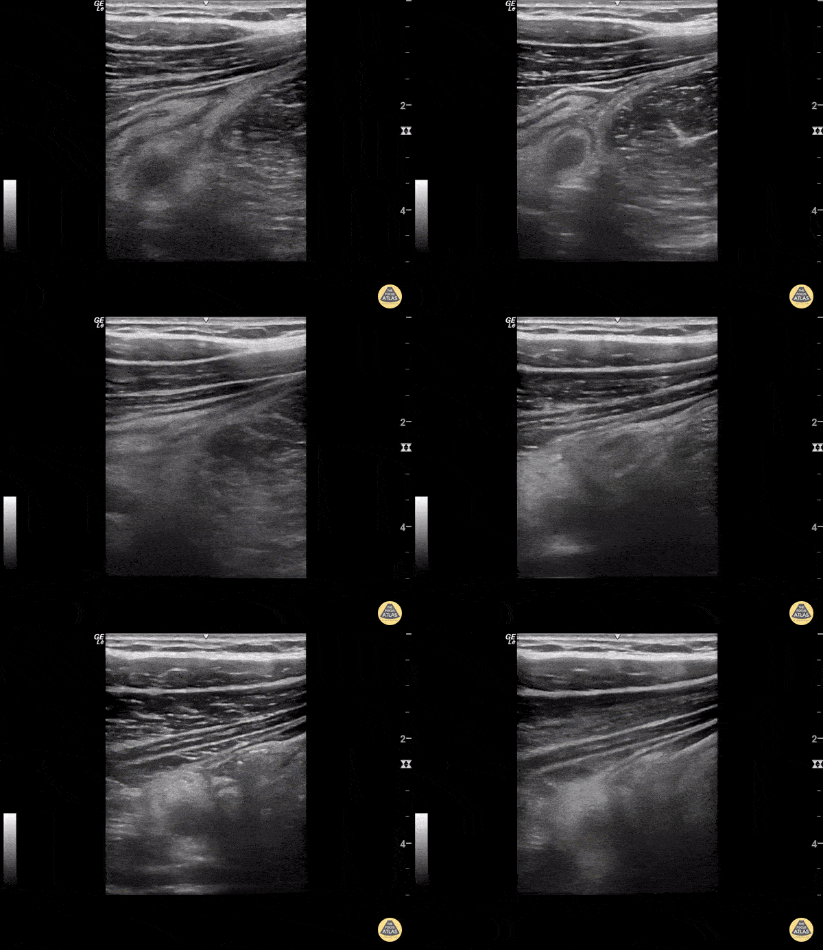
A non-compressible appendix with appendicolith is seen with surrounding fat stranding posterior to a decompressed cecum.
Sukh Singh, MD