Soft Tissue/MSK Evidence
POCUS Evidence Atlas - soft tissue, abscess. EBM review and resource of POCUS for abscess, incision and drainage, I and D diagnosis. Sensitivity Specificity positive and negative likelihood ratios.
The Evidence Atlas: Soft Tissue/MSK
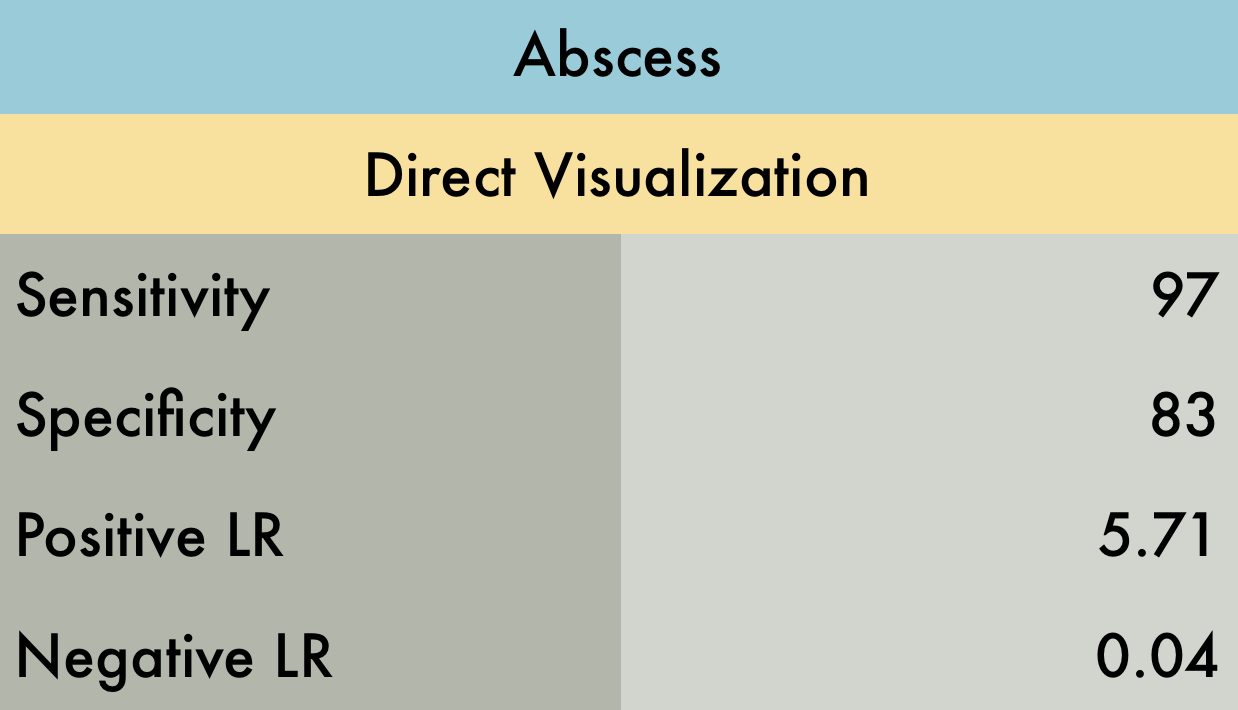
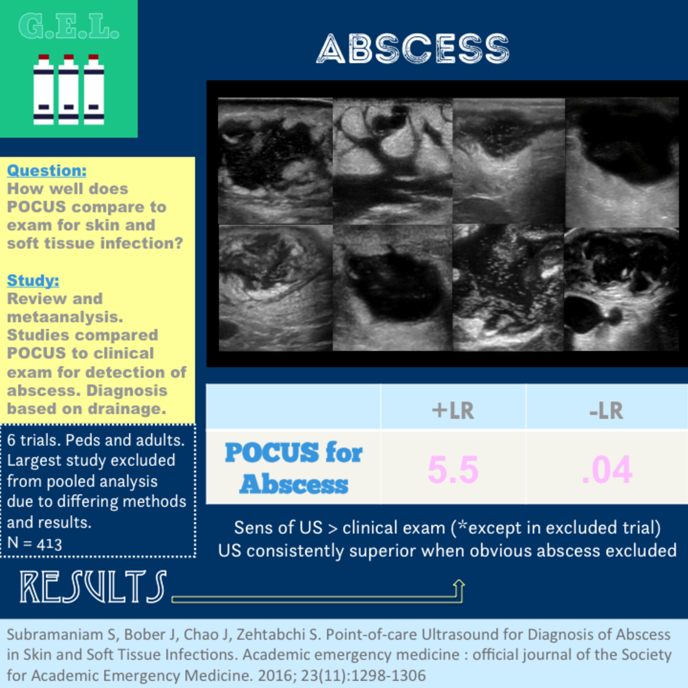
This was a systematic review including 6 prospective observational studies (n=800) comparing physical exam and POCUS to diagnose abscess in both children and adults in the emergency department (ED). Inclusion criteria for all but one study was the presence of a skin & soft tissue infection (SSTI) while once study specifically included patients where abscess was suspected with plan to incise and drain. The reference standard for abscess was pus drainage on initial I&D or at follow up. In many studies, facial and genital/rectal area SSTIs were excluded therefore these results should be interpreted with caution in evaluation of SSTIs in these areas.
PMID: 27770490
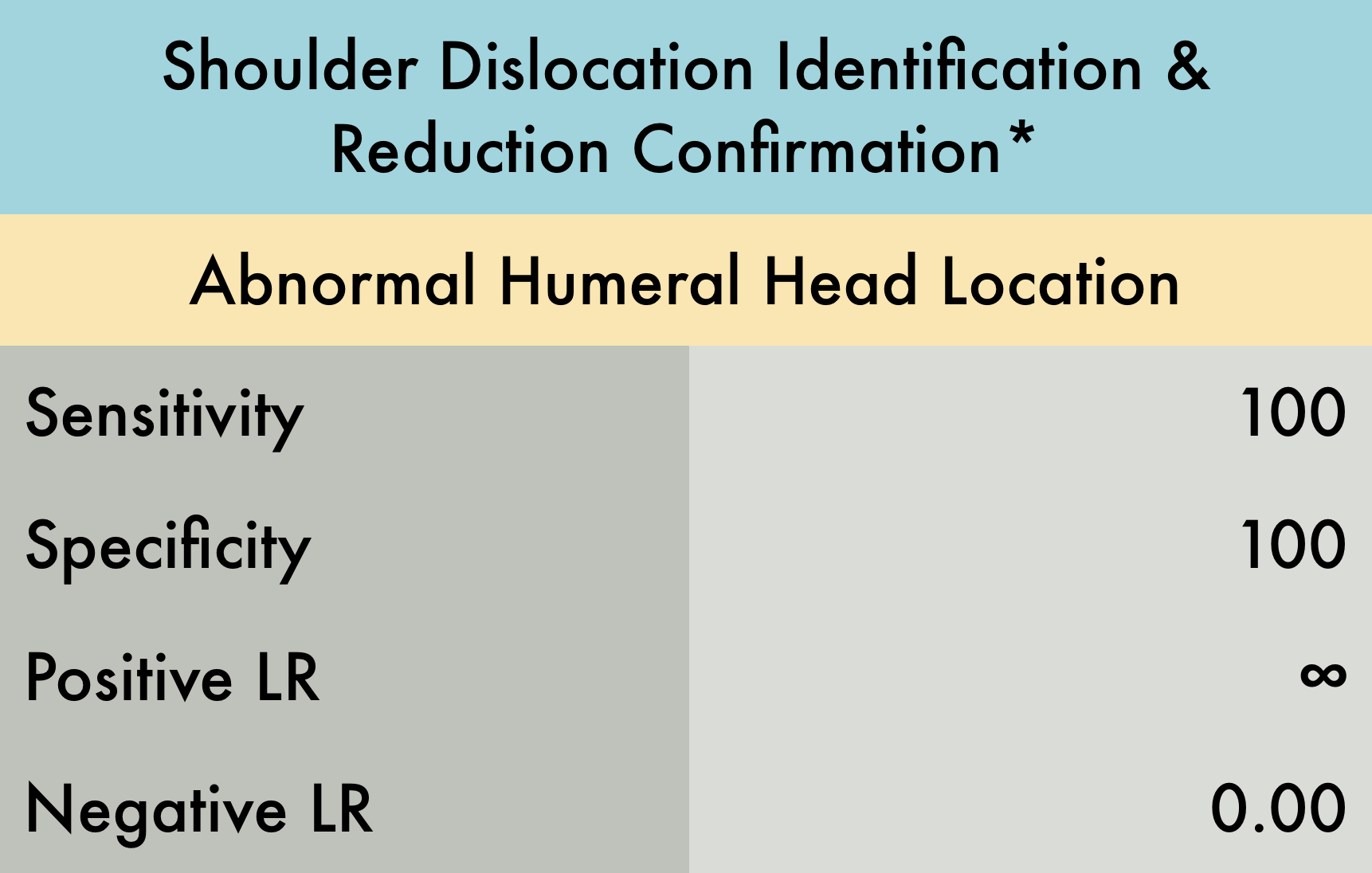
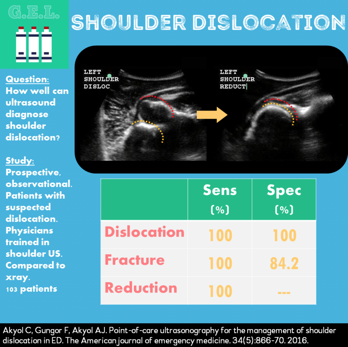
This was a prospective observational study (n= 73) evaluating the diagnostic accuracy of ultrasound for both identification of shoulder dislocation and confirmation of successful reduction. Patients with suspected shoulder dislocation were enrolled, with 69/73 having dislocation confirmed by shoulder radiographs. Patients with multi trauma or decreased level of consciousness were excluded. Both the anterior and lateral approach were used to evaluate for dislocation. Examinations were performed by either an experienced ultrasound trained emergency physician or a senior emergency medicine resident. An ultrasound exam was performed prior to initial shoulder radiographs and after reduction attempt. Ultrasound was found to be 100% accurate with respect to both shoulder dislocation identification and reduction confirmation. *This is a limited study as the sample size is relatively small and this ultrasound examination is dependent on operator experience therefore these results should be interpreted with caution.
PMID: 23489654
Jump to our Soft Tissue or MSK Atlas
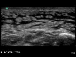
This is the classic appearance of subcutaneous edema which in the appropriate clinical setting (overlying redness and warmth) would be consistent with a cellulitis

While typically an abscess will appear as a hypoechoic fluid filled structure, sometimes it can be missed as its echotexture is similar to other surrounding soft tissue structures. To improve sensitivity, gentle compression should be applied to identify a subtle abscess as seen above.
Image obtained from Ultrasound of the Week.
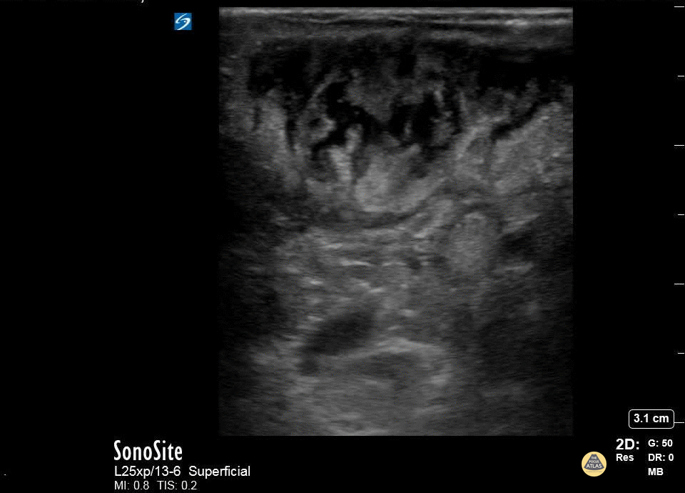
A patient with cerebral palsy and quadriplegia presented with her mother for evaluation of a fluctuant mass in her right axilla. Ultrasound imaging of the mass showed an abscess with multiple septations. The abscess was incised and drained, and the patient was discharged on antibiotics.
Katy Van Donselaar, Emergency Medicine Resident
Christopher Heberer, Emergency Medicine Resident
Simhadri Botta, 4th year Medical Student

Diffuse cobblestoning typically seen with cellulitis.
In this case it is biopsy proven panniculitis.
Dr. Gordon Johnson
@pdxfutebol
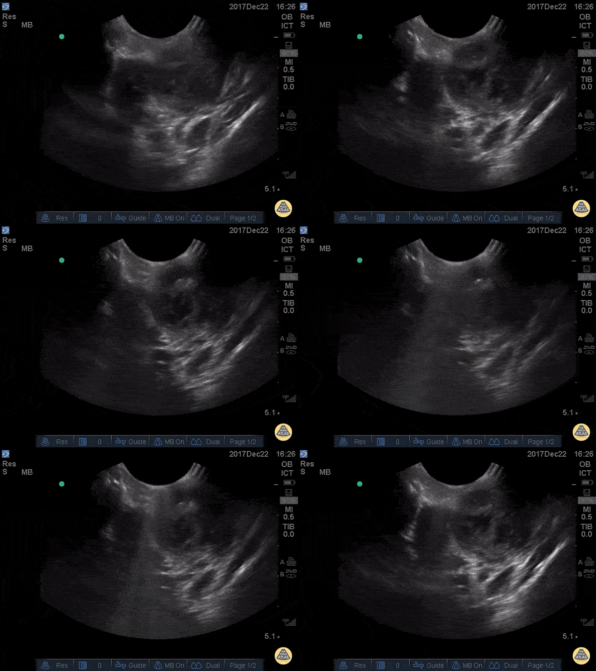
This is an example of a peritonsillar abscess as seen with a high-frequency endocavitary probe. About 3ml of pus was drained with significant relief. Notice that just deep to the abscess is big red. It's not always lateral!
Dr. Jason Tanguay
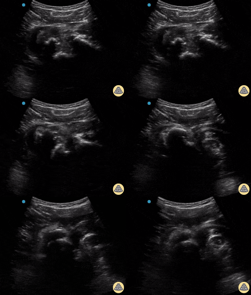
A 30-year-old male presented to the emergency department with an inferior shoulder dislocation (luxatio erecta). Studies have shown that ultrasound has a very high sensitivity and specificity nearing 100% each (Secko 2020) in identification of the same. On ultrasound, the humeral head is shown dislocated anterior to the glenoid fossa, similar to typical anterior shoulder dislocations. This patient's shoulder was reduced with simple traction/countertraction under procedural sedation without any complications.
Mark Zhang
Secko MA, Reardon L, Gottlieb M, et al. Musculoskeletal ultrasonography to diagnose dislocated shoulders: a prospective cohort. Annals of Emergency Medicine. 2020;76(2):119-128.
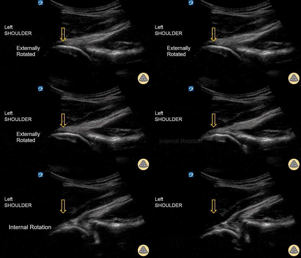
An 18-year-old male with acute left shoulder pain after a physical altercation in which his left arm was pulled. Video clip reveals the left humeral head sliding in and out of the glenoid fossa with internal and external rotation, confirming the diagnosis of shoulder subluxation.
Read more about the case and shoulder POCUS on: https://www.aliem.com/ultrasound-for-win-acute-shoulder-injury-us4tw/
Mark Rivera-Morales, MD, PGY-3
Fernando Rivera, MD, PGY-3
Javier Rosario, MD, FACEP @javimedsimus
UCF/HCA Emergency Medicine Residency Program of Greater Orlando. Osceola Regional Medical Center @UCFEMOrlando
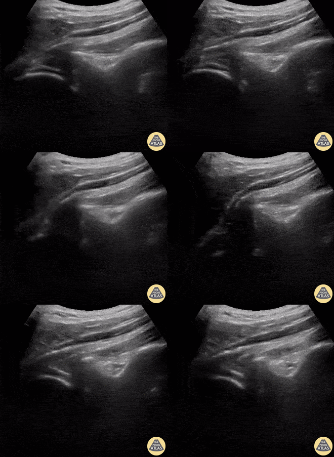
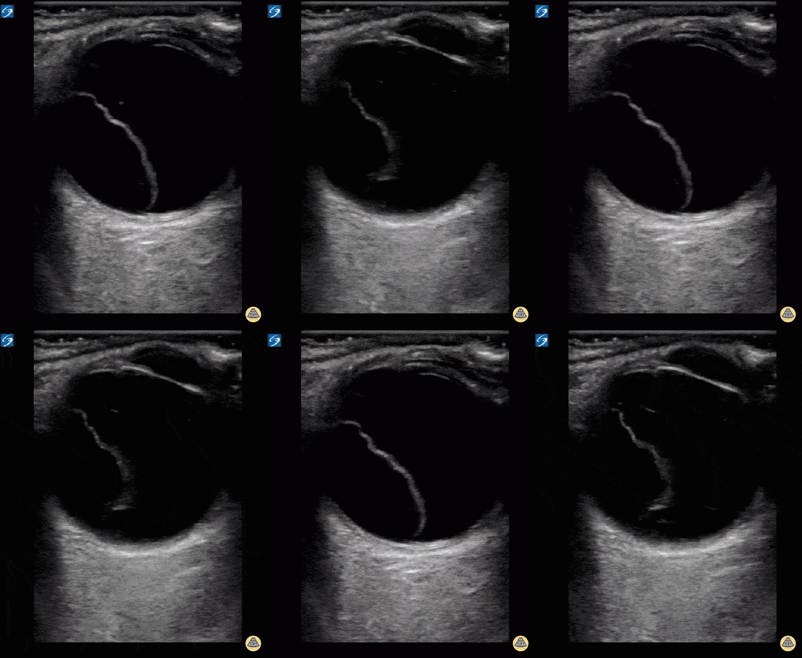
A 9-year-old female presented with left shoulder pain. She has a history of multiple dislocations and, as seen here on POCUS, is able to reduce the dislocation herself.
Julie Klensch, PEM Fellow & Paul Khalil, MD
University of Louisville/Norton Children's Hospital
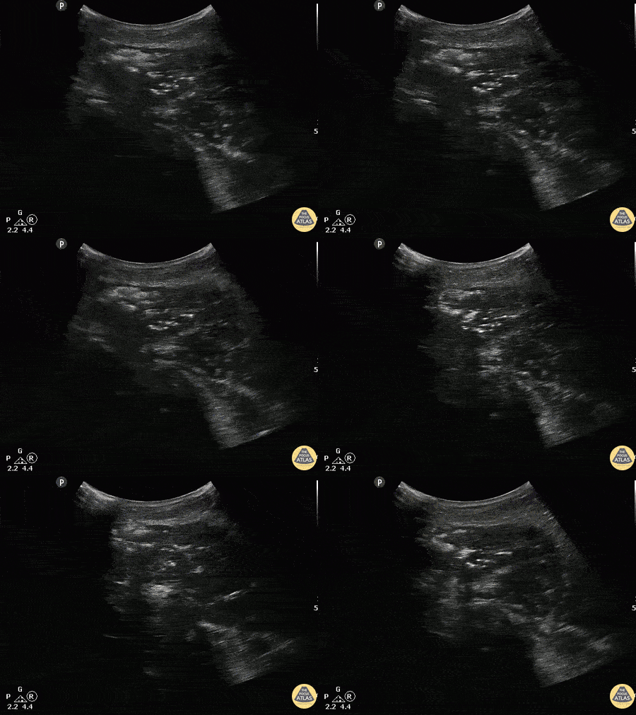
A patient with past medical history of diabetes presented with atraumatic right shoulder pain. Physical exam revealed decreased range of motion without obvious superficial abnormality. The patient was noted to be febrile and tachycardic. A curvilinear ultrasound reveals extensive air (scattered punctate hyperechoic areas) throughout the deltoid muscle indicative of infectious myonecrosis.
Image courtesy of Robert Jones DO, FACEP @RJonesSonoEM
Director, Emergency Ultrasound; MetroHealth Medical Center; Professor, Case Western Reserve Medical School, Cleveland, OH
View his original post here
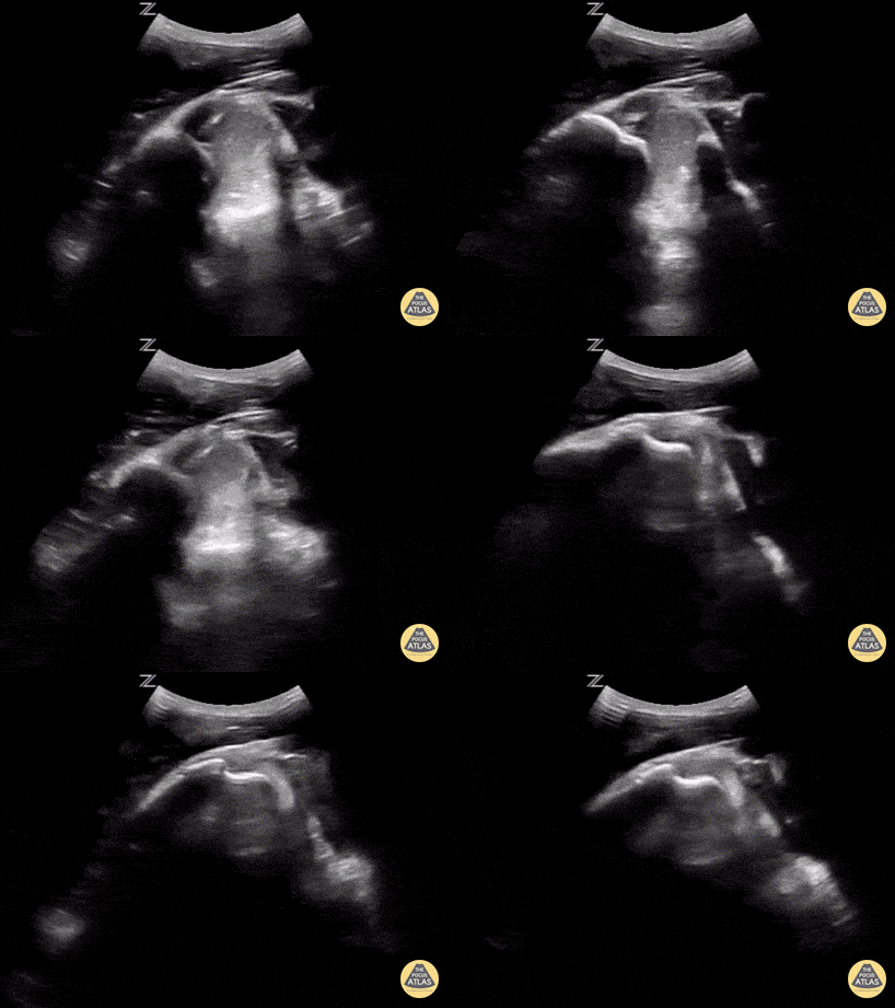
This is a posterior approach looking at the left shoulder showing an anterior dislocation. The dislocation was found before X-ray was shot. While the resident was performing the Cunningham technique to reduce the shoulder, the attending was able to, in real time, watch it go back in place. In between the humeral head and the glenoid rim is a large hematoma where I would aim my needle if I were to do an intra-articular block. It is a helpful technique in those recurrent dislocators. Saves the initial X-ray, but probably should get the post-reduction film at this point.
Matt Rutz, MD - Indiana University Department of Emergency Medicine, Ultrasound Division
For more evidence check out our friends at US G.E.L.
Perfect your Soft Tissue/MSK technique with 5 Minute Sono