DVT Evidence
POCUS Evidence Atlas - DVT, deep vein thrombosis, three point compressions, blood clot. EBM review and resource of POCUS for dvt deep vein thrombosis diagnosis. Sensitivity Specificity positive and negative likelihood ratios.
The Evidence Atlas: DVT
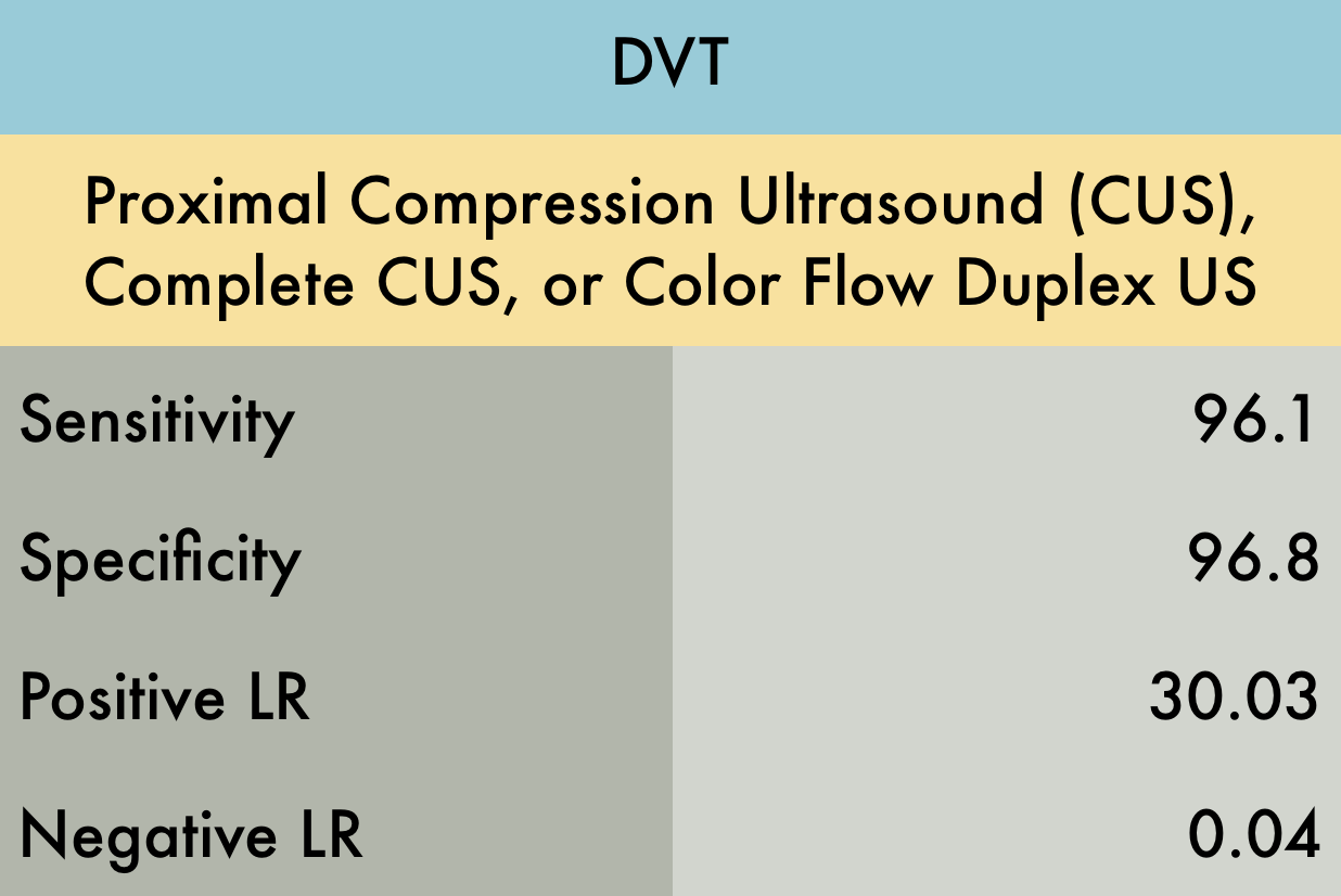
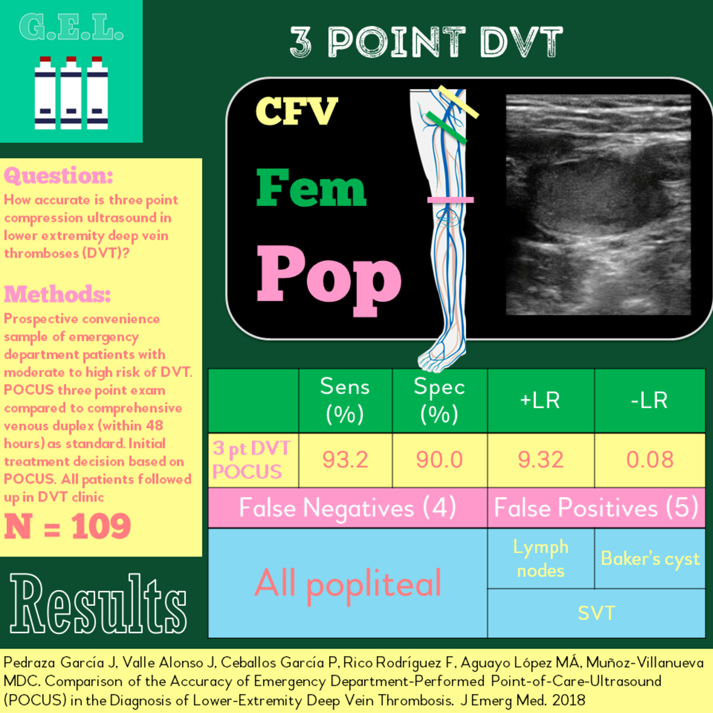
This was a meta-analysis including 16 studies (n=2379) evaluating the operating characteristics of emergency physician (EP) performed ultrasound for the diagnosis of DVT. The providers used color-flow duplex ultrasound in two studies, proximal venous ultrasound in 13 studies (not looking at the calf), and whole-leg venous ultrasound in one study. Reference standard was radiology department ultrasound, vascular lab, or angiography. Considering only high quality studies which met QUADAS-2 Criteria (11 out of 16 initially selected studies), the sensitivity and specificity improved to 97.6% and 96.8% respectively.
PMID: 23138420
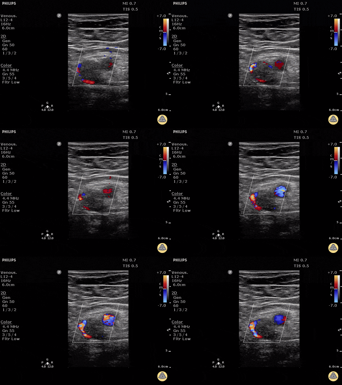
Patient with profound rapid onset lower extremity pain and swelling. POCUS can be used to rapidly assess for DVT with as high as 95% sensitivity and 96% specificity amongst ED providers.
In this image the artery can be seen as the pulsating thick walled vessel with strong pulsations under color doppler. The vein is the large vessel with echogenic material with flow only around the edge of the clot. Inability to compress the vessel would confirm the diagnosis of DVT (not pictured).
Dr. Justin Bowra et al.
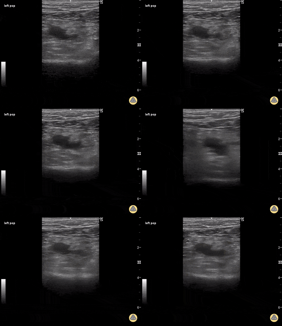
When pressure is applied to the ultrasound probe over blood vessels, veins usually collapse before arteries because of their much thinner walls. In this clip, enough pressure was applied to the ultrasound probe to partially collapse the popliteal artery, which is the pulsatile vessel visualized. Despite such high pressure, the popliteal vein remains fully patent, indicating a deep vein thrombosis within its walls.
Sukh Singh, MD
Patient with pain and swelling of the entire left lower extremity. Three point POCUS carries a sensitivity of 95% and specificity of 96% for detecting DVT when performed by emergency physicians.
Mild external rotation of the hip can help open up the inguinal crease to find the femoral vein. The POCUS should be performed 1-2cm distal to the bifurcation of the common femoral for a complete study.
Dr. Justin Bowra et al.
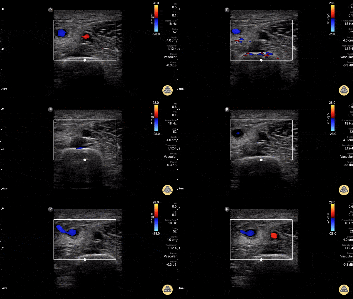
Axillary vein thrombus ruled OUT in a patient with arm swelling. Doppler used to highlight vein and artery.
Dr. Gordon Johnson
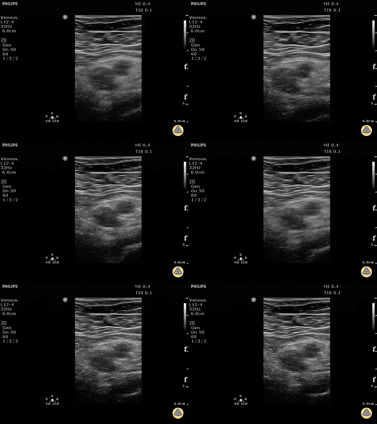
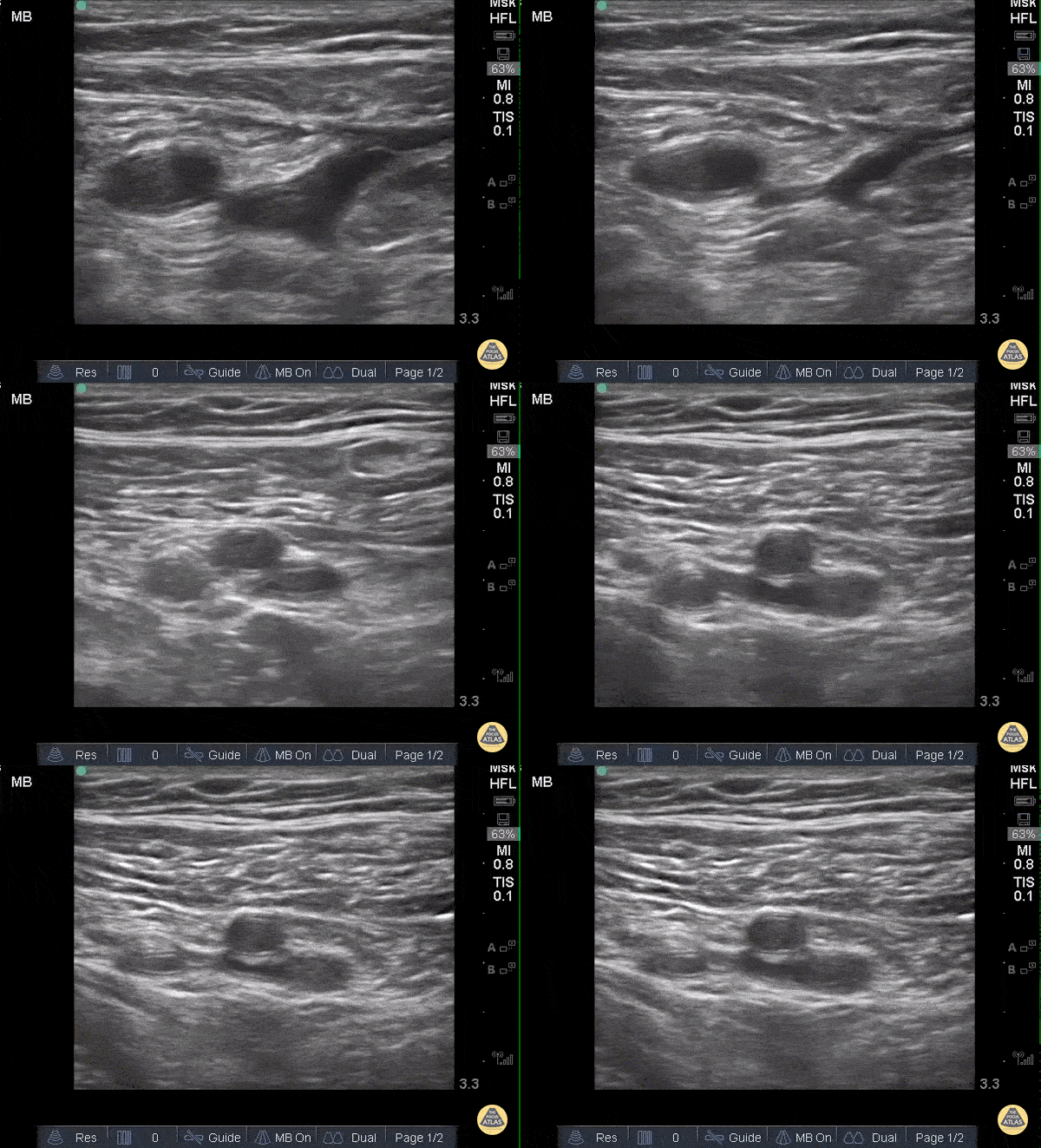
When applying pressure to the ultrasound probe, veins collapse first while arteries remain patent and pulsatile. In this clip one can see that proximally, the large pulsatile vessel (which represents the common femoral artery) sits adjacent to a mostly compressible common femoral vein. As the probe moves distally and both the artery and vein split into deep and superficial branches, the superficial femoral vein is not entirely compressible along its medial aspect, representing a thrombus.
Sukh Singh, MD